Interleukin-6 and Hypoxia Synergistically Promote EMT-Mediated Invasion in Epithelial Ovarian Cancer via the IL-6/STAT3/HIF-1 α Feedback Loop
- PMID: 36814597
- PMCID: PMC9940980
- DOI: 10.1155/2023/8334881
Interleukin-6 and Hypoxia Synergistically Promote EMT-Mediated Invasion in Epithelial Ovarian Cancer via the IL-6/STAT3/HIF-1 α Feedback Loop
Abstract
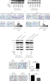
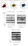
Extensive peritoneal spread and capacity for distant metastasis account for the majority of mortality from epithelial ovarian cancer (EOC). Accumulating evidence shows that interleukin-6 (IL-6) promotes tumor invasion and migration in EOC, although the molecular mechanisms remain to be fully elucidated. Meanwhile, the hypoxic microenvironment has been recognized to cause metastasis by triggering epithelial-mesenchymal transition (EMT) in several types of cancers. Here, we studied the synergy between IL-6 and hypoxia in inducing EMT in two EOC cell lines, A2780 cells and SKOV3 cells. Exogenous recombination of IL-6 and autocrine production of IL-6 regulated by plasmids both induced EMT phenotype in EOC cells characterized by downregulated E-cadherin as well as upregulated expression of vimentin and EMT-related transcription factors. The combined effects of IL-6 and hypoxia were more significant than those of either one treatment on EMT. Suppression of hypoxia-inducible factor-1α (HIF-1α) before IL-6 treatment inhibited the EMT phenotype and invasion ability of EOC cells, indicating that HIF-1α occupies a key position in the regulatory pathway of EMT associated with IL-6. EMT score was found positively correlated with mRNA levels of IL-6, signal transducer and activator of transcription 3 (STAT3), and HIF-1α, respectively, in 489 ovarian samples from The Cancer Genome Atlas dataset. Next, blockade of the abovementioned molecules by chemical inhibitors reversed the alteration in the protein levels of EMT markers induced by either exogenous or endogenous IL-6. These findings indicate a positive feedback loop between IL-6 and HIF-1α, and induce and maintain EMT phenotype through STAT3 signaling, which might provide a novel rationale for prognostic prediction and therapeutic targets in EOC.
Copyright © 2023 Tongshuo Zhang et al.
Conflict of interest statement
The author(s) declare(s) that they have no conflicts of interest.
Figures


Similar articles
-
IL-6 regulates epithelial ovarian cancer EMT, invasion, and metastasis by modulating Let-7c and miR-200c through the STAT3/HIF-1α pathway.Med Oncol. 2024 May 14;41(6):155. doi: 10.1007/s12032-024-02328-2. Med Oncol. 2024. PMID: 38744773
-
GRIM-19 repressed hypoxia-induced invasion and EMT of colorectal cancer by repressing autophagy through inactivation of STAT3/HIF-1α signaling axis.J Cell Physiol. 2019 Aug;234(8):12800-12808. doi: 10.1002/jcp.27914. Epub 2018 Dec 7. J Cell Physiol. 2019. PMID: 30537081
-
IL-6 promotes nuclear translocation of HIF-1α to aggravate chemoresistance of ovarian cancer cells.Eur J Pharmacol. 2021 Mar 5;894:173817. doi: 10.1016/j.ejphar.2020.173817. Epub 2020 Dec 18. Eur J Pharmacol. 2021. PMID: 33345849
-
Hypoxia-induced epithelial to mesenchymal transition in cancer.Cancer Lett. 2020 Sep 1;487:10-20. doi: 10.1016/j.canlet.2020.05.012. Epub 2020 May 26. Cancer Lett. 2020. PMID: 32470488 Free PMC article. Review.
-
The potential roles of HIF-1α in epithelial-mesenchymal transition and ferroptosis in tumor cells.Cell Signal. 2024 Oct;122:111345. doi: 10.1016/j.cellsig.2024.111345. Epub 2024 Aug 10. Cell Signal. 2024. PMID: 39134249 Review.
Cited by
-
Understanding human aging and the fundamental cell signaling link in age-related diseases: the middle-aging hypovascularity hypoxia hypothesis.Front Aging. 2023 Jun 13;4:1196648. doi: 10.3389/fragi.2023.1196648. eCollection 2023. Front Aging. 2023. PMID: 37384143 Free PMC article.
-
Targeting interleukin-6 as a treatment approach for peritoneal carcinomatosis.J Transl Med. 2024 Apr 30;22(1):402. doi: 10.1186/s12967-024-05205-8. J Transl Med. 2024. PMID: 38689325 Free PMC article. Review.
-
Interleukin-6 serves as a critical factor in various cancer progression and therapy.Med Oncol. 2024 Jun 20;41(7):182. doi: 10.1007/s12032-024-02422-5. Med Oncol. 2024. PMID: 38900329 Review.
-
Comparison of Interleukin-6 with Other Markers in Diagnosis of Ovarian Cancer.J Pers Med. 2023 Jun 11;13(6):980. doi: 10.3390/jpm13060980. J Pers Med. 2023. PMID: 37373969 Free PMC article.
-
Predict value of tumor markers combined with interleukins for therapeutic efficacy and prognosis in ovarian cancer patients.Am J Cancer Res. 2024 Oct 15;14(10):4868-4879. doi: 10.62347/GSRD2580. eCollection 2024. Am J Cancer Res. 2024. PMID: 39553206 Free PMC article.
References
MeSH terms
Substances
LinkOut - more resources
Full Text Sources
Medical
Miscellaneous

