. 2023 Feb 21;23(1):172.
doi: 10.1186/s12885-023-10647-2.

Dishevelled 2 regulates cancer cell proliferation and T cell mediated immunity in HER2-positive breast cancer

Affiliations

Dishevelled 2 regulates cancer cell proliferation and T cell mediated immunity in HER2-positive breast cancer

Fahmida Rasha et al. BMC Cancer. .

Abstract

Background: Dishevelled paralogs (DVL1, 2, 3) are key mediators of Wnt pathway playing a role in constitutive oncogenic signaling influencing the tumor microenvironment. While previous studies showed correlation of β-catenin with T cell gene expression, little is known about the role of DVL2 in modulating tumor immunity. This study aimed to uncover the novel interaction between DVL2 and HER2-positive (HER2+) breast cancer (BC) in regulating tumor immunity and disease progression.

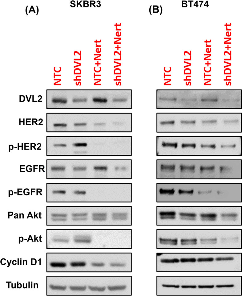
Methods: DVL2 loss of function studies were performed with or without a clinically approved HER2 inhibitor, Neratinib in two different HER2+ BC cell lines. We analyzed RNA (RT-qPCR) and protein (western blot) expression of classic Wnt markers and performed cell proliferation and cell cycle analyses by live cell imaging and flow cytometry, respectively. A pilot study in 24 HER2+ BC patients was performed to dissect the role of DVL2 in tumor immunity. Retrospective chart review on patient records and banked tissue histology were performed. Data were analyzed in SPSS (version 25) and GraphPad Prism (version 7) at a significance p < 0.05.

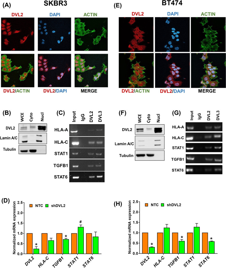
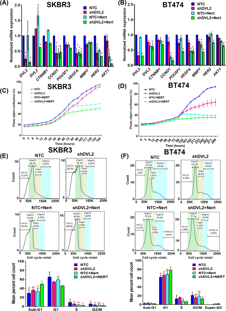
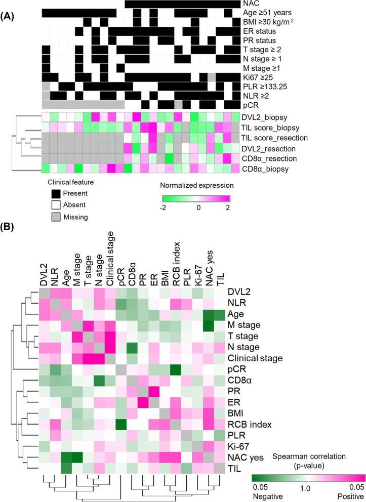
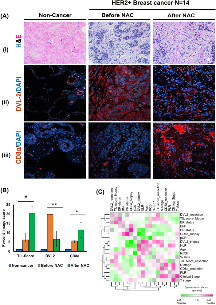
Results: DVL2 regulates the transcription of immune modulatory genes involved in antigen presentation and T cell maintenance. DVL2 loss of function down regulated mRNA expression of Wnt target genes involved in cell proliferation, migration, invasion in HER2+ BC cell lines (±Neratinib). Similarly, live cell proliferation and cell cycle analyses reveal that DVL2 knockdown (±Neratinib) resulted in reduced proliferation, higher growth arrest (G1), limited mitosis (G2/M) compared to non-targeted control in one of the two cell lines used. Analyses on patient tissues who received neoadjuvant chemotherapy (n = 14) further demonstrate that higher DVL2 expression at baseline biopsy pose a significant negative correlation with % CD8α levels (r = - 0.67, p < 0.05) while have a positive correlation with NLR (r = 0.58, p < 0.05), where high NLR denotes worse cancer prognosis. These results from our pilot study reveal interesting roles of DVL2 proteins in regulating tumor immune microenvironment and clinical predictors of survival in HER2+ BC.

Conclusion: Our study demonstrates potential immune regulatory role of DVL2 proteins in HER2+ BC. More in-depth mechanistic studies of DVL paralogs and their influence on anti-tumor immunity may provide insight into DVLs as potential therapeutic targets benefiting BC patients.

Keywords: Breast cancer; Dishevelled; HER2; Tumor infiltrating lymphocytes; Wnt signaling.


Conflict of interest statement

The authors declare no competing interests.

Figures

Fig. 1
Fig. 1
Higher Dishevelled (DVL) 2 expressions may predict lower survival in HER2-positive breast cancer patients. A UALCAN analysis for the comparison between DVL2 protein expressions in breast cancer using CPTAC samples in benign versus primary breast tumors. P-value significant codes: 0 ≤ *** < 0.001 ≤ ** < 0.01 ≤ * < 0.05 ≤ # < 0.1. B UALCAN analysis presenting respective DVL2 proteins expression in different subtypes of breast cancer (luminal, HER2-positive, and triple negative) using CPTAC samples. Z-values represent standard deviations from the median across samples for breast cancer sample/subclass types. Log2 Spectral count ratio values from CPTAC were first normalized within each sample profile, then normalized across samples. P-value significant codes: 0 ≤ *** < 0.001 ≤ ** < 0.01 ≤ * < 0.05 ≤ # < 0.1. C Immunofluorescent staining was performed to compare DVL2 protein expression in non-breast cancer (non-BC) versus different breast cancer subtypes (Luminal, HER2-positive, Triple negative) showing a merge of nuclear staining with DAPI (blue) and DVL2 (red) proteins. Images were taken from a small cohort of breast cancer patients (N = at least 5) in triplicates by a laser scanning confocal microscope Nikon T-1E with a 60x objective and NIS software and analyzed via AdipoGauge software (Windows version 2.0) [25] (N = 3, One-way Anova, different letters denote significance where p < 0.05). D Kaplan-Meier database was used to identify overall survival and distant metastasis-free survival comparing the high and low expressions of DVL2 (57532_at) in HER2-positive breast cancer (n = 276). CPTAC: Clinical Proteomic Tumor Analysis Consortium; N: normal; P: primary tumor; Lum: luminal; HER2: HER2-positive; TNBC: triple negative breast cancer
Fig. 2
Fig. 2
Nuclear DVL2 binds to promoter regions and regulates transcription of infiltrating T-cell genes in HER2+ breast cancer cell lines. A Immunofluorescence staining was performed to analyze DVL2 proteins localization in SKBR3 (ER−/PR−/HER2+) breast cancer cells. The cells were probed with DVL2 (red). The nucleus was stained with DAPI (blue) and the actin filaments (green) were stained with Phalloidin. B Whole (WCE), cytoplasmic (Cyto) and nuclear (Nucl) extracts from SKBR3 breast cancer cells were analyzed using western blots. The blots were probed with DVL2 antibody. Lamin was used as a control for nuclear extract and Tubulin was used as a control for cytosolic proteins. C ChIP PCR was performed for different immune-modulatory genes for Input, IgG, DVL2 and DVL3 in SKBR3 breast cancer cells. D RT-qPCR based mRNA expression analyses of DVL2, HLA-C, STAT1, TGFB1, STAT6 in SKBR3 cells stably expressing NTC vs. shDVL2. Transcript levels were normalized using GAPDH, B2M and GUSB as housekeeping control. All experiments were performed in triplicates and Welch’s independent T-test were performed between NTC and shDVL2 where *p < 0.05 and #p < 0.1. E Immunofluorescence staining was performed to analyze DVL2 proteins localization in BT474 (ER+/PR+/HER2+) breast cancer cells. The cells were probed with DVL2 (red). The nucleus was stained with DAPI (blue) and the actin filaments (green) were stained with Phalloidin. F Whole (WCE), cytoplasmic (Cyto) and nuclear (Nucl) from BT474 breast cancer cells were analyzed using western blots. The blots were probed with DVL2 antibody. Lamin was used as a control for nuclear extract and Tubulin was used as a control for cytosolic proteins. G ChIP PCR was performed for different immune-modulatory genes for Input, IgG, DVL2 and DVL3 in BT474 breast cancer cells. H RT-qPCR based mRNA expression analyses of DVL2, HLA-C, STAT1, TGFB1, STAT6 in BT474 cells stably expressing NTC vs. shDVL2. Transcript levels were normalized using GAPDH, B2M and GUSB as housekeeping control. All experiments were performed in triplicates and Welch’s independent T-test were performed between NTC and shDVL2 where *p < 0.05 and #p < 0.1
Fig. 3
Fig. 3
DVL2 loss of function reduces in vitro Wnt target genes expression, cell proliferation and cell cycle progression ± HER2 inhibition. A, B RT-qPCR based mRNA expression analyses of Wnt target genes (DVL2, DVL3, CTNNB1, CCND1, POU5F1, VEGFA, MMP7, ERBB2, AKT1) respectively in SKBR3 and BT474 cells stably expressing NTC vs. shDVL2 with or without 1 nM Neratinib (Nert) treatments. Transcript levels were normalized using GAPDH, B2M and GUSB as housekeeping control. All experiments were performed in triplicates and Welch’s independent T-test were performed between NTC vs. shDVL2 or NTC vs. NTC + Nert or NTC + Nert vs. shDVL2 + Nert to investigate the additive effect of HER2 inhibition with DVL2 loss of function (n = 3; *p < 0.05). C, D 2D proliferation assay was performed in SKBR3 and BT474 breast cancer cell lines via in vitro live cell image analyses. SKBR3 and BT474 cells stably expressing NTC vs. shDVL2 were seeded in a 96-well plate and upon confluence were treated with 1 nM Nert or DMSO vehicle. Cells were monitored and live cell percentages were quantified in real time via IncuCyte ZOOM (Essen Biosciences) for 144 and 280 h respectively for SKBR3 and BT474 cells. For each time points Mean ± SEM values were shown and considered statistically significant at *p < 0.05. E, F The effect of HER2 inhibition via ± 1 nM Neratinib (Nert) treatment respectively in SKBR3 and BT474 cells stably expressing NTC vs. shDVL2 was analyzed in a cell cycle assay using a Vybrant cell violet dye (Invitrogen) followed by flow cytometry. The live cells are represented in the SSC vs. FSC plots, which were further distributed among different cell cycle stages such as SubG1, G1, S, and G2/M phases. Experiments were performed in triplicates and mean values were shown in the graphs below. One-way Anova with Tukey’s multiple comparison test were performed where different letters denote statistical significance at p < 0.05
Fig. 4
Fig. 4
DVL2 depletion modulates Wnt and HER2 signaling cascades with or without in vitro HER2 inhibition. A, B Immunoblot analyses of lysates respectively from SKBR3 and BT474 cells stably expressing NTC vs. shDVL2 and treated with 1 nM neratinib (Nert) or DMSO control for 24 h and probed with antibodies as indicated with Tubulin as housekeeping control. Please also see Methods and Supplementary Table 1 for additional information
Fig. 5
Fig. 5
Mapping of DVL2 with clinical predictors of survival in a HER2-positive breast cancer patient cohort. A Heatmap showing expression patterns of DVL2, TIL-score and CD8α in association with clinical parameters of breast cancer at biopsy and after resection in 24 HER2+ breast cancer patients who received NAC (n = 14) vs. no NAC (n = 10). B Correlation between DVL2, CD8α and multiple clinical prognostic predictors (TIL, NLR, PLR, pCR, RCB index) and/or clinical demographics of breast cancer in 24 HER2+ breast cancer patients at baseline were evaluated using Spearman’s rank correlation test and graph was generated using Morpheus software (https://software.broadinstitute.org/morpheus). TIL: Tumor infiltrating lymphocytes; NLR: Neutrophil to lymphocyte ratio; PLR: Platelet to lymphocyte ratio; pCR: Pathologic complete response; RCBI: Residual cancer burden index
Fig. 6
Fig. 6
TIL scoring and phenotyping in association with DVL2 and CD8α levels in a HER2-positive breast cancer patient cohort before (biopsy) and after (resection) NAC. A Data shown are for benign tissue and breast cancer specimens before NAC (biopsy) and after NAC (resection); (i) Hemoxylin and Eosin staining for TIL score analysis, (ii) DVL2 (red) and DAPI (blue) immunofluorescent staining, and (iii) CD8α (red) and DAPI (blue) immunofluorescent staining. B Group mean percent image score with SD plotted against TIL-scoring and prevalence normalized fluorescence for each antibody marker before NAC (biopsy) and after NAC (resection) specimens (N = 14; **p < 0.001; *p < 0.05; #p < 0.1). C Correlation between DVL2, CD8α and multiple clinical prognostic predictors (TIL, NLR, PLR, pCR, RCB index) and/or clinical demographics of breast cancer in 14 HER2-positive breast cancer patients who received NAC. Normalized expression levels for each variable after biopsy were evaluated using Spearman’s rank correlation test and graph was generated using Morpheus software (https://software.broadinstitute.org/morpheus). TIL: Tumor infiltrating lymphocytes; NLR: Neutrophil to lymphocyte ratio; PLR: Platelet to lymphocyte ratio; pCR: Pathologic complete response; RCBI: Residual cancer burden index. UALCAN: The University of ALabama at Birmingham CANcer data analysis Portal

Similar articles

Cited by

References

    1. Ferlay J, Soerjomataram I, Dikshit R, Eser S, Mathers C, Rebelo M, Parkin DM, Forman D, Bray F. Cancer incidence and mortality worldwide: sources, methods and major patterns in GLOBOCAN 2012. Int J Cancer. 2015;136(5):E359–E386. doi: 10.1002/ijc.29210. - DOI - PubMed
    1. Schreiber RD, Old LJ, Smyth MJ. Cancer immunoediting: integrating immunity’s roles in cancer suppression and promotion. Science. 2011;331(6024):1565–1570. doi: 10.1126/science.1203486. - DOI - PubMed
    1. Adams S, Gray RJ, Demaria S, Goldstein L, Perez EA, Shulman LN, Martino S, Wang M, Jones VE, Saphner TJ, et al. Prognostic value of tumor-infiltrating lymphocytes in triple-negative breast cancers from two phase III randomized adjuvant breast cancer trials: ECOG 2197 and ECOG 1199. J Clin Oncol. 2014;32(27):2959–2966. doi: 10.1200/JCO.2013.55.0491. - DOI - PMC - PubMed
    1. Carbognin L, Pilotto S, Nortilli R, Brunelli M, Nottegar A, Sperduti I, Giannarelli D, Bria E, Tortora G. Predictive and prognostic role of tumor-infiltrating lymphocytes for early breast cancer according to disease subtypes: sensitivity analysis of randomized trials in adjuvant and neoadjuvant setting. Oncologist. 2016;21(3):283–291. doi: 10.1634/theoncologist.2015-0307. - DOI - PMC - PubMed
    1. Dieci MV, Mathieu MC, Guarneri V, Conte P, Delaloge S, Andre F, Goubar A. Prognostic and predictive value of tumor-infiltrating lymphocytes in two phase III randomized adjuvant breast cancer trials. Ann Oncol. 2015;26(8):1698–1704. doi: 10.1093/annonc/mdv239. - DOI - PMC - PubMed

Substances