. 2023 Jan 31:14:1063069.
doi: 10.3389/fimmu.2023.1063069. eCollection 2023.

The association between altered intestinal microbiome, impaired systemic and ocular surface immunity, and impaired wound healing response after corneal alkaline-chemical injury in diabetic mice

Affiliations

The association between altered intestinal microbiome, impaired systemic and ocular surface immunity, and impaired wound healing response after corneal alkaline-chemical injury in diabetic mice

Yashan Bu et al. Front Immunol. .

Abstract

Purpose: We aim to investigate the effect of sustained hyperglycemia on corneal epithelial wound healing, ocular surface and systemic immune response, and microbiome indices in diabetic mice compared to controls after alkaline chemical injury of the eye.

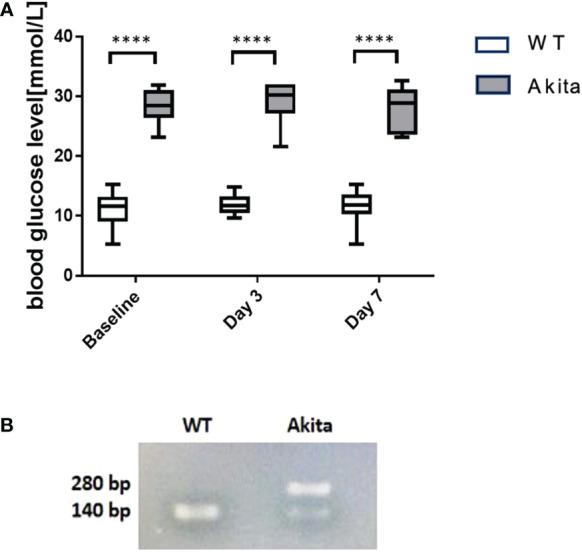
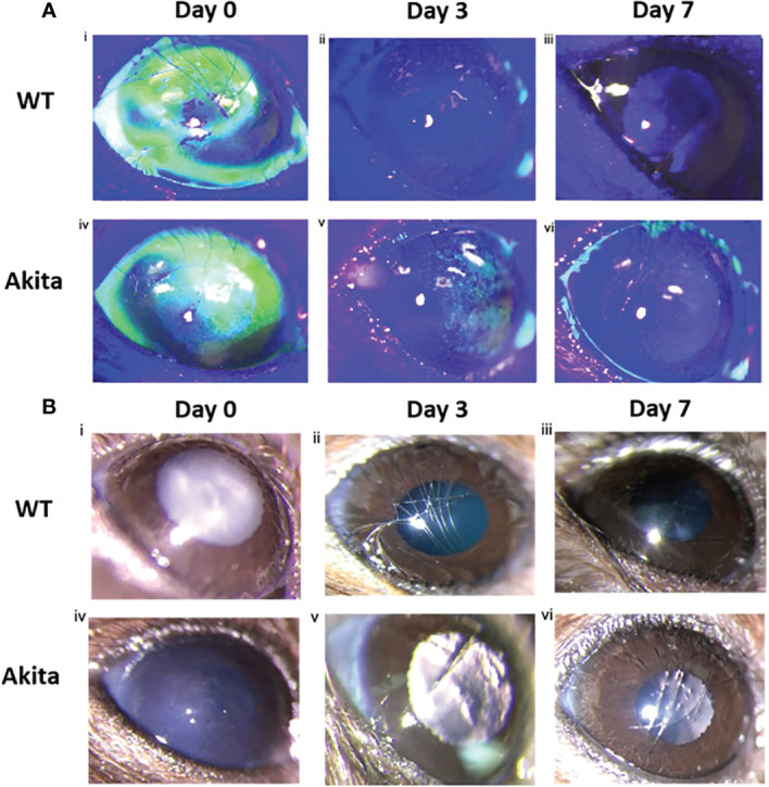
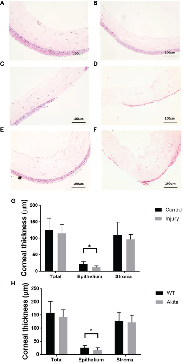
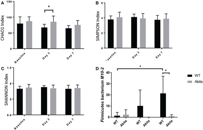
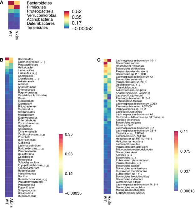
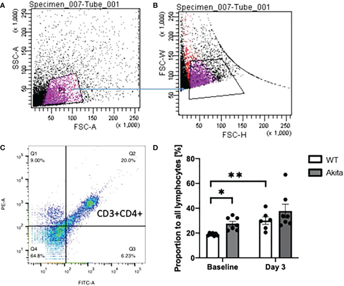
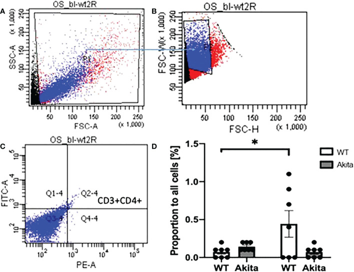
Methods: Corneal alkaline injury was induced in the right eye of Ins2Akita (Akita) mice and wild-type mice. The groups were observed at baseline and subsequently days 0, 3, and 7 after injury. Corneal re-epithelialization was observed under slit lamp with fluorescein staining using a cobalt blue light filter. Enucleated cornea specimens were compared at baseline and after injury for changes in cornea thickness under hematoxylin and eosin staining. Tear cytokine and growth factor levels were measured using protein microarray assay and compared between groups and time points. Flow cytometry was conducted on peripheral blood and ocular surface samples to determine CD3+CD4+ cell count. Fecal samples were collected, and gut microbiota composition and diversity pattern were measured using shotgun sequencing.

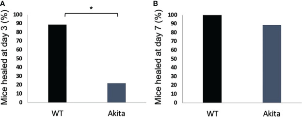
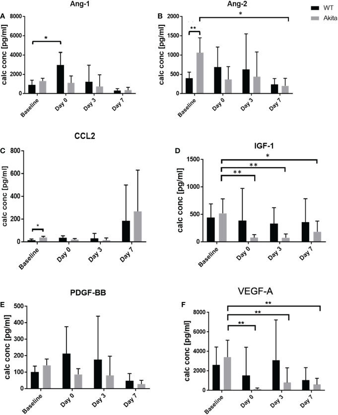
Results: Akita mice had significantly delayed corneal wound healing compared to controls. This was associated with a reduction in tear levels of vascular endothelial growth factor A, angiopoietin 2, and insulin growth factor 1 on days 0, 3, and 7 after injury. Furthermore, there was a distinct lack of upregulation of peripheral blood and ocular surface CD3+CD4+ cell counts in response to injury in Akita mice compared to controls. This was associated with a reduction in intestinal microbiome diversity indices in Akita mice compared to controls after injury. Specifically, there was a lower abundance of Firmicutes bacterium M10-2 in Akita mice compared to controls after injury.

Conclusion: In diabetic mice, impaired cornea wound healing was associated with an inability to mount systemic and local immune response to ocular chemical injury. Baseline and post-injury differences in intestinal microbial diversity and abundance patterns between diabetic mice and controls may potentially play a role in this altered response.

Keywords: T-cell mediated immunity; alkaline chemical injury; corneal wound healing; diabetes; intestinal microbiome; ocular surface.


Conflict of interest statement

The authors declare that the research was conducted in the absence of any commercial or financial relationships that could be construed as a potential conflict of interest.

Figures

Figure 1
Figure 1
Result of blood glucose level monitoring of WT and Akita mice. (A) Result of blood glucose level monitoring of WT and Akita mice. (A) Blood glucose measurement at baseline, day 3, and day 7. Unpaired t-test shows that Akita mice have significantly higher blood glucose level as compared to WT mice at baseline (before injury), day 3, and day 7 after injury. (B) Confirmation of WT and Akita mice by genotyping with PCR gel electrophoresis;, two DNA strands of WT mice are both able to be digested by enzyme Fnu4HI while heterozygous Akita mice have one mutated DNA strand that cannot be digested; thus, one band is present in the lane containing WT mice DNA sample whereas two bands present in that containing Akita mice DNA samples (n = 50 in WT, n = 50 in Akita). ****p<0.0001.
Figure 2
Figure 2
Wound healing analysis for WT and Akita mice with slit lamp images. (A) Injured cornea from WT and Akita mice at days 0, 3, and 7 after injury using 0.1 M NaOH and 10 s injury time, with fluorescein stain under cobalt blue light. Photos of the corneas taken immediately after injury showed significantly impaired corneal epithelium for both WT and Akita mice. (i, iv) The cornea from WT mouse was fully re-epithelized at day 3 after injury (ii); the cornea from Akita mouse was not fully re-epithelized at day 3 (v) and day 7 (vi) after injury. Magnification:40×. (B) Bright-field images of the right (injured) cornea from WT and Akita mice taken at day 0 (i, iv), day 3 (ii, v), and day 7 (iii, vi) after corneal alkaline injury (n = 55 in WT, n = 55 in Akita).
Figure 3
Figure 3
Percentage of diabetic and WT mice healed at day 3 after the corneal alkaline injury. (A) Pearson chi-squared test was performed (confidence level = 95% and p =0.0044, n = 9) and indicated that a significantly greater percentage of WT mice were healed than diabetic mice at day 3. (B) Pearson chi-squared test was performed and indicated no significant difference between the percentage of WT mice and diabetic mice healed at day 7 (p > 0.9999, n = 9 for both the WT and Akita groups). *p<0.05.
Figure 4
Figure 4
H&E staining of the cornea cross section and thickness measurements. Representative images of corneas were selected from (A) a WT mouse at baseline, (B) an Akita mouse at baseline, (C) a WT mouse immediately after corneal alkaline injury, (D) an Akita mouse immediately after corneal alkaline injury, (E) a WT mouse immediately at day 3 after corneal alkaline injury, and (F) an Akita mouse immediately at day 3 after corneal alkaline injury. (G) Corneal thickness measurement for total cornea, corneal epithelium, and corneal stroma for mouse corneas without injury (control group) and those immediately after injury; significantly reduced cornea thicknesses were found in the injured group compared with controls (n = 7, p = 0.0129). (H) Corneal thickness measurement for total cornea, corneal epithelium, and corneal stroma for corneas from WT mice and Akita mice at day 3 after injury. Significantly reduced cornea thickness was found for Akita mice at day 3 after injury compared to the WT group (n = 7 for both the WT and Akita groups, p = 0.0498). *p<0.05.
Figure 5
Figure 5
Comparison of tear protein concentration of the right (injured) eye from WT and Akita mice measured at baseline (before injury) with day 0, day 3, and day 7 after corneal alkaline injury. (A) Tear level of Ang-1 showed significantly elevated level immediately after injury WT mice (n = 6, p = 0.0412). (B) Tear level of Ang-2 showed significantly higher tear level at baseline and has significantly decreased at day 7 after injury for Akita mice (n = 6, p = 0.0243). (C) Tear level of CCL2 showed significantly elevated level in Akita mice at baseline. (D) Tear level of IGF-1 has significantly decreased immediately after injury (n = 8, p = 0.0018), and on post-injury day 3 (n = 8, p = 0.0026) and day 7 (n = 8, p = 0.0147) in Akita mice. (E) Tear level of PDGF-BB showed a trend of decrease from baseline level to the post-injury period in tear levels of Akita mice; however, there was no statistical significance. (F) Tear level of VEGF-A for Akita mice has significantly decreased immediately after injury (n = 8, p = 0.0016), on day 3 (n = 8, p = 0.0063), and on post-injury day 7 (n = 8, p = 0.0032) compared to baseline level (n number means the number of eyes/mice from both the WT and Akita groups, same for all measurements). *p<0.05; **p<0.01.
Figure 6
Figure 6
Comparison of intestinal microbiome diversity pattern of WT and Akita mice measured at baseline (before injury) with day 0, day 3, and day 7 aftercorneal alkaline injury. (A) Comparison of alpha diversity index at baseline, day 3, and day 7 after injury. Akita mice were found to have higher abundance ofmicrobiota composition as compared to WT mice from the CHAO1 index at day 3 after injury (n = 7, p = 0.0376). (B, C) Comparison of alpha diversity index at baseline, day 3, and day 7 after injury by the SIMPSON index (B) and SHANNON index (C) at day 3 after injury. No significance difference was found at baseline, day 3, or day 7 after injury after one-way ANOVA test (n = 7). (D) Firmicutes bacterium M10-2 had higher abundance in WT mice as compared to diabetic mice at day 7 after cornea alkaline injury (n = 5, p = 0.0164); there was a significant increase of Firmicutes bacterium M10-2 at day 7 after injury as compared to baseline level for WT mice (n = 5, p = 0.0215) (n number means the number of mice from both the WT and Akita groups, same for all measurements). *p<0.05.
Figure 7
Figure 7
Heat maps for relative abundance of bacteria present in the intestinal microbiome of WT and Akita mice at the phylum, species, and genus level. (A) Heat map of bacteria present in intestinal microbiome of WT and Akita mice generated at the phylum level based on mean relative abundance. (B) Heat map of bacteria present in the intestinal microbiome of WT and Akita mice generated at the genus level based on mean relative abundance. (C) Heat map of bacteria present in the intestinal microbiome of WT and Akita mice generated at the species level based on mean relative abundance. Values of relative abundance are presented with corresponding colors as shown in the legend for B to D (n = 5 in WT and Akita).
Figure 8
Figure 8
Proportion of CD3+CD4+ T cells in lymphocytes in peripheral blood samples of WT mice and Akita mice at baseline and at day 3 after corneal alkaline injury (A) Gating of lymphocytes (P1). (B) Gating of single cells against double or multiple cells (P2). (C) Gating of CD3+CD4+ T cells. (D) Akita mice had a higher abundance of CD3+CD4+ T cells in peripheral blood as compared to WT mice at baseline as indicated by an unpaired t-test (n = 7, p = 0.0146). WT mice had a significant increase of CD3+CD4+ cells in the peripheral blood at day 3 after the injury as indicated by a paired t-test (n = 7 for both the WT and Akita groups, p = 0.0022). *p<0.05; **p<0.01.
Figure 9
Figure 9
Proportion of CD3+CD4+ T cells in lymphocytes in ocular surface samples of WT and Akita mice at baseline and day 3 after corneal alkaline injury. (A) Gating of lymphocytes (P1). (B) Gating of single cells against double or multiple cells (P2). (C) Gating of CD3+CD4+ T cells (Q2–4). (D) Using a paired t-test, WT mice had a significant increase of CD3+CD4+ T cells on the ocular surface at day 3 after CABI as compared to baseline (n = 7, p = 0.0471), whereas no significant change was observed in CD3+CD4+ T cells on Akita mice ocular surface after injury (n = 7 for both the WT and Akita groups, p = 0.0941). *p<0.05.

Similar articles

Cited by

References

    1. Schultz RO, Van Horn DL, Peters MA, Klewin KM, Schutten WH. Diabetic keratopathy. Trans Am Ophthalmol Soc (1981) 79:180–99. - PMC - PubMed
    1. Vieira-Potter VJ, Karamichos D, Lee DJ. Ocular complications of diabetes and therapeutic approaches. BioMed Res Int (2016) 2016:3801570. doi: 10.1155/2016/3801570 - DOI - PMC - PubMed
    1. Shih KC, Lam KS, Tong L. A systematic review on the impact of diabetes mellitus on the ocular surface. Nutr Diabetes (2017) 7(3):e251. doi: 10.1038/nutd.2017.4 - DOI - PMC - PubMed
    1. Ljubimov AV. Diabetic complications in the cornea. Vision Res (2017) 139:138–52. doi: 10.1016/j.visres.2017.03.002 - DOI - PMC - PubMed
    1. Yang L, Di G, Qi X, Qu M, Wang Y, Duan H, et al. . Substance p promotes diabetic corneal epithelial wound healing through molecular mechanisms mediated via the neurokinin-1 receptor. Diabetes (2014) 63(12):4262–74. doi: 10.2337/db14-0163 - DOI - PubMed

Substances