. 2023 Feb 14;13(1):2595.
doi: 10.1038/s41598-023-28857-w.

LAMP3 transfer via extracellular particles induces apoptosis in Sjögren's disease

Affiliations

LAMP3 transfer via extracellular particles induces apoptosis in Sjögren's disease

Tsutomu Tanaka et al. Sci Rep. .

Abstract

Sjögren's disease (SjD) is an autoimmune disease that affects exocrine tissues and is characterized by increased apoptosis in salivary and lacrimal glands. Although the pathogenic mechanism triggering SjD is not well understood, overexpression of lysosome-associated membrane protein 3 (LAMP3) is associated with the disease in a subset of SjD patients and the development of SjD-like phenotype in mice. In this study, histological analysis of minor salivary glands of SjD patients suggested that LAMP3-containing material is being ejected from cells. Follow-on in vitro experiments with cells exposed to extracellular particles (EPs) derived from LAMP3-overexpressing cells showed increased apoptosis. Proteomics identified LAMP3 as a major component of EPs derived from LAMP3-overexpressing cells. Live-cell imaging visualized release and uptake of LAMP3-containing EPs from LAMP3-overexpressing cells to naïve cells. Furthermore, experiments with recombinant LAMP3 protein alone or complexed with Xfect protein transfection reagent demonstrated that internalization of LAMP3 was required for apoptosis in a caspase-dependent pathway. Taken together, we identified a new role for extracellular LAMP3 in cell-to-cell communication via EPs, which provides further support for targeting LAMP3 as a therapeutic approach in SjD.


Conflict of interest statement

The authors declare no competing interests.

Figures

Figure 1
Figure 1
LAMP3-positive material is released in minor salivary glands from SjD patients. A representative immunofluorescent image of minor salivary glands from SjD patients (n = 11) stained with anti-LAMP3 antibody (scale bar: 50 μm). The image shows accumulation of LAMP3 in the lumen of the gland (the arrows) and in the epithelial cells (the arrowheads).
Figure 2
Figure 2
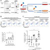
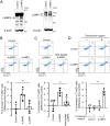
Culture medium of LAMP3-overexpressing A253 cells induce apoptosis in naïve A253 cells. A253 cells were transfected with control empty plasmid, LAMP3-encoding plasmid or LAMP1-encoding plasmid. (A) Representative Western blots of transfected A253 cells (uncropped images are provided in Supplementary Fig. 3) and schematic methods of the following in vitro assays. (B) Naïve A253 cells were treated with culture medium of control or LAMP3-overexpressing A253 cells for 48 h (n = 3). (C) Naïve A253 cells were treated with culture medium of control, LAMP1-overexpressing, or LAMP3-overexpressing A253 cells or with post-ultracentrifugation supernatant or post-ultracentrifugation pellet from LAMP3-overexpressing A253 cells for 48 h (n = 4). Apoptotic cells in naïve cell culture were determined by flow cytometry using APC Annexin V/7-AAD. Graphs show difference in mean (± SD) number of Annexin V+/7-AAD+ cells in cell culture compared with control. **p < 0.01 (one-way ANOVA).
Figure 3
Figure 3
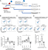
A fraction of extracellular particles from LAMP3-overexpressing A253 cells induces apoptosis in naïve A253 cells. (A) Schematic methods. Two fractions of extracellular particles (EPs) were separated from culture medium of control, LAMP3-overexpressing, or LAMP1-overexpressing A253 cells using centrifugation in combination with or without total exosome isolation reagent. Naïve A253 cells were treated with (B) EP fraction 2, (C) EP fraction 1 or (D) heat-denatured (heat-treated) EP fraction 1. Apoptotic cells in naïve cell culture were determined by flow cytometry using APC Annexin V/7-AAD 72 h after incubation. Graphs show difference in mean (± SD) number of Annexin V+/7-AAD+ cells in cell culture compared with control (n = 3 for all the experiments). **p < 0.01 (one-way ANOVA).
Figure 4
Figure 4
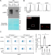
Extracellular particles from LAMP3-overexpressing cells contain LAMP3. (A) Western blot analysis of EPs separated from the culture medium of control, LAMP3-overexpressing, and LAMP1-overexpressing A253 cells using centrifugation in combination with or without total exosome isolation reagent. Equal volume of protein loaded in each lane. Lower panel shows same membrane as the one used in Western blot analysis but stained with Reversible Protein Stain Kit. Uncropped images are provided in Supplementary Fig. 3. (B) Cell surface LAMP3 expression was analyzed by flow cytometry (bold line: LAMP3 staining, filled area with dashed line: isotype control) and under an immunofluorescent microscopy (scale bar: 100 μm) in control, LAMP1-overexpressing, and LAMP3-overexpressing A253 cells 48 h post-transfection. (C) EPs derived from control and LAMP3-overexpressing A253 cells were immunoprecipitated using anti-LAMP3 monoclonal antibody-conjugated beads. Presence of LAMP3 protein on EP membrane surface was determined by analyzing beads stained with anti-LAMP3 polyclonal antibody using flow cytometry. Graph showing difference in mean (± SD) percentage of LAMP3-positive beads compared with beads bound to isotype control (n = 4). **p < 0.01 (one-way ANOVA).
Figure 5
Figure 5
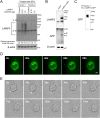
Extracellular particles from LAMP3-overexpressing transfer LAMP3 to naïve cells. (A) Western blot analysis of naïve A253 cells treated with EPs from control or LAMP3-overexpressing A253 cells. “※” indicates non-specific bands. (B) Western blot analysis of LAMP3-GFP expression in A253 cells transfected with empty plasmid (control) or pLenti-LAMP3-mGFP plasmid (A253-LAMP3-mGFP cells). (C) Western blot analysis of EPs derived from A253-LAMP3-mGFP cells. (D) A253-LAMP3-mGFP cells were imaged in real time to visualize EP release. A particle containing LAMP3-mGFP is seen being released from the cell into the extracellular environment (scale bar: 5 μm). (E) Naïve A253 cells were treated with LAMP3-mGFP–containing particle and imaged in real time to visualize particle uptake. Uptake of a LAMP3-mGFP–containing particle by a naïve cell and subsequent membrane blebbing are shown (scale bar: 10 μm). Uncropped images of Western blots are provided in Supplementary Fig. 3.
Figure 6
Figure 6
Transfection of recombinant LAMP3 protein induces apoptosis in A253 cells. (A) Representative Western blots of A253 cells transfected with recombinant LAMP3 (rLAMP3), recombinant LAMP1 (rLAMP1), or control FLAG peptide (uncropped images are provided in Supplementary Fig. 3). (B) Apoptotic cells were determined in A253 cell culture by flow cytometry using APC Annexin V/7-AAD 14 h after transfection. Graph showing difference in mean (± SD) number of Annexin V+/7-AAD+ cells in A253 cell culture transfected with rLAMP3 or rLAMP1 compared with control. (C) A253 cells were transfected with rLAMP3, heat-treated rLAMP3, or FLAG peptide. Apoptotic cells were determined in A253 cell culture by flow cytometry using APC Annexin V/7-AAD 14 h after transfection. Graph showing difference in mean (± SD) number of Annexin V+/7-AAD+ cells in A253 cell culture transfected with rLAMP3 or heat-treated rLAMP3 compared with control. (D) A253 cells were treated with control FLAG peptide or rLAMP3 alone (without transfection reagent) or transfected with control FLAG peptide or rLAMP3 with transection reagent. After 14 h, number of apoptotic cells was determined by flow cytometry using APC Annexin V/7-AAD. Graph showing difference in mean (± SD) number of Annexin V + /7-AAD + cells in cell culture treated with rLAMP3 alone or transfected with FLAG peptide or rLAMP3 compared with FLAG peptide alone. **p < 0.01 (one-way ANOVA, n = 4 for all the experiments).
Figure 7
Figure 7
LAMP3 transfer via extracellular particles induces caspase-dependent apoptosis. (A) Naïve A253 cells were treated with EPs from control and LAMP3-overexpressing A253 cells in combination with or without zVAD-fmk (zVAD), a pan-caspase inhibitor. Apoptotic cells were determined by flow cytometry using APC Annexin V/7-AAD 72 h after incubation. Graph showing difference in mean (± SD) number of Annexin V+/7-AAD+ cells in naïve A253 cell culture compared with control (n = 5). (B) Naïve A253 cells were treated with EPs from control and LAMP3-overexpressing A253 cells for 6 h. Galectin-3 puncta formation in treated A253 cell culture was visualized by immunofluorescence (scale bar: 50 μm). Graph showing percentage of galectin-3 puncta–positive cells per 100 treated A253 cells (n = 3). **p < 0.01 (one-way ANOVA).
Figure 8
Figure 8
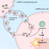
Graphical summary. Extracellular particles containing LAMP3 are released by LAMP3-overexpressing cells and taken up by a neighboring LAMP3-naïve cell. This results in LAMP3 protein transfer to the cytoplasm of the naïve cell. Here, LAMP3 induces lysosomal membrane permeabilization (LMP), leading to apoptotic cell death.

Similar articles

References

    1. Odani T, Chiorini JA. Targeting primary Sjögren's syndrome. Mod. Rheumatol. 2019;29(1):70–86. doi: 10.1080/14397595.2018.1546268. - DOI - PubMed
    1. Manganelli P, Fietta P. Apoptosis and Sjögren syndrome. Semin. Arthritis Rheum. 2003;33(1):49–65. doi: 10.1053/sarh.2003.50019. - DOI - PubMed
    1. Baban B, et al. Reciprocal relation between GADD153 and Del-1 in regulation of salivary gland inflammation in Sjögren syndrome. Exp. Mol. Pathol. 2013;95(3):288–297. doi: 10.1016/j.yexmp.2013.09.002. - DOI - PubMed
    1. Tanaka T, et al. LAMP3 induces apoptosis and autoantigen release in Sjögren's syndrome patients. Sci. Rep. 2020;10(1):15169. doi: 10.1038/s41598-020-71669-5. - DOI - PMC - PubMed
    1. Salaun B, et al. CD208/dendritic cell-lysosomal associated membrane protein is a marker of normal and transformed type II pneumocytes. Am. J. Pathol. 2004;164(3):861–871. doi: 10.1016/S0002-9440(10)63174-4. - DOI - PMC - PubMed

Publication types

Substances