Review
. 2023 Jan 28;28(3):1283.
doi: 10.3390/molecules28031283.

Role of Nanomedicine-Based Therapeutics in the Treatment of CNS Disorders

Affiliations
Review

Role of Nanomedicine-Based Therapeutics in the Treatment of CNS Disorders

Zi-Hua Guo et al. Molecules. .

Abstract

Central nervous system disorders, especially neurodegenerative diseases, are a public health priority and demand a strong scientific response. Various therapy procedures have been used in the past, but their therapeutic value has been insufficient. The blood-brain barrier (BBB) and the blood-cerebrospinal fluid barrier is two of the barriers that protect the central nervous system (CNS), but are the main barriers to medicine delivery into the CNS for treating CNS disorders, such as brain tumors, Parkinson's disease, Alzheimer's disease, and Huntington's disease. Nanotechnology-based medicinal approaches deliver valuable cargos targeting molecular and cellular processes with greater safety, efficacy, and specificity than traditional approaches. CNS diseases include a wide range of brain ailments connected to short- and long-term disability. They affect millions of people worldwide and are anticipated to become more common in the coming years. Nanotechnology-based brain therapy could solve the BBB problem. This review analyzes nanomedicine's role in medication delivery; immunotherapy, chemotherapy, and gene therapy are combined with nanomedicines to treat CNS disorders. We also evaluated nanotechnology-based approaches for CNS disease amelioration, with the intention of stimulating the immune system by delivering medications across the BBB.

Keywords: blood–brain barrier; central nervous system disorders; immunotherapy; nanomedicine; nanotechnology.


Conflict of interest statement

The authors declare no conflict of interest.

Figures

Figure 1
Figure 1
The cervical lymph nodes through the CNS’s lymphatic and lymphatic drainage pathways. (A) The meningeal lymphatic vascular system is depicted schematically in the mouse brain. Along with the venous sinuses, arteries, and cranial nerves, the dural lymphatic vessels leave the cranium through the foramina in alignment with the dural blood vessels and cranial nerves. Along with the olfactory nerves, certain lymphatic veins may be seen crisscrossing the cribriform plate. Tracers injected into the SAS or brain parenchyma drain into the dcLNs adjacent to the jugular vein through the dural lymphatic arteries. (B) ISF and CSF circulation up close. CSF and solutes are transported into the brain via the perivascular glymphatic drainage system through the periarterial pathway, whereas ISF and solutes are transported out of the brain by the perivenous glymphatic pathway. CSF macromolecules and immune cells are mostly carried by the dural lymphatic channels into the lymph nodes and extracranial systemic circulation, and CSF can reach the venous system via arachnoid granulations. (C) Routes for antigens and antigen-presenting cells to exit the central nervous system (APCs). Dendritic cells, in particular, may migrate along the rostral migratory stream (RMS) to enter the lymphatics via the olfactory bulb’s SAS. Alternatively, antigens and APCs are proposed to leave the CNS via the glymphatic pathway (as demonstrated for antigens), reaching the SAS and entering the meningeal lymphatic vasculature via SAS and trafficking to the dcLNs. APCs in the meningeal spaces may also go to the dcLNs through meningeal lymphatic channels. It is still unknown how much each mechanism contributes to cell and antigen outflow. Reproduced with permission from [47].
Figure 2
Figure 2
BBB’s structure and composition. The brain endothelium, astrocytes, extracellular matrix, and endothelial cells form tight junctions, which make up the majority of the BBB. Image reproduced with permission from [91].
Figure 3
Figure 3
An overview of the immunotherapeutic techniques currently under investigation for the treatment of GBM. (a) CAR T cells identify antigens via a genetically designed extracellular receptor that, following antigen binding, induces intracellular T cell activation and degranulation. (b) Immunological checkpoint protein inhibitors limit the dampening of immune responses during activation and exhaustion. (c) Vaccines expose antigen-presenting cells to tumor antigens, inducing an immune response specific to the target antigens. Therapeutic targets or mediators being pursued for each modality are denoted in the boxes. CAR: chimeric antigen receptor; CTL: cytotoxic T cell. Reproduced with permission from [191].
Figure 4
Figure 4
Immune-modulating effect of a PCB-based zwitterionic nanoparticle for AD therapy. (A) MCPZFS NPs for AD therapy: structure, preparation, and mechanism. MCPZFS NPs can penetrate the BBB and endocytose into microglia cells to normalized malfunctioning microglia. Pro-inflammatory mediators were reduced. Microglia’s phagocytic capacity was restored when BDNF production rose. The injured neuron is then healed in numerous ways. (1) NPs endocytosed amyloid-β into microglia. (2) After perturbing the endosome/lysosome membrane, NPs leaked into the cytoplasm. (4) Finally, ROS-mediated release of fingolimod, siSTAT3, ZnO, and amyloid-β. (B) The effect of NPs on microglial phagocytosis and Aβ degradation after 2 h co-incubation with FITC-A42 and NPs. Flow cytometry was utilized to detect the degradation of A42 of BV2 in BafA1 or MG132 (top right) and the phagocytosis and degradation behavior mediated by MEPZFS NPs and MCPZFS NPs. (C) Representing the effect of NPs on the inflammatory regulation of microglia via the ELISA method was applied to determine the levels of TNF-α, IL-1β, and BDNF in the supernatants. Samples: (I) PBS, II) Aβ42, (III) Aβ42 + fingolimod, (IV) Aβ42 + siSTAT3, (V) Aβ42 + CPFS, (VI) Aβ42 + CPZS, (VII) Aβ42 + CPZF-siNC, (VIII) Aβ42 + CPZFS, (IX) Aβ42 + MCPZFS, and (X) Aβ42 + MEPZFS. Copyright, 2019, Wiley and Sons IncReproduced with permission from [265]. * p < 0.05, ** p < 0.01.
Figure 5
Figure 5
Illustration of glycosylated “triple-interaction” stabilized siRNA nanomedicine (Gal-NP@siRNA) and strategy of treating AD pathology in APP/PS1 transgenic mice. (A) Schematic of Gal NP@siRNA manufacturing. (B,C) How Gal-NP@siRNA enters the brain and accumulates. 24 h fasting increases BBB luminal Glut1 expression. After treatment with Gal-NP@siRNA, glucose replenishment in fasting mice leads to Glut1 recycling from the BBB luminal to the abluminal membrane. (D) Gal-NP@siRNA-mediated BACE1 mRNA knockdown reduces amyloid plaques. Adapted with permission from [266].
Figure 6
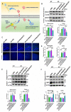
Figure 6
Gal-NP@siBACE1 treatment modulates AD hallmarks in APP/PS1 mice. (A) A mechanism for siBACE1′s actions. (B) BACE1 protein expression in the hippocampus and cortex of nanocarrier-treated APP/PS1 mice, control APP/PS1 groups, and WT mice. BACE1 expression was quantified relative to actin (n = 3, mean ± SEM, * p < 0.05, ** p < 0.01). CLSM imaging data to assess amyloid plaque load. A plaque (green) in APP/PS1 transgenic and WT mice hippocampus and cortex. DAPI staining nuclei (blue), 100 µm scale bars. (C) Representative confocal laser scanning microscopy (CLSM) imaging data are assessing the amyloid plaque burden. Immunofluorescence of Aβ plaques (green) in the hippocampus and cortex from APP/PS1 transgenic and WT mice. Nuclei were stained by DAPI (blue). Scale bars, 100 µm. (D) Amyloid plaques were measured in the hippocampus (left) and cortex [181] (n = 4, ** p < 0.01; mean ± SEM). (E) p-tau and (F) MBP expression in the hippocampus and cortex for nanocarrier-treated APP/PS1 mice, control APP/PS1 groups, and WT mice (top). Quantification of Western blotting analysis was relative to β-actin (bottom) (n = 3, mean with SEM, * p < 0.05, ** p < 0.01). All samples were collected after 10 administrations of nanomedicine. Adapted with permission from [266].

Similar articles

Cited by

References

    1. Lammers T., Aime S., Hennink W.E., Storm G., Kiessling F. Theranostic nanomedicine. Acc. Chem. Res. 2011;44:1029–1038. doi: 10.1021/ar200019c. - DOI - PubMed
    1. Zheng M., Tao W., Zou Y., Farokhzad O.C., Shi B. Nanotechnology-based strategies for siRNA brain delivery for disease therapy. Trends Biotechnol. 2018;36:562–575. doi: 10.1016/j.tibtech.2018.01.006. - DOI - PubMed
    1. Tayyba T. Role of extracellular vesicles in human diseases. Biomed. Lett. 2019;5:67–68.
    1. Kim B.Y., Rutka J.T., Chan W.C. Nanomedicine. N. Engl. J. Med. 2010;363:2434–2443. doi: 10.1056/NEJMra0912273. - DOI - PubMed
    1. Song S., Gao P., Sun L., Kang D., Kongsted J., Poongavanam V., Zhan P., Liu X. Recent developments in the medicinal chemistry of single boron atom-containing compounds. Acta Pharm. Sin. B. 2021;11:3035–3059. doi: 10.1016/j.apsb.2021.01.010. - DOI - PMC - PubMed

LinkOut - more resources