. 2023 Jan 11:13:1081094.
doi: 10.3389/fmicb.2022.1081094. eCollection 2022.

Structural insights into catalytical capability for CPT11 hydrolysis and substrate specificity of a novel marine microbial carboxylesterase, E93

Affiliations

Structural insights into catalytical capability for CPT11 hydrolysis and substrate specificity of a novel marine microbial carboxylesterase, E93

Yang Li et al. Front Microbiol. .

Abstract

Introduction: CPT11 (Irinotecan; 7-ethyl-10-[4-(1-piperidino)-1-piperidino] carbonyloxycamptothecin) is an important camptothecin-based broad-spectrum anticancer prodrug. The activation of its warhead, SN38 (7-ethyl-10-hydroxycamptothecin), requires hydrolysis by carboxylesterases. NPC (7-ethyl-10-[4-(1-piperidino)-1-amino] carbonyloxycamptothecin) is a metabolic derivative of CPT11 and is difficult to be hydrolyzed by human carboxylesterase. Microbial carboxylesterase with capability on both CPT11 and NPC hydrolysis is rarely reported. A marine microbial carboxylesterase, E93, was identified to hydrolyze both substrates in this study. This enzyme was an appropriate subject for uncovering the catalytic mechanism of carboxylesterases to CPT11 and NPC hydrolysis.

Methods: X-ray diffraction method was applied to obtain high-resolution structure of E93. Molecular docking was adopted to analyze the interaction of E93 with p-NP (p-nitrophenyl), CPT11, and NPC substrates. Mutagenesis and enzymatic assay were adopted to verify the binding pattern of substrates.

Results: Three core regions (Region A, B, and C) of the catalytic pocket were identified and their functions on substrates specificity were validated via mutagenesis assays. The Region A was involved in the binding with the alcohol group of all tested substrates. The size and hydrophobicity of the region determined the binding affinity. The Region B accommodated the acyl group of p-NP and CPT11 substrates. The polarity of this region determined the catalytic preference to both substrates. The Region C specifically accommodated the acyl group of NPC. The interaction from the acidic residue, E428, contributed to the binding of E93 with NPC.

Discussion: The study analyzed both unique and conserved structures of the pocket in E93, for the first time demonstrating the discrepancy of substrate-enzyme interaction between CPT11 and NPC. It also expanded the knowledge about the substrate specificity and potential application of microbial Family VII carboxylesterases.

Keywords: crystal structure; enzyme catalysis; marine bacterial carboxylesterase; p-nitrophenyl; prodrug; substrate specificity.


Conflict of interest statement

The authors declare that the research was conducted in the absence of any commercial or financial relationships that could be construed as a potential conflict of interest.

Figures

Figure 1
Figure 1
Structure and relationship demonstration of CPT11, NPC and SN38. CPT11 is oxidized by cytochrome P450 3A4 (CYP3A4) isoenzyme to produce 7-ethyl-10-[4-(1-piperidino)-1-amino]-carbonyloxycamptothecin (NPC). CPT11 are metabolized by carboxylesterases (CES) to produce active metabolite, 7-ethyl-10-hydroxycamptothecin (SN38). The only structural difference between CPT11 and NPC is marked with the green box.
Figure 2
Figure 2
Catalytical features of E93 with p-NP esters as substrates. (A) Gel filtration chromatography and SDS-PAGE results of E93. High-purity protein samples were collected for crystallization (in the red box). (B) Enzymatic activities to substrates with various fatty acid chain of p-Nitrophenyl (p-NP) esters. The catalytic activity of E93 to p-NP hexanoate (C6) was 100%. (C) Effects of temperature on enzyme activities. Enzymatic activities were measured in a series of temperatures. The activity obtained at 45°C was taken as 100%. (D) Effects of different pH on enzyme activities. Enzymatic activities were determined under a series of pH. The activity obtained at pH 7.5 was taken as 100%. (E) Tolerance of ions. The reaction without additional ions was used as a control. The C6 substrate was used for measurements. (F) Effects of organic solvents on the enzymatic activity. The reaction without any organic solvent was used as a control. The solvents with no inhibition effect on the enzyme activity were labeled with light green color in panels (E,F). The data showed in panels (B–F) is from triplicate experiments (mean ± SD). *p < 0.05, **p < 0.01, ***p < 0.001, ****p < 0.0001, and ns, not significant.
Figure 3
Figure 3
Crystal structure of E93 and structure comparison with microbial family VII carboxylesterases. (A) Global structure of E93. The structure contains three domains, including the catalytical domain (yellow), α/β domain (blue), and regulatory domain (green). (B) Conformational comparison of the catalytic triad of E93 (S189, E314, and H414; yellow) with that of pnbCE (S189, E310, H399; cyan). (C) Crystal structure comparison of E93 (yellow, PDB code: 7X8L) with pnbCE (cyan, PDB code: 1QE3). E93 and pnbCE are structurally homologous proteins. The RMSD value of E93 and pnbCE is 1.074 (Cα 328). The unique loop (271aa-277aa in E93 and 268aa-274aa in pnbCE) was shown in red box. (D) Structural topology of E93. The color scheme is consistent with that of Figure 2A.
Figure 4
Figure 4
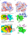
Comparison of the catalytic pockets of E93 and pnbCE. (A,B) The electrostatic surfaces of E93 (A) and pnbCE (B). Sticks show catalytic triad. The positive charged regions are shown in blue and the negative charged regions are shown in red. Regions A–C are specifically noted. (C) The detailed exhibition of catalytic pocket of E93 and the amino acid composition of each region. The magenta area indicates the catalytic triad. (D) Specific features of the Region B in E93. The amino acids that constitute Region B are shown as green sticks. The catalytic triad of E93 is precisely immerged in an electronic density map contoured to 1.0 σ at the 2Fo-Fc map closing to the Region B. (E) The detailed exhibition of catalytic pocket of pnbCE and the amino acid composition of each region. The magenta area indicates the catalytic triad. Amino acids that comprise different regions are shown. (F) Specific features of the Region B in pnbCE. The catalytic triad is shown with magenta stick. The amino acids that constitute Region B are shown with green sticks.
Figure 5
Figure 5
Binding patterns of E93 with different substrates in the Region A of catalytic pocket, simulated by AutoDock. (A–C) The docking of CPT11 (A), NPC (B), and p-NP hexanoate (C6) (C) into the catalytic pocket of E93. Magenta sticks represent the catalytic triad. The large-sized amino acids (Orange) involved in forming the tunnel structure of the Region A in E93 are shown with orange sticks. The interactions of E93_W107 and E93_Y318 with the alcoholic group of CPT11, NPC, and C6 are shown. The interaction between the catalytic residue S189 and the carbonyl carbon of substrate is also demonstrated. The black dash line denotes hydrogen bond, and the blue dash line denotes π–π interaction. The electronic density map is contoured to 1.0 σ at the 2Fo-Fc map. (D) Structural superposition of the large amino acids within Region A of E93 (yellow) and the corresponding amino acids of pnbCE (cyan). The Region A of E93 has a width of 7.2 Å. The Special loop structure with significant conformational difference is specifically labeled. The distance between catalytic S189 and M273 in the special loop is 4.9 Å. (E) The large-sized amino acids involved in forming the tunnel structure of the Region A in pnbCE are shown with orange sticks. The catalytic triad of pnbCE is highlighted by the magenta sticks. The Region A of pnbCE has a width of 8.3 Å. The distance between pnbCE_I270, corresponding to E93_M273, and the catalytic resideue, S189 in pnbCE, is 9.4 Å. The dash line in panels (D,E) was used to show the distance between two amino acids. (F,G) B-factor analysis of E93 (F) and pnbCE (G). The thickness of coil represents the flexibility of the structure. The thicker the coil is, the higher flexibility the structure is. The catalytic triad residues of E93 and pnbCE are shown as sticks. The loop with high flexibility in catalytic center of two enzymes is labeled with red box.
Figure 6
Figure 6
Details of the binding of CPT11 and C6 in Region B, and NPC in Region C. (A) Interaction of Q193 and T375 with the 4PP group of CPT11 in Region B. The dashed lines illustrate hydrogen bonds. (B) Interaction of E428 with the acyl groups of NPC within Region C. Dashed lines illustrate hydrogen bonds. (C) The relative locations of p-NP hexanoate (C6), Q193, and T375 in Region B. The dashed lines illustrate interaction. The electronic density map is contoured to 1.0 σ at the 2Fo-Fc map.
Figure 7
Figure 7
Mutagenesis analysis of key residues involved in hydrolyzing CPT11, NPC, and p-NP hexanoate (C6). Comparison of the hydrolytic activities of each mutant for CPT11 (A), NPC (B) and p-NP hexanoate (C6) (C). The activity of wild-type E93 was taken as 100% for panel (C). (D) Catalytic activity of wild-type E93, E93_T375A, and E93_T375I on p-NP ester substrate hydrolysis. The data showed in panels (A–D) is from triplicate experiments (mean ± SD). *p < 0.05, **p < 0.01, ***p < 0.001, ****p < 0.0001, and ns, not significant.

Similar articles

Cited by

References

    1. Adams P. D., Afonine P. V., Bunkoczi G., Chen V. B., Davis I. W., Echols N., et al. . (2010). PHENIX: a comprehensive Python-based system for macromolecular structure solution. Acta Crystallogr. D Biol. Crystallogr. 66, 213–221. doi: 10.1107/S0907444909052925, PMID: - DOI - PMC - PubMed
    1. Arpigny J., Jaeger K.-E. (1999). Bacterial lipolytic enzymes: classification and properties. Biochem. J. 343, 177–183. doi: 10.1042/0264-6021:3430177 - DOI - PMC - PubMed
    1. Barber R. D. (2021). Software to visualize proteins and perform structural alignments. Curr. Protoc. 1:e292. doi: 10.1002/cpz1.292, PMID: - DOI - PubMed
    1. Emsley P., Cowtan K. (2004). Coot: model-building tools for molecular graphics. Acta Crystallogr. D Biol. Crystallogr. 60, 2126–2132. doi: 10.1107/S0907444904019158, PMID: - DOI - PubMed
    1. Fukuchi M., Kuwabara K., Ishiguro T., Kumagai Y., Ishibashi K., Mochiki E., et al. . (2020). Efficacy of irinotecan as third-line chemotherapy for unresectable or recurrent gastric cancer. In Vivo 34, 903–908. doi: 10.21873/invivo.11856, PMID: - DOI - PMC - PubMed