. 2023 Apr;30(4):1005-1017.
doi: 10.1038/s41418-023-01117-0. Epub 2023 Feb 8.

Lymphoma cells lacking pro-apoptotic BAX are highly resistant to BH3-mimetics targeting pro-survival MCL-1 but retain sensitivity to conventional DNA-damaging drugs

Affiliations

Lymphoma cells lacking pro-apoptotic BAX are highly resistant to BH3-mimetics targeting pro-survival MCL-1 but retain sensitivity to conventional DNA-damaging drugs

Sarah T Diepstraten et al. Cell Death Differ. 2023 Apr.

Abstract

BH3-mimetic drugs are an anti-cancer therapy that can induce apoptosis in malignant cells by directly binding and inhibiting pro-survival proteins of the BCL-2 family. The BH3-mimetic drug venetoclax, which targets BCL-2, has been approved for the treatment of chronic lymphocytic leukaemia and acute myeloid leukaemia by regulatory authorities worldwide. However, while most patients initially respond well, resistance and relapse while on this drug is an emerging and critical issue in the clinic. Though some studies have begun uncovering the factors involved in resistance to BCL-2-targeting BH3-mimetic drugs, little focus has been applied to pre-emptively tackle resistance for the next generation of BH3-mimetic drugs targeting MCL-1, which are now in clinical trials for diverse blood cancers. Therefore, using pre-clinical mouse and human models of aggressive lymphoma, we sought to predict factors likely to contribute to the development of resistance in patients receiving MCL-1-targeting BH3-mimetic drugs. First, we performed multiple whole genome CRISPR/Cas9 KO screens and identified that loss of the pro-apoptotic effector protein BAX, but not its close relative BAK, could confer resistance to MCL-1-targeting BH3-mimetic drugs in both short-term and long-term treatment regimens, even in lymphoma cells lacking the tumour suppressor TRP53. Furthermore, we found that mouse Eµ-Myc lymphoma cells selected for loss of BAX, as well as upregulation of the untargeted pro-survival BCL-2 family proteins BCL-XL and A1, when made naturally resistant to MCL-1 inhibitors by culturing them in increasing doses of drug over time, a situation mimicking the clinical application of these drugs. Finally, we identified therapeutic approaches which could overcome these two methods of resistance: the use of chemotherapeutic drugs or combined BH3-mimetic treatment, respectively. Collectively, these results uncover some key factors likely to cause resistance to MCL-1 inhibition in the clinic and suggest rational therapeutic strategies to overcome resistance that should be investigated further.


Conflict of interest statement

All authors are employees of WEHI which receives milestone and royalty payments related to venetoclax. AS and GLK have received research funding from Servier.

Figures

Fig. 1
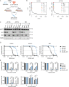
Fig. 1. Whole genome CRISPR/Cas9 KO screens reveal that loss of BAX confers resistance to BH3-mimetic drugs targeting MCL-1 in Eµ-Myc mouse lymphoma cell lines with Trp53 KO.
A Western blot to validate CRISPR/Cas9-mediated KO of Trp53 in AH15A Eµ-Myc mouse lymphoma cells. Cells were treated with the TRP53-activating drug Nutlin-3a for 24 h to enable visualisation of stabilised wildtype TRP53 protein in Trp53 wildtype cells. Probing for β-ACTIN served as a loading control. B Dose–response curves for control AH15A Eµ-Myc lymphoma cells (containing Cas9 and a non-targeting control sgRNA) and isogenic Trp53 KO cells treated with the MCL-1 inhibitor S63845 for 24 h. Live cells were identified as Annexin V/PI double negative by flow cytometry. Trp53 KO cells showed a ~2-fold increase in IC50 for S63845 treatment. C Schematic of CRISPR/Cas9 screen performed in the AH15A Trp53 KO mouse Eµ-Myc lymphoma cell line. Cells were treated with DMSO (vehicle, control) or 1 µM of S63845 (~IC99 dose) for 24 h and surviving cells were collected for next-generation sequencing. D MAGeCK analysis to identify top enriched sgRNAs in S63845-resistant cell populations. Significant hits (FDR < 0.05) are indicated with black text. Loss of Bax was identified as the top, and only significant, hit that promoted drug resistance in the AH15A Trp53 KO Eµ-Myc lymphoma cell line.
Fig. 2
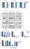
Fig. 2. Whole genome CRISPR/Cas9 KO screens reveal loss of Bax as the top factor conferring resistance to BH3-mimetic drugs targeting MCL-1 in Eµ-Myc mouse lymphoma cell lines.
A Schematic of CRISPR/Cas9 screen performed in Eµ-Myc lymphoma cells with wildtype Trp53. Cells were treated with DMSO (vehicle, control) or 100 nM of S63845 (~IC50 dose) for 2 weeks and surviving cells were collected for next-generation sequencing. B MAGeCK analysis to identify top enriched sgRNAs in S63845-resistant cell populations. Significant hits (FDR < 0.05) are indicated with black text. Loss of Bax was identified as the top hit in the two independent Eµ-Myc lymphoma cell lines used. C Western blots to validate CRISPR/Cas9-mediated loss of BAK, BAX or both BAK and BAX in three independent Eµ-Myc lymphoma cell lines. Probing for HSP70 served as a loading control. D Dose–response curves for control Eµ-Myc lymphoma cells (containing Cas9 and a non-targeting control sgRNA) and isogenic Bak as well as Bax single KO or Bak/Bax double KO cells treated with the MCL-1 inhibitor S63845 for 24 h. Live cells were identified as Annexin V/PI double negative by flow cytometry. E Isogenic WT, Bak KO, Bax KO or Bak/Bax double KO AF47A lymphoma cells treated with the cytotoxic drugs doxorubicin, vincristine, cisplatin, etoposide or ionomycin. All data are presented as mean ± SD for 3 independent experiments. One-way ANOVA was used to measure statistical significance (*p < 0.05, **p < 0.01, ***p < 0.001, ****p < 0.0001, ns = not significant).
Fig. 3
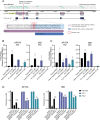
Fig. 3. Lymphomas arising from Eµ-Myc/Bax KO mice show resistance to MCL-1-targeting BH3-mimetics but retain sensitivity to etoposide.
A Cell lines were derived from lymphomas arising in Eµ-Myc/Bak KO and Eµ-Myc/Bax KO mice. Western blotting was used to validate the loss of BAK or BAX protein in these cell lines, respectively. Probing for HSP70 was used as a loading control. B Dose–response curves for Eµ-Myc/Bak KO and Eµ-Myc/Bax KO lymphoma cell lines treated with the MCL-1 inhibitor S63845 for 24 h. Live cells were identified as Annexin V/PI double negative by flow cytometry. Three independent cell lines were tested for each genotype. C, D Survival of Eµ-Myc/Bak KO and Eµ-Myc/Bax KO lymphoma cell lines treated with the cytotoxic drugs etoposide (C) or ionomycin (D). Loss of BAX provided no protection from these drugs. One Eµ-Myc/Bax KO cell line (171) showed resistance to etoposide treatment, likely due to the presence of an additional mutation in Trp53. All data are presented as mean ± SD for 3 independent experiments.
Fig. 4
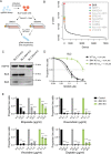
Fig. 4. Generation and characterisation of MCL-1 inhibitor-resistant Eµ-Myc lymphoma cell lines.
To generate MCL-1 inhibitor-resistant cells, three independent Eµ-Myc lymphoma cell lines were cultured in increasing amounts of S63845 over time, until they were resistant to 1 µM S63845 (10× IC50). Cell lines were then either collected for analysis (resistant cell lines R1.1 and R2.1) or continued to be cultured until they were resistant to 5 µM S63845 (resistant cell lines R1.2 and R2.2). Two independent culturing campaigns were undertaken. A Cell viability assay following treatment of parental and drug-resistant cell lines with 1 µM S63845 for 24 h. Live cells were identified as Annexin V/PI double negative by flow cytometry. B Western blotting for TRP53 as well as for pro-apoptotic and pro-survival BCL-2 family proteins in parental and drug-resistant cell lines. Probing for HSP70 was used as a loading control. * indicates non-specific bands. C Parental and drug-resistant cell lines derived from three independent Eµ-Myc lymphoma cell lines were treated with the chemotherapeutic drug etoposide for 24 h. Live cells were identified as Annexin V/PI double negative by flow cytometry. D Treatment of Eµ-Myc lymphoma cell lines exhibiting upregulated pro-survival proteins BCL-XL or A1 (AH15A R1.2, R2.2) with the BH3-mimetic drugs S63845 (MCL-1 inhibitor), A-1331852 (BCL-XL inhibitor) and venetoclax/ABT-199 (BCL-2 inhibitor), either alone or in the indicated combinations for 24 h. Live cells were identified as Annexin V/PI double negative by flow cytometry. In (D), significance is measured versus the DMSO-treated sample for each cell line. All data are presented as mean ± SD for 2–3 independent experiments. One-way ANOVA was used to measure statistical significance (*p < 0.05, **p < 0.01, ***p < 0.001, ****p < 0.0001, ns = not significant).
Fig. 5
Fig. 5. Various defects suppressing Bax expression were identified in MCL-1 inhibitor-resistant Eµ-Myc lymphoma cells.
A Next-generation sequencing of the Bax coding regions revealed a 504G>A substitution downstream of the transcriptional start site in two drug-resistant Eµ-Myc lymphoma cell lines which lack BAX protein expression (AF47A R1.2 and 560 R1.2). This mutation disrupts the mRNA splicing acceptor site and would likely result in the retention of intron 1 in the consensus mouse Bax transcript Bax-201 (ENSEMBL ENSMUST00000033093.10). Gene sequencing coverage is indicated by green bars. B qRT-PCR of parental and drug-resistant Eµ-Myc lymphoma cells treated with DMSO (vehicle, negative control) or the TRP53-activating drug Nutlin-3a for 24 h, of which Bax is a transcriptional target. Data are shown normalised to the housekeeping gene Hmbs, and relative to the parental Eµ-Myc lymphoma cell line treated with DMSO. C qRT-PCR of parental and drug-resistant Eµ-Myc lymphoma cells treated with DMSO or the hypomethylating agent 5’azacytidine (inhibitor of DNMT1) for 24 h. Data are shown normalised to the housekeeping gene Hmbs, and relative to the parental cell line treated with DMSO. D Parental and drug-resistant Eµ-Myc lymphoma cell lines exhibiting loss of BAX protein expression were treated with 5’azacytidine for 24 h, alone or in combination with S63845. Live cells were identified as Annexin V/PI double negative by flow cytometry. All flow cytometry data are presented as mean ± SD for 3 independent experiments. One-way ANOVA was used to determine statistical significance (**p < 0.01; ****p < 0.0001, ns = not significant). All qRT-PCR data are presented as mean ± SD for 2 independent experiments.
Fig. 6
Fig. 6. Whole genome CRISPR/Cas9 KO screens reveal loss of BAX as the top factor conferring resistance to BH3-mimetic drugs targeting MCL-1 in human Burkitt lymphoma cells.
A Schematic of CRISPR/Cas9 KO screen performed in the human Burkitt lymphoma cell line BL2. Cells were treated with DMSO (vehicle, negative control) or 100 nM of S63845 (~IC99 dose) for 24 h and surviving cells were sorted by flow cytometry and collected for next-generation sequencing. B MAGeCK analysis to identify top enriched sgRNAs in drug-resistant cell populations. Significant hits (FDR < 0.05) are indicated with black text. Loss of BAX was identified as the top factor conferring resistance to S63845 in BL2 cells. C Western blot analysis to validate CRISPR/Cas9-mediated loss of BAK or BAX in BL2 cells. Probing for HSP70 was used as a loading control. D Dose–response curves for control BL2 cells (containing Cas9 and a non-targeting control sgRNA) and isogenic BAK KO or BAX KO BL2 lymphoma cells treated with the MCL-1 inhibitor S63845 for 24 h. Live cells were identified as Annexin V/PI double negative by flow cytometry. E Isogenic WT, BAK KO or BAX KO BL2 cells were treated with etoposide, doxorubicin, vincristine or cisplatin for 24 h and cell survival was determined as described above. In (E), significance is measured versus the DMSO-treated sample for each cell line. One-way ANOVA was used to determine statistical significance (****p < 0.0001). All data are presented as mean ± SD for 3 independent experiments.

Similar articles

Cited by

References

    1. Singh R, Letai A, Sarosiek K. Regulation of apoptosis in health and disease: the balancing act of BCL-2 family proteins. Nat Rev Mol Cell Biol. 2019;20:175–93.. doi: 10.1038/s41580-018-0089-8. - DOI - PMC - PubMed
    1. Cory S, Adams JM. The Bcl2 family: regulators of the cellular life-or-death switch. Nat Rev Cancer. 2002;2:647–56. doi: 10.1038/nrc883. - DOI - PubMed
    1. Riedl SJ, Salvesen GS. The apoptosome: signalling platform of cell death. Nat Rev Mol Cell Biol. 2007;8:405–13. doi: 10.1038/nrm2153. - DOI - PubMed
    1. Roberts AW, Wei AH, Huang DCS. BCL2 and MCL1 inhibitors for hematologic malignancies. Blood. 2021;138:1120–36. doi: 10.1182/blood.2020006785. - DOI - PubMed
    1. Diepstraten ST, Anderson MA, Czabotar PE, Lessene G, Strasser A, Kelly GL. The manipulation of apoptosis for cancer therapy using BH3-mimetic drugs. Nat Rev Cancer. 2022;22:45–64. doi: 10.1038/s41568-021-00407-4. - DOI - PubMed

Publication types

MeSH terms

Substances