. 2022 Dec 16;4(6):fcac315.
doi: 10.1093/braincomms/fcac315. eCollection 2022.

Allele-specific silencing as therapy for familial amyotrophic lateral sclerosis caused by the p.G376D TARDBP mutation

Affiliations

Allele-specific silencing as therapy for familial amyotrophic lateral sclerosis caused by the p.G376D TARDBP mutation

Roberta Romano et al. Brain Commun. .

Abstract

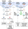
Amyotrophic lateral sclerosis is a neurodegenerative disease characterized by the degeneration of motor neurons. There is no treatment for this disease that affects the ability to move, eat, speak and finally breathe, causing death. In an Italian family, a heterozygous pathogenic missense variant has been previously discovered in Exon 6 of the gene TARDBP encoding the TAR DNA-binding protein 43 protein. Here, we developed a potential therapeutic tool based on allele-specific small interfering RNAs for familial amyotrophic lateral sclerosis with the heterozygous missense mutation c.1127G>A. We designed a small interfering RNA that was able to diminish specifically the expression of the exogenous Green Fluorescent Protein (TAR DNA-binding protein 43G376D mutant protein) in HEK-293T cells but not that of the Green Fluorescent Protein (TAR DNA-binding protein 43 wild-type). Similarly, this small interfering RNA silenced the mutated allele in fibroblasts derived from patients with amyotrophic lateral sclerosis but did not silence the wild-type gene in control fibroblasts. In addition, we established that silencing the mutated allele was able to strongly reduce the pathological cellular phenotypes induced by TAR DNA-binding protein 43G376D expression, such as the presence of cytoplasmic aggregates. Thus, we have identified a small interfering RNA that could be used to silence specifically the mutated allele to try a targeted therapy for patients carrying the p.G376D TAR DNA-binding protein 43 mutation.

Keywords: ALS; TDP-43; allele-specific silencing; antisense oligonucleotides; siRNA therapy.


Figures

Graphical abstract
Graphical abstract
Fig. 1
Fig. 1
Pedigree of the family affected by ALS for the TDP-43G376D mutation. Square indicates male; circle female. Arrows indicate patients from which fibroblasts were obtained. Circles and squares crossed by a line indicate deceased female and male, respectively. (A) Family tree showing the first generation in which the first case of ALS was reported. (B) Family tree of Patient 1. (C) Family tree of Patient 2.
Fig. 2
Fig. 2
An allele-specific siRNA downregulates the GFP-tagged TDP-43G376D mutant allele. (A) HEK-293T cells were transfected with plasmids encoding the GFP-TDP-43WT or GFP-TDP-43G376D proteins and with control RNA, TDP-43 siRNA or putative allele-specific siRNAs (M9, M10, M9/10 and M10/9). Cells were then lysed and analysed by Western blotting using specific anti-TDP-43 and anti-tubulin antibodies, in order to analyse GFP-TDP-43 levels and equal loading, respectively. See Supplementary Fig. 1 for uncropped blots. (B) Quantification of the amount of GFP-TDP-43 wild-type and mutant proteins upon transfection of the different siRNAs. Data are the mean ± SEM of at least three independent experiments. Statistical analysis was performed using one-way ANOVA followed by Dunnett’s multiple comparisons test. *P < 0.05; ** < 0.01; ***P < 0.001; F = 17.92 (for GFP-TDP-43WT); F = 8.488 (for GFP-TDP-43G376D). (C) The amount of GFP-TDP-43 transcript was quantified, compared with the Rplp0 transcript as control, using real-time PCR in HEK-293T cells transfected with GFP-TDP-43WT or GFP-TDP-43G376D plasmids and treated with control RNA, TDP-43 siRNA or allele-specific siRNA M10. Data represent the mean ± SEM of at least three experiments. Statistical analysis was performed using one-way ANOVA followed by Sidak’s multiple comparisons test. *P < 0.05; ** P < 0.01; ***P < 0.001; F = 6.606.
Fig. 3
Fig. 3
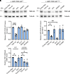
Allele-specific siM10 decreases TDP-43G376D abundance in ALS fibroblasts. (A) Fibroblasts isolated from two controls and two ALS patients carrying the TDP-43G376D mutation (ALS1, ALS2) were transfected with siM10, TDP-43i and control RNA for 6 days, lysed and subjected to Western blot analysis using anti-TDP-43 antibody and Stain-Free Imaging Technology (Bio-Rad) for loading control. See Supplementary Fig. 2 for uncropped blots. (B) Quantification of the amount of TDP-43 upon transfection with control RNA, TDP43 siRNA and allele-specific siRNA M10. Statistical analysis was performed using one-way ANOVA followed by Sidak’s multiple comparisons test. Data represent the mean ± SEM of at least three experiments. *P < 0.05; ** < 0.01; ***P < 0.001; F = 35.65 (for control cells); F = 12.77 (for ALS cells). (C) The amount of TDP-43 wild-type transcript was quantified, compared with the Rplp0 transcript as control, using real-time PCR in control and ALS fibroblasts treated with control RNA, TDP-43 siRNA or allele-specific siRNA M10. Data represent the mean ± SEM of at least three experiments. Statistical analysis was performed using one-way ANOVA followed by Sidak’s multiple comparisons test. *P < 0.05; ** < 0.01; ***P < 0.001; F = 16.03. (D) The amount of TDP-43 mutant transcript was quantified, compared with the Rplp0 transcript as control, using real-time PCR in ALS fibroblasts treated with control RNA, TDP-43 siRNA or allele-specific siRNA M10. Data represent the mean ± SEM of at least three experiments. Statistical analysis was performed using one-way ANOVA followed by Sidak’s multiple comparisons test. *P < 0.05; ** < 0.01; ***P < 0.001; F = 16.68.
Fig. 4
Fig. 4
Allele-specific siM10 decreases TDP-43 cytoplasmic inclusions in ALS fibroblasts. (A) Control and ALS fibroblasts were fixed and immunolabelled with anti-C-terminal TDP-43 antibody followed by Alexa-568-conjugated secondary antibody. Bar 50 µM. (B) Quantification of the percentage of cells showing TDP-43 cytoplasmic aggregates. At least 50 cells per sample were analysed. Data represent the mean ± SEM of at least three experiments. Statistical analysis was performed using one-way ANOVA followed by Tukey’s multiple comparisons test. *P < 0.05; ** < 0.01; ***P < 0.001; F = 84.83. (C) Control and ALS fibroblasts treated with control RNA, TDP-43 siRNA or allele-specific siRNA M10 were fixed and immunolabelled with anti-C-terminal TDP-43 antibody followed by Alexa-568-conjugated secondary antibody. Nuclei were stained with DAPI. Bar 10 µM. Quantification of the percentage of cells with TDP-43 aggregates is also shown. At least 50 cells per sample were analysed. Data represent the mean ± SEM of at least three experiments. Statistical analysis was performed using one-way ANOVA followed by Dunnett’s multiple comparisons test. *P < 0.05; ** < 0.01; ***P < 0.001; F = 474.4 (for ALS1A); F = 209.6 (for ALS2A).
Fig. 5
Fig. 5
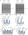
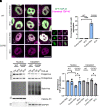
Neuro2A cells transfected with GFP-TDP-43G376D show aggregates which are no longer visible following siM10 treatment. (A) Neuro2A cells were transfected with plasmids encoding the GFP-TDP-43WT or GFP-TDP-43G376D proteins and with control RNA or M10 siRNA. Seventy-two hours after transfection, cells were fixed and immunolabelled with anti-C-terminal TDP-43 antibody followed by Alexa-568-conjugated secondary antibody. Bar 10 µM. (B) Quantification of the percentage of cells showing TDP-43 cytoplasmic aggregates. At least 50 cells per sample were analysed. Data represent the mean ± SEM of at least three experiments. Statistical analysis was performed using one-way ANOVA followed by Dunnett’s multiple comparisons test. *P < 0.05; ** < 0.01; ***P < 0.001; F = 142.3. (C) HEK-293T cells were transfected with plasmids encoding the GFP-TDP-43WT or GFP-TDP-43G376D proteins and with control RNA or M10 siRNA. Seventy-two hours after transfection cells were lysed and cytoplasmic and nuclear fractions were separated. Samples were analysed by Western blotting using anti-TDP-43, anti-GAPDH, anti-histone H3 antibodies. Stain-Free Imaging Technology (Bio-Rad) was used for loading control. GAPDH and histone H3 were used. See Supplementary Figure 3 for uncropped blots. (D) Quantification of endogenous TDP-43 was shown. Data represent the mean ± SEM of at least three experiments. Statistical analysis was performed using one-way ANOVA followed by Tukey’s multiple comparisons test. *P < 0.05; ** < 0.01; ***P < 0.001; F = 4.945 (for nucleus); F = 3.710 (for cytoplasm).
Fig. 6
Fig. 6
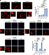
M10 treatment rescues cell viability and oxidative stress. (A) Neuro2A cells were transfected with plasmids encoding the GFP-TDP-43WT or GFP-TDP-43G376D proteins and with control RNA or M10 siRNA. Seventy-two hours after transfection, cells were subjected to SRB assay to measure cell viability. Data represent the mean ± SEM of at least three experiments. Statistical analysis was performed using one-way ANOVA followed by Tukey’s multiple comparisons test. *P < 0.05; ** < 0.01; ***P < 0.001; F = 4.060. (B) Fibroblasts isolated from two controls and two ALS patients carrying the TDP-43G376D mutation (ALS1A, ALS2A) were transfected with control RNA and M10 for 6 days, treated with 10 µM DCFH-DA for 30 min and then lysed. Fluorescence intensity was read at 490 nm in a microplate reader. Data represent the mean ± SEM of at least three experiments. Statistical analysis was performed using one-way ANOVA followed by Tukey’s multiple comparisons test. *P < 0.05; ** < 0.01; ***P < 0.001; F = 5.946.

Similar articles

Cited by

  • The new missense G376V-TDP-43 variant induces late-onset distal myopathy but not amyotrophic lateral sclerosis.
    Zibold J, Lessard LER, Picard F, da Silva LG, Zadorozhna Y, Streichenberger N, Belotti E, Osseni A, Emerit A, Errazuriz-Cerda E, Michel-Calemard L, Menassa R, Coudert L, Wiessner M, Stucka R, Klopstock T, Simonetti F, Hutten S, Nonaka T, Hasegawa M, Strom TM, Bernard E, Ollagnon E, Urtizberea A, Dormann D, Petiot P, Schaeffer L, Senderek J, Leblanc P. Zibold J, et al. Brain. 2024 May 3;147(5):1768-1783. doi: 10.1093/brain/awad410. Brain. 2024. PMID: 38079474 Free PMC article.

References

    1. Gentile F, Scarlino S, Falzone YM, et al. . The peripheral nervous system in amyotrophic lateral sclerosis: Opportunities for translational research. Front Neurosci. 2019;13:601. - PMC - PubMed
    1. Jo M, Lee S, Jeon YM, Kim S, Kwon Y, Kim HJ. The role of TDP-43 propagation in neurodegenerative diseases: Integrating insights from clinical and experimental studies. Exp Mol Med. 2020;52(10):1652–1662. - PMC - PubMed
    1. Prasad A, Bharathi V, Sivalingam V, Girdhar A, Patel BK. Molecular mechanisms of TDP-43 misfolding and pathology in amyotrophic lateral sclerosis. Front Mol Neurosci. 2019;12:25. - PMC - PubMed
    1. Pinarbasi ES, Cağatay T, Fung HYJ, Li YC, Chook YM, Thomas PJ. Active nuclear import and passive nuclear export are the primary determinants of TDP-43 localization. Sci Rep. 2018;8(1):7083. - PMC - PubMed
    1. Barmada SJ, Skibinski G, Korb E, Rao EJ, Wu JY, Finkbeiner S. Cytoplasmic mislocalization of TDP-43 is toxic to neurons and enhanced by a mutation associated with familial amyotrophic lateral sclerosis. J Neurosci. 2010;30(2):639–649. - PMC - PubMed

LinkOut - more resources