

Tumor-infiltrating T cells as a risk factor for lymph node metastasis in patients with submucosal colorectal cancer
- PMID: 36746991
- PMCID: PMC9902519
- DOI: 10.1038/s41598-023-29260-1
Tumor-infiltrating T cells as a risk factor for lymph node metastasis in patients with submucosal colorectal cancer
Abstract
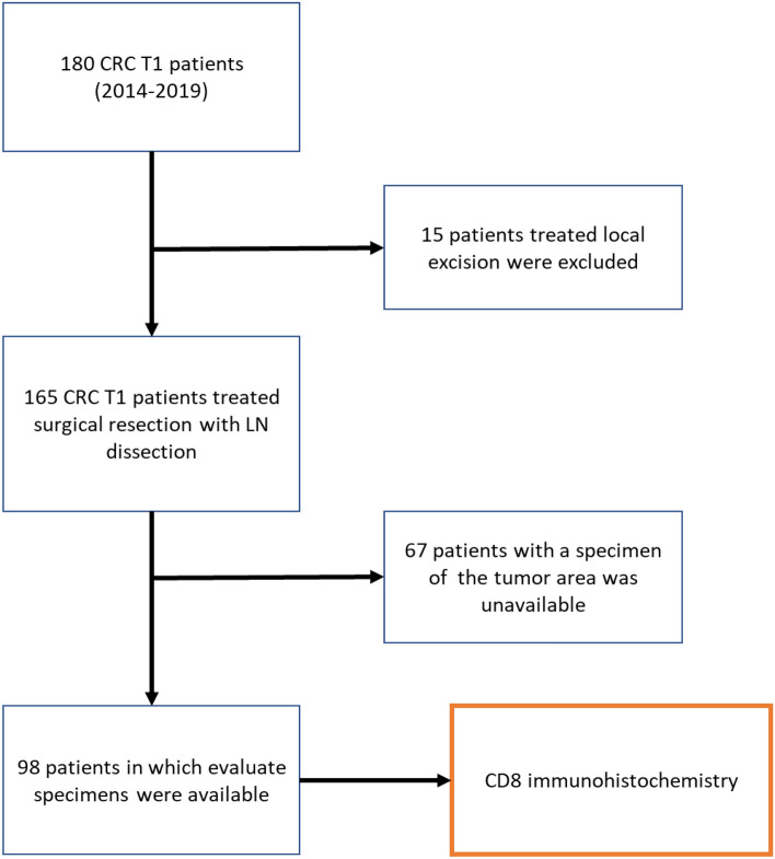
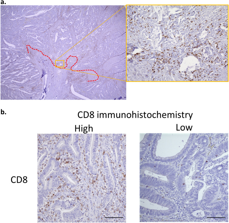
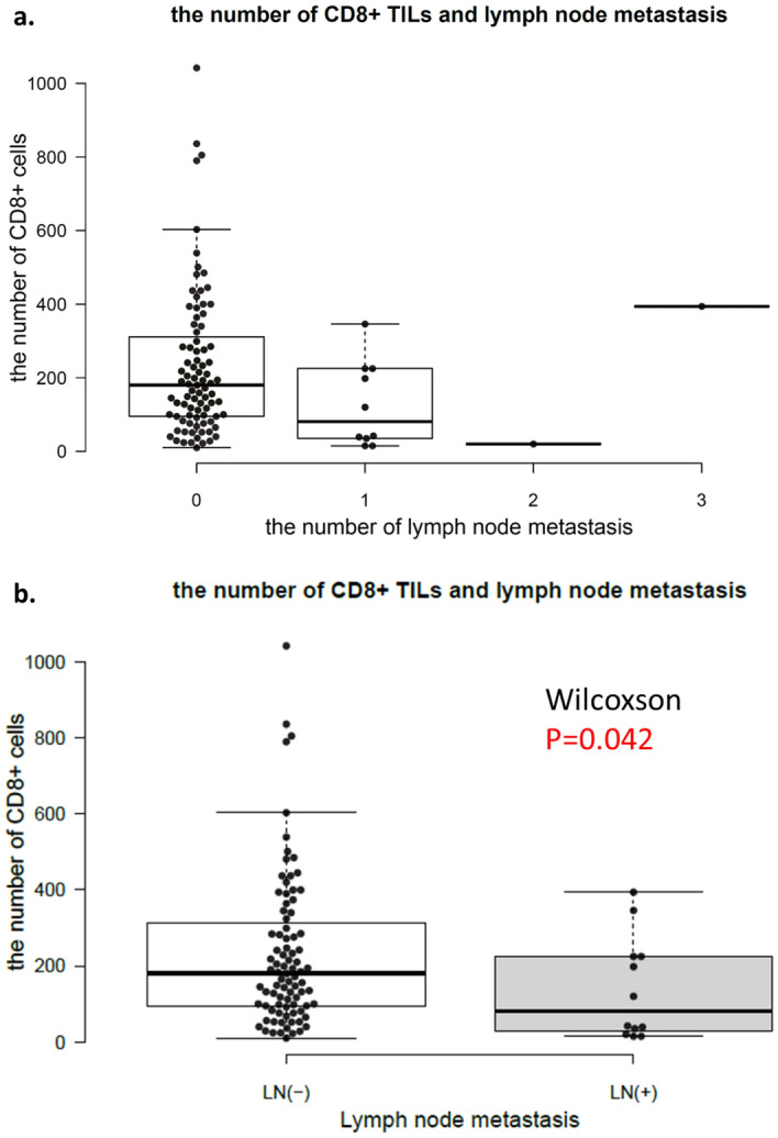
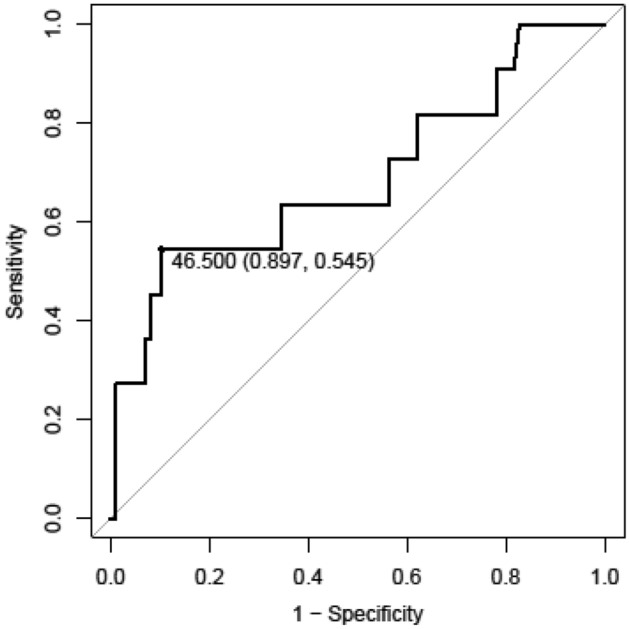
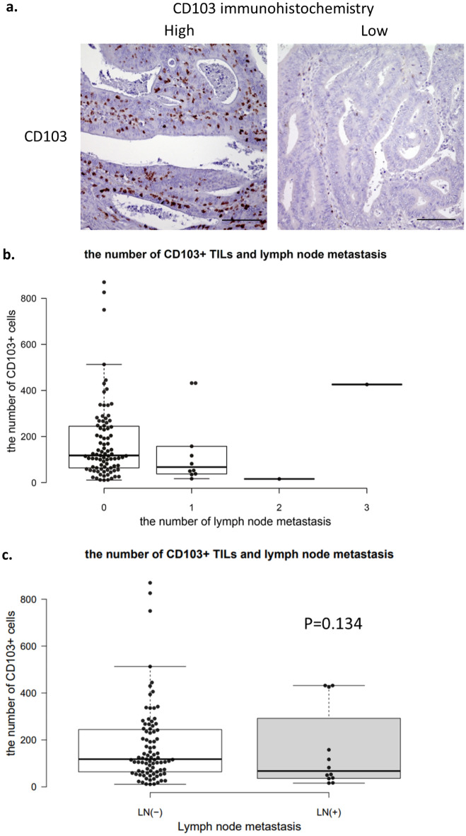
Approximately 10% of patients with colorectal cancer with submucosal invasion have lymph node metastasis. Pathological risk factors for lymph node metastasis have varying sensitivities and specificities. To predict the risk of lymph node metastasis, the identification of new risk factors is vital. Tumor-infiltrating T cells have been reported to improve the prognosis of many solid tumors. Therefore, the purpose of this study was to examine the relationship between lymph node metastasis and tumor-infiltrating T cells in patients with colorectal cancer with submucosal invasion. We examined CD8+ tumor-infiltrating T cells level as a risk factor for lymph node metastasis in patients with colorectal cancer with submucosal invasion. Using immunohistochemical staining, we identified CD8 + T cells in surgically resected specimens from 98 patients with SM-CRC. We showed that low CD8+ tumor-infiltrating T cells levels are positively correlated with lymph node metastasis. Furthermore, by combining the number of CD8+ tumor-infiltrating T cell and the number of CD103+ tumor-infiltrating T cells, the results showed a high positive predictive value for lymph node metastasis in cases with low numbers of both types of tumor-infiltrating T cells and a high negative predictive value in cases with high numbers of both types of tumor-infiltrating T cells.
© 2023. Crown.
Conflict of interest statement
The authors declare no competing interests.
Figures

Similar articles
-
Evaluation of tumor cell dissociation as a predictive marker of lymph node metastasis in submucosal invasive colorectal carcinoma.Dis Colon Rectum. 2005 May;48(5):938-45. doi: 10.1007/s10350-004-0883-6. Dis Colon Rectum. 2005. PMID: 15785891
-
A three-tier classification system based on the depth of submucosal invasion and budding/sprouting can improve the treatment strategy for T1 colorectal cancer: a retrospective multicenter study.Mod Pathol. 2015 Jun;28(6):872-9. doi: 10.1038/modpathol.2015.36. Epub 2015 Feb 27. Mod Pathol. 2015. PMID: 25720321
-
Prognostic and diagnostic significance of tumor budding associated with β-catenin expression in submucosal invasive colorectal carcinoma.Tohoku J Exp Med. 2013 Jan;229(1):53-9. doi: 10.1620/tjem.229.53. Tohoku J Exp Med. 2013. PMID: 23238650
-
Current problems and perspectives of pathological risk factors for lymph node metastasis in T1 colorectal cancer: Systematic review.Dig Endosc. 2022 Jul;34(5):901-912. doi: 10.1111/den.14220. Epub 2022 Jan 11. Dig Endosc. 2022. PMID: 34942683 Review.
-
Endoscopic and surgical resection of T1a/T1b esophageal neoplasms: a systematic review.World J Gastroenterol. 2013 Mar 7;19(9):1424-37. doi: 10.3748/wjg.v19.i9.1424. World J Gastroenterol. 2013. PMID: 23539431 Free PMC article. Review.
Cited by
-
Prognostic Value of Combined LMR and CEA Dynamic Monitoring in Postoperative Colorectal Cancer Patients.J Inflamm Res. 2023 Sep 22;16:4229-4250. doi: 10.2147/JIR.S422500. eCollection 2023. J Inflamm Res. 2023. PMID: 37772275 Free PMC article.
References
-
- Fujino S, Miyoshi N, Ohue M, et al. A nomogram for predicting lymph node metastasis in submucosal colorectal cancer. Int. Surg. 2017;102:102–108. doi: 10.9738/INTSURG-D-16-00210.1. - DOI
MeSH terms
LinkOut - more resources
Full Text Sources
Medical
Research Materials
