. 2023 Feb 28;97(2):e0000923.
doi: 10.1128/jvi.00009-23. Epub 2023 Feb 6.

p38 Mitogen-Activated Protein Kinase Signaling Enhances Reovirus Replication by Facilitating Efficient Virus Entry, Capsid Uncoating, and Postuncoating Steps

Affiliations

p38 Mitogen-Activated Protein Kinase Signaling Enhances Reovirus Replication by Facilitating Efficient Virus Entry, Capsid Uncoating, and Postuncoating Steps

Adil Mohamed et al. J Virol. .

Abstract

Mammalian orthoreovirus serotype 3 Dearing is an oncolytic virus currently undergoing multiple clinical trials as a potential cancer therapy. Previous clinical trials have emphasized the importance of prescreening patients for prognostic markers to improve therapeutic success. However, only generic cancer markers such as epidermal growth factor receptor (EGFR), Hras, Kras, Nras, Braf, and p53 are currently utilized, with limited benefit in predicting therapeutic efficacy. This study aimed to investigate the role of p38 mitogen-activated protein kinase (MAPK) signaling during reovirus infection. Using a panel of specific p38 MAPK inhibitors and an inactive inhibitor analogue, p38 MAPK signaling was found to be essential for establishment of reovirus infection by enhancing reovirus endocytosis, facilitating efficient reovirus uncoating at the endo-lysosomal stage, and augmenting postuncoating replication steps. Using a broad panel of human breast cancer cell lines, susceptibility to reovirus infection corresponded with virus binding and uncoating efficiency, which strongly correlated with status of the p38β isoform. Together, results suggest p38β isoform as a potential prognostic marker for early stages of reovirus infection that are crucial to successful reovirus infection. IMPORTANCE The use of Pelareorep (mammalian orthoreovirus) as a therapy for metastatic breast cancer has shown promising results in recent clinical trials. However, the selection of prognostic markers to stratify patients has had limited success due to the fact that these markers are upstream receptors and signaling pathways that are present in a high percentage of cancers. This study demonstrates that the mechanism of action of p38 MAPK signaling plays a key role in establishment of reovirus infection at both early entry and late replication steps. Using a panel of breast cancer cell lines, we found that the expression levels of the MAPK11 (p38β) isoform are a strong determinant of reovirus uncoating and infection establishment. Our findings suggest that selecting prognostic markers that target key steps in reovirus replication may improve patient stratification during oncolytic reovirus therapy.

Keywords: breast cancer; oncolytic viruses; p38 MAPK; prognostic indicators; reovirus; virus entry.


Conflict of interest statement

The authors declare no conflict of interest.

Figures

FIG 1
FIG 1
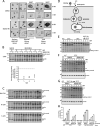
p38 MAPK signaling promotes establishment of reovirus infection. (A) Summary of p38 MAPK activation and downstream signaling. (B) L929 cells were treated with 10 μM p38 MAPK inhibitors (SB202190 and SB203580) or inactive analogue (SB202474) for 1 h, followed by treatment with 500 nM p38 MAPK activator (anisomycin) for 30 min. Prototypic proteins of the p38 MAPK signaling pathway (A) were assessed using Western blot analysis. (C) L929 cells were treated with SB202190, SB203580, SB202474, or BIRB-796 at 20 μM, 10 μM, or 5 μM and respective DMSO for 12 h, followed by a medium change and additional incubation for 12 h. Cells were incubated in the presence of hydrogen peroxide (H2O2; 5 mM, 1 mM, or 0.2 mM) for 24 h without a 12-h medium change. Cell viability was assessed using resazurin fluorescence (n = 3 independent experiments). (D to G) Cells were treated with indicated compound dilutions for 1 h at 37°C. Cells were then prechilled at 4°C for 1 h, followed by reovirus (MOI, 3) addition and incubation at 4°C for 1 h. Following extensive washing to remove unbound virus, medium was replenished with the indicated compound dilutions and incubated at 37°C for 12 h. (D) L929 cell lysates were assessed for reovirus proteins using Western blot analysis, representative image of 2 independent experiments. A background nonspecific band was used as a loading control. (E and F) L929 cells were fixed at 12 hpi, and virus-infected cells were stained using reovirus-specific antibodies, followed by either processing for immunohistochemistry using BCIP/NBT substrate, representative of 3 independent experiments (E), or flow cytometric analysis using Alexa Fluor 647 secondary antibodies, with 2 to 5 independent experiments depending on the inhibitor (F). (G) Similar to panel E, except that a panel of cell lines was used and SB202190 was used at 10 μM. Image is representative of 3 independent experiments. H1299 (MOI, 1), T-47D (MOI, 1), B16-F10 (MOI, 3), and NIH 3T3 (MOI, 20) cells were used.
FIG 2
FIG 2
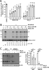
Reovirus entry but not binding is attenuated upon inhibition of p38 MAPK signaling. (A) L929 cells were treated with the indicated compounds (SB202190, 10 μM; anisomycin, 500 nM) for 1 h at 37°C. Cells were then prechilled at 4°C for 1 h, followed by reovirus (MOI, 1 or 3) addition and incubation at 4°C for 1 h. Following extensive washing to remove unbound virus, cell lysates were collected and assessed for reovirus proteins using Western blot analysis. Western blots are representative of 3 independent experiments. (B) Cells were treated similarly to those for panel A, except that 35S-radiolabeled reovirus (MOI, 1, 3, or 9) was used, and cell lysates were collected and run on a scintillation counter. Each point on the bar graphs represents an independent experiment. (C) (Left) Cells were treated similarly to those for panel A, except that Alexa Fluor 546-labeled reovirus was used at an MOI of 20. Cells were fixed at 0 h immediately after binding or after 2 h of incubation at 37°C. External virions were stained using reovirus-specific antibodies without cell permeabilization. Cells were imaged using confocal microscopy, and external (Alexa Fluor 647) and total (Alexa Fluor 546) virions were quantified using the spot identification tool on Harmony high-content imaging and analysis software. Each point on the bar graph represents an individual cell, pooled from 2 independent experiments. ****, P < 0.0001; ns, not significant (P > 0.05) (unpaired t test). (Right) Diagrammatical depiction of the external and internal virion staining strategy.
FIG 3
FIG 3
Reovirus uncoating is mediated by p38 MAPK signaling. (A to C) Cells were treated with indicated compounds for 1 h at 37°C. Cells were then prechilled at 4°C for 1 h, followed by reovirus addition and incubation at 4°C for 1 h. Following extensive washing to remove unbound virus, medium was replenished with the indicated compounds and incubated at 37°C. (A) L929 cells were infected with Alexa Fluor 546-labeled reovirus (MOI, 20) and fixed at the indicated time points, followed by staining of external virions using reovirus-specific antibodies and Alexa Fluor 647-conjugated secondary antibodies. Nuclei were labeled using Hoechst 33342. Cells were imaged using confocal microscopy. Microscopy images are representative of 2 independent experiments. White arrows indicate aggregated virus compartments. (B) (Top) At the indicated time points, cell lysates were collected and assessed for reovirus proteins using Western blot analysis to monitor outer capsid uncoating. (Bottom) Quantification of capsid uncoating [(μ1C/δ)/β-actin], with each point representing an independent experiment (n = 3 total). (C) Similar to panel B but on the indicated cancer cell lines. Representative blots for 2 experiments for H1299 cells and a single experiment for T-47D and B16-F10 cells are shown. An MOI of 3 was used for all cell lines. (D) Summary of temperature effects on key steps of endosomal trafficking based on previous studies referenced in the text. (E to G) Reovirus (MOI, 3) was bound to L929 cells at 4°C cells similarly to what is shown in panel A, and cells were incubated at indicated temperature in the presence or absence of SB202190 at 10 μM. Cell lysates were collected at the indicated time points, and Western blot analysis for reovirus proteins was performed. (H) Quantification summary of capsid uncoating [(δ × 100/(δ + μ1C)] from Western blot analysis in panels E to G. Each circle represents an independent experiment (n = 2 to 5).
FIG 4
FIG 4
p38 MAPK signaling facilitates postuncoating steps of reovirus replication. (A) L929 cells were prechilled at 4°C for 1 h, followed by reovirus (MOI, 3) addition and incubation at 4°C for 1 h. Following extensive washing to remove unbound virus, medium was replenished and cells were incubated at 37°C. At the indicated time points, NH4Cl or SB202190 was added, and cells were collected at 12 hpi for flow cytometric analysis to assess the percentage of cells positive for productive reovirus infection. (B) Similar to panel A, except that NH4Cl with or without SB202190 was added at the indicated time points. Cell lysates were collected at 12 hpi and assessed for reovirus proteins using Western blot analysis. Quantification of 2 independent experiments is provided below the blot. (C) Similar to panel A, except that cells were left uninfected (UI), or infected with either reovirus virions or ISVPs followed by treatment with or without NH4Cl and with or without SB202190 as indicated. Cell lysates were collected at 12 hpi and assessed for reovirus proteins using Western blot analysis. (Left) Representative blot of 3 independent experiments. (Right) Average de novo σ3 and μ1 plus μ1C band intensities relative to untreated control are quantified for 3 independent experiments. (A to C) In all experiments, SB202190 was used at 20 μM and NH4Cl at 10 mM. *, P < 0.05; **, P < 0.01; ***, P < 0.001; ****, P < 0.0001; ns, P > 0.05 (one-way analysis of variance with Dunnett’s multiple-comparison test with DMSO).
FIG 5
FIG 5
Differential susceptibility of breast cancer cell lines to reovirus infection. (A) (Top) Breast cancer cell lines were infected with the indicated doses of reovirus for 12 h. Cells were fixed and virus-infected cells stained using reovirus-specific antibodies and BCIP/NBT substrate. MOIs were calculated as per titers on L929 cells. Microscopy images are representative of 2 independent experiments. (Bottom) A panel of cancer cell lines (H1299, T-47D, HCT116, MDA-MB-468, MDA-MB-231, MCF-7, BT549, and Hs578T) were infected with various doses of reovirus, fixed at 18 hpi, and stained using reovirus-specific antibodies and pNPP substrate. Absorbance at 405 was measured using a plate reader. Error bars depict SD, n = 3 for breast cancer cells, n = 4 for HCT116, and n = 6 for H1299 cells. (B) Breast cancer cell lines were exposed to reovirus (MOI, 0.3, 1, or 3) at 4°C for 1 h to permit binding and washed extensively to remove unbound virus, and cell lysates were collected at 0 hpi for detection of cell-bound viruses by Western blotting. “Input” indicates virus inoculum. (C) Basal transcript levels of F11R were obtained from the EMBL-EBI Expression Atlas database (https://www.ebi.ac.uk/gxa/home) and compared between cell lines. (D to E) The indicated cells were exposed to reovirus (MOI, 3) for 1 h at 4°C for 1 h, washed extensively to remove unbound virus, and incubated at 37°C. Cell lysates were collected at the indicated time points and assessed for reovirus uncoating using Western blot analysis. (F) Quantification of percent capsid uncoating at 3 hpi for all cell lines tested. Each point on the bar graph represents an independent experiment. (G) Correlation plot of reovirus uncoating at 3 hpi versus overall susceptibility (1/CCID50 calculated from panel E).
FIG 6
FIG 6
Reovirus uncoating correlates with expression of p38β isoform. (A) Western blot analysis of whole virions and ISVPs using polyclonal anti-reovirus antibodies. (B, C) MCF7 or MDA-MB-231 cells were exposed to serial dilutions (initial MOI, 10) of whole virions or ISVPs, normalized for equal amounts of viral particles. At 12 hpi, reovirus-infected cells were identified by immunofluorescence staining (B) or flow cytometry (C). (D) (Top) Heat map representation of correlations between indicated RNA/protein expression and reovirus uncoating/infectivity (1/CCID50) in H1299, T-47D, HCT116, MDA-MB-468, MDA-MB-231, and MCF-7 cells. RNA1, log2(TPM + 1) from Broad Institute DepMap; RNA2, TPM from EMBL-EBI Expression Atlas; Protein, normalized protein expression from Broad Institute DepMap. (Bottom) Representative correlation plots for MAPK11 (p38β) from the top. (E) The indicated cell lines were counted and normalized to 1 × 106 cells. Equal volumes of cell lysates were processed by Western blotting for p38 MAPK isoforms. Western blots are representative of 2 independent experiments.

Similar articles

Cited by

References

    1. Mohamed A, Clements DR, Gujar SA, Lee PW, Smiley JR, Shmulevitz M. 2020. Single amino acid differences between closely related reovirus T3D lab strains alter oncolytic potency in vitro and in vivo. J Virol 94:e01688-19. 10.1128/JVI.01688-19. - DOI - PMC - PubMed
    1. Muller L, Berkeley R, Barr T, Ilett E, Errington-Mais F. 2020. Past, present and future of oncolytic reovirus. Cancers (Basel) 12:3219. 10.3390/cancers12113219. - DOI - PMC - PubMed
    1. Bernstein V, Ellard SL, Dent SF, Tu D, Mates M, Dhesy-Thind SK, Panasci L, Gelmon KA, Salim M, Song X, Clemons M, Ksienski D, Verma S, Simmons C, Lui H, Chi K, Feilotter H, Hagerman LJ, Seymour L. 2018. A randomized phase II study of weekly paclitaxel with or without pelareorep in patients with metastatic breast cancer: final analysis of Canadian Cancer Trials Group IND.213. Breast Cancer Res Treat 167:485–493. 10.1007/s10549-017-4538-4. - DOI - PubMed
    1. Gong J, Sachdev E, Mita AC, Mita MM. 2016. Clinical development of reovirus for cancer therapy: an oncolytic virus with immune-mediated antitumor activity. World J Methodol 6:25–42. 10.5662/wjm.v6.i1.25. - DOI - PMC - PubMed
    1. Marcato P, Shmulevitz M, Lee PW. 2005. Connecting reovirus oncolysis and Ras signaling. Cell Cycle 4:556–559. 10.4161/cc.4.4.1600. - DOI - PubMed

Publication types

Substances

Grants and funding