. 2023 May 3;31(5):1365-1382.
doi: 10.1016/j.ymthe.2023.01.025. Epub 2023 Feb 2.

Extracellular vesicle-transmitted miR-671-5p alleviates lung inflammation and injury by regulating the AAK1/NF-κB axis

Affiliations

Extracellular vesicle-transmitted miR-671-5p alleviates lung inflammation and injury by regulating the AAK1/NF-κB axis

Jie Lian et al. Mol Ther. .

Abstract

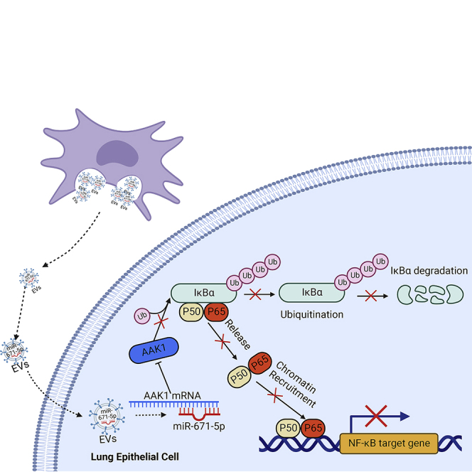
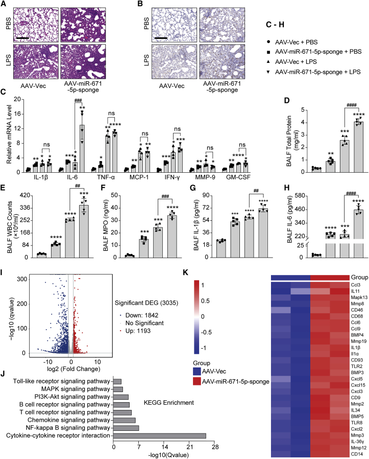
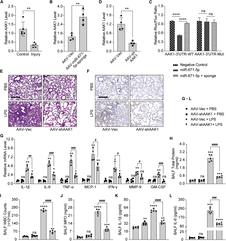
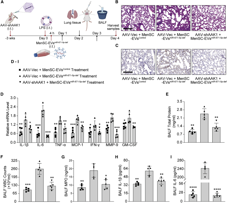
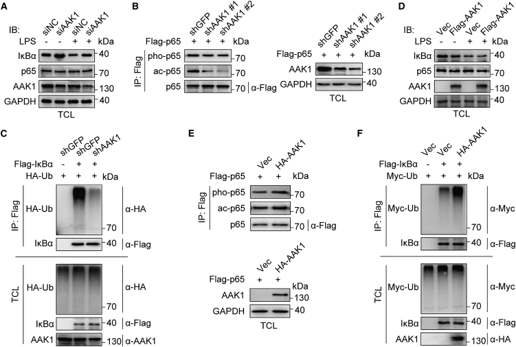
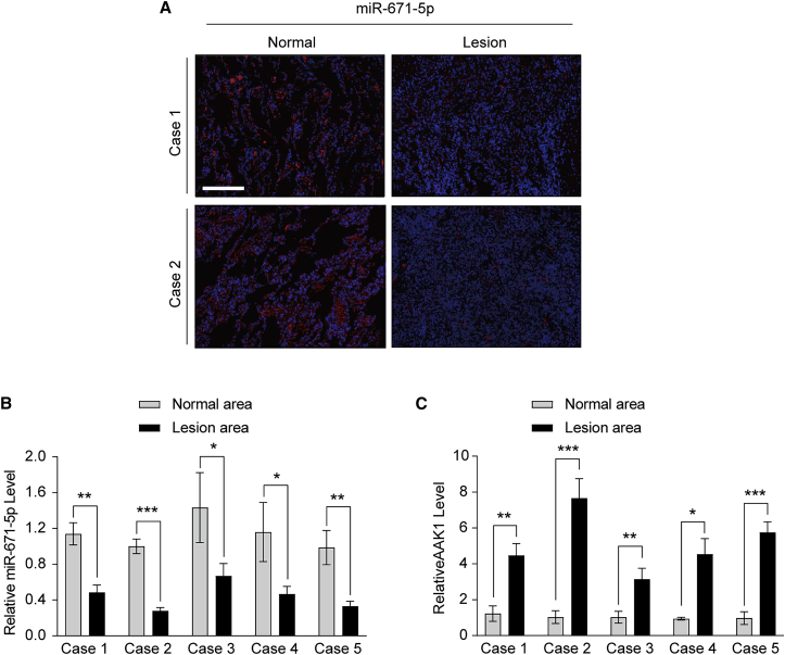
Mesenchymal stem cells regulate remote intercellular signaling communication via their secreted extracellular vesicles. Here, we report that menstrual blood-derived stem cells alleviate acute lung inflammation and injury via their extracellular vesicle-transmitted miR-671-5p. Disruption of this abundantly expressed miR-671-5p dramatically reduced the ameliorative effect of extracellular vesicles released by menstrual blood-derived stem cells on lipopolysaccharide (LPS)-induced pulmonary inflammatory injury. Mechanistically, miR-671-5p directly targets the kinase AAK1 for post-transcriptional degradation. AAK1 is found to positively regulate the activation of nuclear factor κB (NF-κB) signaling by controlling the stability of the inhibitory protein IκBα. This study identifies a potential molecular basis of how extracellular vesicles derived from mesenchymal stem cells improve pulmonary inflammatory injury and highlights the functional importance of the miR-671-5p/AAK1 axis in the progression of pulmonary inflammatory diseases. More importantly, this study provides a promising cell-based approach for the treatment of pulmonary inflammatory disorders through an extracellular vesicle-dependent pathway.

Keywords: AAK1; NF-κB signaling; extracellular vesicles; menstrual blood-derived stem cells; miR-671-5p; pulmonary inflammatory injury.


Conflict of interest statement

Declaration of interests The authors declare no competing interests.

Figures

None
Graphical abstract
Figure 1
Figure 1
MenSC-EVs, as well as their parent cells, significantly improve LPS-induced ALI mice by i.t. transplantation (A) Similar to the effect of MenSCs, MenSC-EVs administration dramatically improves ALI in mice, as demonstrated by histological analysis. Lung sections harvested from mice on day 4 post LPS stimulation (4 mg/kg) were subjected to H&E staining. Scale bar, 200 μm. (B) Decreased accumulation of pulmonary macrophages in mice subjected to MenSCs or MenSC-EVs treatment was detected by CD68 IHC staining of lung sections. The mice were sacrificed on day 4 after LPS stimulation (4 mg/kg). Scale bar, 200 μm. (C) Relative mRNA expression of proinflammatory factors in lungs of mice administered MenSCs, MenSC-EVs, or NHLF-EVs in the presence of LPS (4 mg/kg) was detected by qRT-PCR assay. PBS was used as a negative control. (D) Total protein concentration in BALF isolated from mice treated with MenSCs, MenSC-EVs, or NHLF-EVs was measured by BCA to examine the changes in pulmonary vascular and epithelial permeability. The mice were sacrificed on day 4 after LPS stimulation (4 mg/kg). (E and F) WBC counts (E) and MPO activity (F) in BALF isolated from mice treated with MenSCs, MenSC-EVs, or NHLF-EVs was measured to examine the effect of MenSCs or MenSC-EVs on the influx of inflammatory cells. The mice were sacrificed on day 4 after LPS stimulation (4 mg/kg). (G and H) The effect of MenSCs or MenSC-EVs on production of the proinflammatory cytokines IL-1β (G) and IL-6 (H) in BALF from mice treated with MenSCs, MenSC-EVs, or NHLF-EVs. The mice were sacrificed on day 4 after LPS stimulation (4 mg/kg). n = 5 per group (C–H); data are presented as mean ± SD. ∗p < 0.05, ∗∗p < 0.01, ∗∗∗p < 0.001, and ∗∗∗∗p < 0.0001 versus the injury group; #p < 0.05, ##p < 0.01, ###p < 0.001, and ####p < 0.0001 versus the NHLF-EVs group. All p values in this figure were obtained by one-way ANOVA. NHLF, normal human lung fibroblasts; BALF, bronchoalveolar lavage fluid; WBC, white blood cell; MPO, myeloperoxidase; BCA, bicinchoninic acid.
Figure 2
Figure 2
Comprehensive analysis of genes and pathways involved in MenSCs and MenSC-EVs treatment based on mRNA high-throughput sequencing (A) Volcano plot of RNA-seq transcriptome data displaying the pattern of the gene expression profile in the lungs of mice with or without MenSCs or MenSC-EVs treatment. The mice were sacrificed on day 4 post LPS stimulation (4 mg/kg). Red and blue dots indicate significantly up- or down-regulated genes, respectively. p < 0.05, |log2FC| > 1. (B) Kyoto Encyclopedia of Genes and Genomes (KEGG) enrichment analysis showing the inflammatory pathways involved in MenSC-based improvement of ALI. Total RNAs isolated from the lungs of mice with or without MenSCs or MenSC-EVs treatment in the presence of LPS stimulation (4 mg/kg) were subjected to RNA-seq analysis. (C) Representative heatmap of key factors involved in regulation of the inflammatory response based on the above mRNA-seq data. Red, relatively up-regulated expression; blue, relatively down-regulated expression. Each column represents one individual sample, and each row represents one single gene. n = 3 per group. DEG, differentially expressed gene. FC: fold change.
Figure 3
Figure 3
The improvement of ALI by MenSC-EVs depends on miR-671-5p transmitted by an EV-dependent pathway (A) qRT-PCR analysis showing a robust elevation in miR-671-5p expression in the lungs of mice subjected to LPS stimulation (4 mg/kg). (B) Uptake assay showing the transfer of DiO-labeled MenSC-EVs into BEAS-2B cells. The MenSC-EVs were pre-labeled with DiO for 15 min before incubation with BEAS-2B cells. (C) miR-671-5p is transferred from MenSC-EVs to pulmonary epithelial cells by an EV-dependent pathway. MenSCs overexpressing the miR-671-5p mimic were pre-treated with GW4869 (10 μM) or a vehicle control for 24 h and then incubated with BEAS-2B cells in a Transwell plate. (D and E) Depletion of miR-671-5p in MenSCs reduced the therapeutic effect of MenSC-EVs on ALI, as detected by H&E staining (D) and CD68 IHC staining (E) of lung sections from mice administered EVs isolated from MenSCs stably expressing a sponge depleting miR-671-5p or an empty vector control. The mice were sacrificed on day 4 after LPS stimulation (4 mg/kg). Scale bar, 200 μm. (F) The relative mRNA levels of proinflammatory cytokines in lungs of mice administrated EVs isolated from MenSCs stably expressing a sponge depleting miR-671-5p or an empty vector control were measured by qRT-PCR. (G–K) Total protein concentration (G), WBC counts (H), MPO activity (I), and production of the proinflammatory factors IL-1β (J) and IL-6 (K) in BALF from mice administered EVs isolated from MenSCs stably expressing a sponge depleting miR-671-5p or an empty vector control were measured to determine the effect of miR-671-5p deficiency on EV-mediated amelioration of ALI. n = 3 per group (C); data are presented as mean ± SD. ∗p < 0.05, ∗∗p < 0.01, ∗∗∗p < 0.001, and ∗∗∗∗p < 0.0001; p value was calculated by one-way ANOVA compared with the MenSCs-NC + BEAS-2B group. #p < 0.05, ##p < 0.01, ###p < 0.001, and ####p < 0.0001, p value was calculated by Student’s t test. n = 5 per group (A and F–K); data are presented as mean ± SD. ∗p < 0.05, ∗∗p < 0.01, ∗∗∗p < 0.001, and ∗∗∗∗p < 0.0001; all p values in this figure were obtained by Student’s t test. NC, negative control; mimic, miR-671-5p-mimic; def, deficient.
Figure 4
Figure 4
miR-671-5p negatively regulates acute lung inflammation and injury (A) The effect of miR-671-5p depletion on ALI, as assayed by H&E staining of lung sections from mice with or without AAV-mediated miR-671-5p knockdown. The mice were treated with LPS (4 mg/kg) or a vehicle control after 3 weeks of AAV infection. Scale bar, 200 μm. (B) The effect of AAV-mediated miR-671-5p depletion on the accumulation of pulmonary macrophages, as detected by CD68 IHC staining of lung sections from mice with or without AAV-mediated miR-671-5p knockdown. Scale bar, 200 μm. (C) The effect of AAV-mediated miR-671-5p depletion on the expression of proinflammatory factors in the lungs of mice treated with or without LPS, as determined by qRT-PCR assay. (D–H) Total protein concentration (D), WBC counts (E), MPO activity (F), and production of the proinflammatory factors IL-1β (G) and IL-6 (H) in BALF from mice with or without miR-671-5p depletion were detected to examine the influence of miR-671-5p on pulmonary inflammatory injury. The mice were treated with LPS (4 mg/kg) or a vehicle control after 3 weeks of AAV infection. (I–K) Lung tissues isolated from mice receiving the AAV-expressing miR-671-5p sponge or an empty vector control were subjected to high-throughput mRNA sequencing analysis to detect the effect of miR-671-5p depletion on downstream gene expression profiles and signaling pathways. A representative graph shows the up- or down-regulated genes (I), a KEGG enrichment analysis shows the downstream pathways regulated by miR-671-5p (J), and a representative heatmap shows the expression changes of the indicated factors (K). n = 4 per group (C), n = 5 per group (D–H); data are presented as mean ± SD. ∗p < 0.05, ∗∗p < 0.01, ∗∗∗p < 0.001, and ∗∗∗∗p < 0.0001 compared with the AAV-vector (Vec) + PBS group; ns, not significant, #p < 0.05, ##p < 0.01, ###p < 0.001, and ####p < 0.0001 for comparisons between the indicated groups. A Student’s t test and one-way ANOVA were used to compare two groups and multiple groups. n = 3 per group (I–K). AAV, adeno-associated virus.
Figure 5
Figure 5
AAK1, a direct target of miR-671-5p, positively regulates lung inflammation and injury in LPS-induced ALI mice (A) qRT-PCR analysis of lung tissues from mice treated with or without LPS (4 mg/kg), showing the markedly declined AAK1 RNA level in response to LPS induction. (B) qRT-PCR analysis showing the effect of AAV-mediated miR-671-5p depletion on AAK1 expression in lung tissues of mice subjected to 3 weeks of AAV infection. (C) Dual-luciferase reporter assay showing that AAK1 is a direct target of miR-671-5p. The seed region (nt 685–692) within the 3′ UTR of AAK1 or its Mut form was cloned into an SV40 promoter-driven luciferase reporter. (D) qRT-PCR analysis showing the AAV-mediated knockdown efficiency of AAK1 shRNA in lung tissues of mice receiving 3 weeks of AAV-shAAK1 infection. (E and F) AAV-mediated AAK1 disruption alleviates LPS-induced pulmonary inflammatory injury. Mice administered the AAV-expressing AAK1 shRNA or a mock control were subjected to histological analysis after LPS or PBS treatment. Shown are H&E staining analysis (E) and CD68 IHC histological analysis (F). Scale bar, 200 μm. (G) The effect of AAV-mediated AAK1 shRNA knockdown on expression of inflammatory mediators in lungs of mice injured with LPS (4 mg/kg) or a vehicle PBS control. The mice were injured with LPS 3 weeks after AAV infection. (H–L) Total protein concentration (H), WBC counts (I), MPO activity (J), as well as production of the proinflammatory factors IL-1β (K) and IL-6 (L) in BALF from mice with or without AAK1 disruption were detected to examine the influence of AAK1 disruption on pulmonary inflammatory injury. The lungs of mice were treated with LPS (4 mg/kg) or a vehicle PBS control after 3 weeks of AAV infection. n = 5 per group (A and H–L), n = 4 per group (B, D, and G), and n = 3 (C); data are presented as mean ± SD. ∗p < 0.05, ∗∗p < 0.01, ∗∗∗p < 0.001, and ∗∗∗∗p < 0.0001 compared with the AAV-Vec + PBS group; #p < 0.05, ##p < 0.01, ###p < 0.001, and ####p < 0.0001 for comparisons between the indicated groups. A Student’s t test and one-way ANOVA were used to compare two groups and multiple groups. WT, wild type; Mut, mutant; sponge, miR-671-5p-sponge.
Figure 6
Figure 6
Disruption of AAK1 reverses the reduced protective effect of MenSC-EVs on ALI caused by miR-671-5p deficiency (A) Schematic showing the detailed procedure for the combined AAV-shAAK1 and MenSC-EVs treatment mouse model study; created with BioRender. (B and C) Representative H&E (B) and CD68 IHC (C) staining of lung sections from mice administered the combined EVs isolated from MenSCs stably expressing a sponge depleting miR-671-5p or an empty Vec control and AAK1 shRNA or a mock control. The mice were sacrificed on day 4 after LPS stimulation (4 mg/kg). Scale bar, 200 μm. (D) qRT-PCR analysis of inflammatory mediator expression levels in lungs of mice treated with the combined EVs isolated from MenSCs stably expressing a sponge depleting miR-671-5p or an empty Vec control and AAK1 shRNA or a mock control. The mice were sacrificed on day 4 after LPS stimulation (4 mg/kg). (E–I) Total protein concentration (E), WBC counts (F), MPO activity (G), and production of the proinflammatory factors IL-1β (H) and IL-6 (I) in BALF from mice administered the combined EVs isolated from MenSCs stably expressing a sponge depleting miR-671-5p or an empty Vec control and AAK1 shRNA or a mock control. The mice were sacrificed on day 4 after LPS stimulation (4 mg/kg). n = 5 per group; data are presented as mean ± SD. ∗p < 0.05, ∗∗p < 0.01, ∗∗∗p < 0.001, and ∗∗∗∗p < 0.0001 compared with the AAV-Vec + MenSC-EVsmiR−671−5p-def treatment group. One-way ANOVA was used to obtain all p values in this figure.
Figure 7
Figure 7
AAK1 activates the NF-κB signaling pathway by controlling IκBα stability (A) Immunoblot analysis of IκBα and p65 expression in total lysates of BEAS-2B cells transfected with AAK1 siRNA or an NC in the presence or absence of LPS (100 ng/mL, 24 h). (B) Immunoblot analysis of p65 S536 phosphorylation (pho) and K310 acetylation (ac) levels in the immunoprecipitates from BEAS-2B cells transduced with a lentivirus expressing AAK1 shRNA or the shGFP control by anti-p65 immunoprecipitation. The knockdown efficiency of AAK1 in total lysates of BEAS-2B cells is shown on the right. (C) Immunoblot analysis of IκBα ubiquitination in total lysates of HEK293T cells expressing the indicated combinations of FLAG-tagged IκBα, HA-tagged ubiquitin, and AAK1 shRNA or the shGFP control. The cells were treated with MG-132 (20 μM, 4 h) before collection. (D) Immunoblot analysis of IκBα and p65 expression in total lysates of BEAS-2B cells transduced with a lentivirus expressing FLAG-tagged AAK1 or an empty Vec control in the presence or absence of LPS (100 ng/mL, 24 h). (E) Immunoblot analysis of p65 S536 phosphorylation and K310 acetylation levels in the immunoprecipitates from BEAS-2B cells transduced with a lentivirus overexpressing AAK1 or an empty Vec control by anti-p65 immunoprecipitation. AAK1 reintroduction in BEAS-2B cells was verified by immunoblot analysis of the total lysates (bottom panel). (F) Immunoblot analysis of IκBα ubiquitination in total lysates of HEK293T cells expressing the indicated combinations of FLAG-tagged IκBα, Myc-tagged ubiquitin, and HA-tagged AAK1 or empty Vec. The cells were treated with MG-132 (20 μM, 4 h) before collection.
Figure 8
Figure 8
miR-671-5p has a significant negative correlation with AAK1 in pulmonary inflammatory disease (A) Representative images of lung sections from lesion areas or corresponding adjacent normal areas of 2 different patients with organizing pneumonia, showing the expression levels of miR-671-5p, as determined by FISH assay. Scale bar, 200 μm. (B and C) qRT-PCR analysis of miR-671-5p (B) and AAK1 (C) expression in inflammatory lesion areas or corresponding adjacent normal areas of lung tissues from 5 different patients with organizing pneumonia. n = 3 (B and C), representing 3 pairs of samples from one individual patient; data are presented as mean ± SD. ∗p < 0.05, ∗∗p < 0.01, and ∗∗∗p < 0.001 for comparisons between the indicated groups. All p values in this figure were obtained by Student’s t test.

Similar articles

Cited by

References

    1. Lian J., Lin J., Zakaria N., Yahaya B.H. Acute lung injury: disease modelling and the therapeutic potential of stem cells. Adv. Exp. Med. Biol. 2020;1298:149–166. doi: 10.1007/5584_2020_538. - DOI - PubMed
    1. Huang C., Wang Y., Li X., Ren L., Zhao J., Hu Y., Zhang L., Fan G., Xu J., Gu X., et al. Clinical features of patients infected with 2019 novel coronavirus in Wuhan, China. Lancet. 2020;395:497–506. doi: 10.1016/S0140-6736(20)30183-5. - DOI - PMC - PubMed
    1. Tzotzos S.J., Fischer B., Fischer H., Zeitlinger M. Incidence of ARDS and outcomes in hospitalized patients with COVID-19: a global literature survey. Crit. Care. 2020;24:516. doi: 10.1186/s13054-020-03240-7. - DOI - PMC - PubMed
    1. Xu Z., Shi L., Wang Y., Zhang J., Huang L., Zhang C., Liu S., Zhao P., Liu H., Zhu L., et al. Pathological findings of COVID-19 associated with acute respiratory distress syndrome. Lancet Respir. Med. 2020;8:420–422. doi: 10.1016/S2213-2600(20)30076-X. - DOI - PMC - PubMed
    1. Galipeau J., Sensébé L. Mesenchymal stromal cells: clinical challenges and therapeutic opportunities. Cell Stem Cell. 2018;22:824–833. doi: 10.1016/j.stem.2018.05.004. - DOI - PMC - PubMed

Publication types