Review
. 2023 Jan-Dec;15(1):2167189.
doi: 10.1080/19420862.2023.2167189.

The emerging landscape of novel 4-1BB (CD137) agonistic drugs for cancer immunotherapy

Affiliations
Review

The emerging landscape of novel 4-1BB (CD137) agonistic drugs for cancer immunotherapy

Christina Claus et al. MAbs. 2023 Jan-Dec.

Abstract

The clinical development of 4-1BB agonists for cancer immunotherapy has raised substantial interest during the past decade. The first generation of 4-1BB agonistic antibodies entering the clinic, urelumab (BMS-663513) and utomilumab (PF-05082566), failed due to (liver) toxicity or lack of efficacy, respectively. The two antibodies display differences in the affinity and the 4-1BB receptor epitope recognition, as well as the isotype, which determines the Fc-gamma-receptor (FcγR) crosslinking activity. Based on this experience a very diverse landscape of second-generation 4-1BB agonists addressing the liabilities of first-generation agonists has recently been developed, with many entering clinical Phase 1 and 2 studies. This review provides an overview focusing on differences and their scientific rationale, as well as challenges foreseen during the clinical development of these molecules.

Keywords: 4-1BB; 4-1BB agonists; CD137; TNFRSF9; bispecific antibodies; cancer immunotherapy; costimulatory agonist.


Conflict of interest statement

All authors are Roche employees and declare ownership of Roche stock options. Authors are inventors on patent applications (WO2016075278, WO2018114754, and WO2018114748) held/submitted by F. Hoffmann La Roche AG that cover tumor-targeted 4-1BBLs and their combination therapy.

Figures

Shown are the characteristics of urelumab and utomilumab of 4–1BB receptor epitope binding (4–1BBL non-blocking or blocking), dependency of FcgRIIB hypercross-linking and competition for binding with endogenous IgGs as well as soluble 4–1BB.
Figure 1.
Differences between urelumab and utomilumab. (a) Urelumab binds to CRD1 of the 4–1BB receptor in a non-4-1BBL competing way. If three 4–1BB receptors are trimerized by 4–1BBL, additional binding of urelumab can lead to increased clustering of 4–1BB receptors in a super-agonistic way. This activation will be systemic but can further be potentiated by hyper-crosslinking via FcγRIIB binding. However, urelumab will compete with endogenous IgGs for FcγRIIB binding during this process. (b) Utomilumab binds to CRD2 and CRD3 and therefore competes with 4–1BBL. Only if utomilumab is hyper-crosslinking via FcγRIIb, it will lead to sufficient hyper-crosslinking and activation of 4–1BB receptors. Therefore, utomilumab will compete with soluble and membrane-bound 4–1BBL for binding to 4–1BB receptors as well as with endogenous IgGs for FcγRIIB binding.
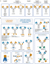
Display the structure of the different molecules as pictures. 4–1BB binding sites are highlighted in yellow and targeting binding sites are highlighted in blue. If molecules are tri- or tetraspecific additional binding domains are indicated in dark gray and dark green. The molecules are clustered by ratio of binding sites (4–1BB binding sites to other binding sites). Molecule names as well as important structural characteristics are indicated.
Figure 2.
Different molecule designs of second-generation 4–1BB agonistic drugs. (a) Second generation agonistic human 4–1BB IgGs featuring isotypes as indicated. Sometimes an improved or attenuated binding to FcγRIIB is integrated. Some molecules show controlled 4–1BB binding depending on ATP concentration or de-masking of the binding sites. All IgG-like molecules display bivalent agonistic binding to 4–1BB (yellow). EU101 is not included as the isotype is not disclosed. (b) Most of the 4–1BB agonistic drugs display bi-, tri- or tetra-specificity, e.g., in addition to the specificity to 4–1BB (in yellow) they also recognize at least another binding site (blue, green, and dark gray), whereby the main crosslinking target site is highlighted in blue. If molecules implement an antibody-like format, typically mutations are introduced to abolish Fcγ-receptor and complement binding. Other molecules without a Fc-fragment binding to FcRn display binding to human serum albumin (HSA) to improve in vivo half-life. Molecules are sorted by their ratio of the 4–1BB agonistic binding sites (yellow) and the crosslinking target sites (blue). The format of HLX35/BNA035 and BI 765179 are assumptions based on pictures of the companies’ webpages. BGB-B16 is not implemented as the structure of the bispecific antibody is not disclosed. Abbreviation: 4–1BBL = trimeric 4–1BB Ligand ectodomain, DARPins = Designed ankyrin repeat proteins, Fcab = Fc-region with antigen binding, FcγRIIB = Fc-gamma-receptor IIB, VHH = antigen-binding domain of camelid dimeric heavy chain antibodies, scFv = single-chain variable antibody fragment, sdAb = single monomeric variable antibody domain, SIRPα = Signal regulatory protein alpha ectodomain, VH = variable domain of heavy antibody chain, VHH = antigen-binding domain of camelid dimeric heavy chain antibodies, VH/VL = variable domain of heavy and light antibody chain.
Shows schematically how 4–1BBL and agonistic 4–1BB molecules are interacting with the 4–1BB receptors. The epitopes can be in the CRD1-CDR4 domains and the epitope can be distal or proximal to the effector cell membrane.
Figure 3.
Schematic binding epitopes to the 4–1BB receptor (if disclosed). (a) The trimeric 4–1BBL interacts with CRD2 and CDR3 of three 4–1BB receptors, a type I transmembrane protein. (b) The epitope location of different antibody-based 4–1BB agonists is indicated. Non-4-1BBL-blocking binders are indicated in red, 4–1BBL-blocking binders are indicated in blue.
The figure indicates the needed amounts of molecules and targeting receptors for optimal 4–1BB clustering depending on the valency of the molecule.
Figure 4.
Theoretical impact on hyper-crosslinking based on the different 4–1BB receptor to crosslinking-target binding site ratios. The number of 4–1BB receptors (yellow) is fixed to six receptors, whereas the number of crosslinking targets (blue) and number of drug molecules varies to demonstrate the impact of different binding site ratios. (a) In case of even ratios of crosslinking target to 4–1BB receptor binding sites (1 + 1, 2 + 2 or 3 + 3), a higher number of molecules is needed to gain equal 4–1BB receptor hyper-clustering. (b) An amplification occurs if the ratio of crosslinking target to 4–1BB receptor binding sites is uneven and in favor of 4–1BB binding sites (1 + 2 or 1 + 3). In this case a lower number of crosslinking targets is needed to elicit the same 4–1BB receptor hyper-crosslinking.
The figure is ordering the different molecule designs by size in kDa on a scale. The smallest molecule has a molecular weight of 7.2 kDa the biggest molecules display a molecular weight of around 296 kDa.
Figure 5.
Molecular weight of 4–1BB agonistic drugs. The molecular weight of each molecule was taken from literature or estimated from their formats, and molecules were sorted by their molecular weight in kilo-Dalton (kDa) starting with the smallest molecule (BT7480) on the left and the biggest molecules (~296 kDa) on the right. 4–1BB agonistic binding sites are highlighted in yellow and targeting binding sites are indicated in blue. For the tri-specific molecules binding to HSA is shown in dark gray. For the tetra-specific molecules PD-L1 binding sites are shown in green and CD3 binding sites in dark gray. Abbreviation: 4–1BBL = trimeric 4–1BB Ligand ectodomain, CH/CL = constant domain of heavy and light antibody chain, DARPins = Designed ankyrin repeat proteins, Fcab = Fc-region with antigen binding, scFv = single-chain variable antibody fragment, sdAb = single monomeric variable antibody domain, SIRPα = Signal regulatory protein alpha ectodomain, VH = variable domain of heavy antibody chain, VHH = antigen-binding domain of camelid dimeric heavy chain antibodies, VH/VL = variable domain of heavy and light antibody chain, * = Fc-fragment contains mutations to increase Fcγ-receptor IIB-binding, # = Fc fragment contains mutations to attenuate Fcγ-receptor-binding.
The figure displays the expected locations of 4–1BB activation depending on the crosslinking mechanism (e.g., tumor and lymphoid organs). Further it is indicated which cells are involved in 4–1BB-mediated T cell activation leading to different cell settings. They are clustered into cis-, trans- and autocrine-settings. The names of the molecules are listed in correlation to the expected involved cells, organs, and settings.
Figure 6.
Expected MoA of 4–1BB agonists based on their targeting modality. (a) Summarize the main organs (tumor and lymphoid tissue) where 4–1BB agonists can improve the anti-tumoral immune response as well as the main targeted cells (shown in blue) and 4–1BB+ T cells (shown in yellow). (b) The predicted cellular interaction and MoA are outlined. 4–1BB crosslinking can occur in a cis-, trans- or autocrine-setting. Crosslinking cells can be tumor cells, fibroblasts, macrophages, and dendritic cells (shown in blue) or T cells themselves in an autocrine setting. (c) Names of 4–1BB agonistic drugs in clinical trials, which implement the predicted and outlined MoA. The tetra-specific molecules GNC-035, GNC-038 and GNC-039 are not included. Abbreviations: CAFs = Cancer Associated Fibroblasts, CEACAM5 = Carcinoembryonic Antigen Cell Adhesion Molecule 5, DCs = Dendritic Cells, EGFRvIII = epidermal growth factor receptor variant III, FcγR = Fc-gamma-receptors, FAP = Fibroblast Activating Protein alpha, FRCs = Fibroblastic reticular cells, Her2 = human epidermal growth factor receptor 2, PD-L1 = Programmed Death-Ligand 1, ROR1 = Receptor Tyrosine Kinase Like Orphan Receptor 1, PSMA = Prostate-specific membrane antigen, Mϕ = Macrophages, TAMs = Tumor Associated Macrophages, tdLN = tumor draining lymph node.
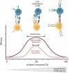
Shows schematically the expected bell shape curved optimal dose curve. The optimal dose window (maximal efficacy) depends on the receptor occupancy and the receptor expression.
Figure 7.
Predicted optimal dose dependent on receptor occupancy leading to a bell-shaped activity curve. Increased concentration of bispecific 4–1BB agonist will lead to saturation of both binding sites while abolishing optimal 4–1BB clustering. Max = maximum.
Display planned or ongoing combination partners which are combined with indicated molecules. Each combination is also linked to the clinical trial number. The combination partners are clustered in groups by function.
Figure 8.
Ongoing or planned combinations in clinical trials. Reported combination partners were set against the clinical trial number and clustered by the used 4–1BB agonist. Most clinical trials combine the 4–1BB agonist with a checkpoint inhibitor (e.g., PD1, PD-L1 or CTLA-4 inhibitor) and/or chemotherapy.

Similar articles

Cited by

References

    1. Jeong S, Park SH.. Co-stimulatory receptors in cancers and their implications for cancer immunotherapy. Immune Netw. 2020;20(e3). doi:10.4110/in.2020.20.e3. - DOI - PMC - PubMed
    1. Kwon BS, Weissman SM. cDNA sequences of two inducible T-cell genes. Proc Natl Acad Sci U S A. 1963-1967:86. doi:10.1073/pnas.86.6.1963. - DOI - PMC - PubMed
    1. Hurtado JC, Kim SH, Pollok KE, Lee ZH, Kwon BS. Potential role of 4-1BB in T cell activation. Comparison with the costimulatory molecule CD28. J Immunol. 1995;155(7):3360–22. doi:10.4049/jimmunol.155.7.3360. - DOI - PubMed
    1. Pollok KE, Kim YJ, Zhou Z, Hurtado J, Kim KK, Pickard RT, Kwon BS. Inducible T cell antigen 4-1BB. Analysis of expression and function. J Immunol. 1993;150(3):771–81. doi:10.4049/jimmunol.150.3.771. - DOI - PubMed
    1. Vinay DS, Kwon BS. 4-1BB signaling beyond T cells. Cell Mol Immunol. 2011;8(281–284):281–84. doi:10.1038/cmi.2010.82. - DOI - PMC - PubMed

Substances

Grants and funding

The author(s) reported there is no funding associated with the work featured in this article.