. 2023 Jul;44(7):1391-1403.
doi: 10.1038/s41401-022-01037-8. Epub 2023 Jan 25.

CB2R agonist GW405833 alleviates acute liver failure in mice via inhibiting HIF-1α-mediated reprogramming of glycometabolism and macrophage proliferation

Affiliations

CB2R agonist GW405833 alleviates acute liver failure in mice via inhibiting HIF-1α-mediated reprogramming of glycometabolism and macrophage proliferation

Sheng-Lan Cai et al. Acta Pharmacol Sin. 2023 Jul.

Abstract

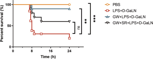
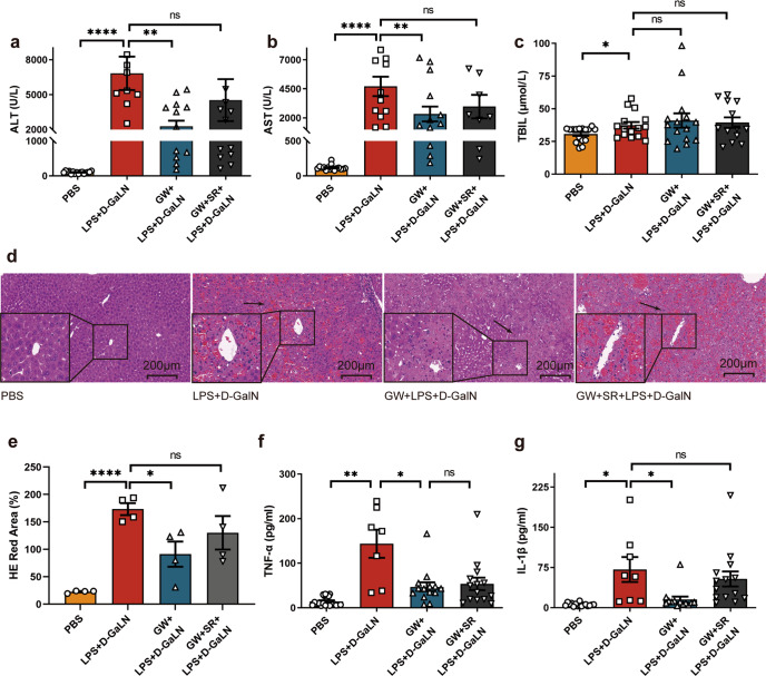
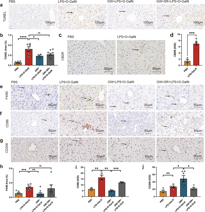
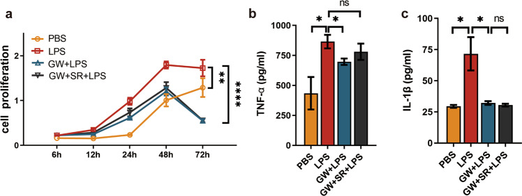
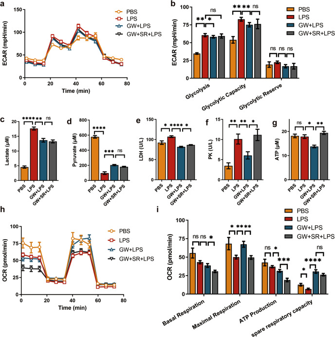
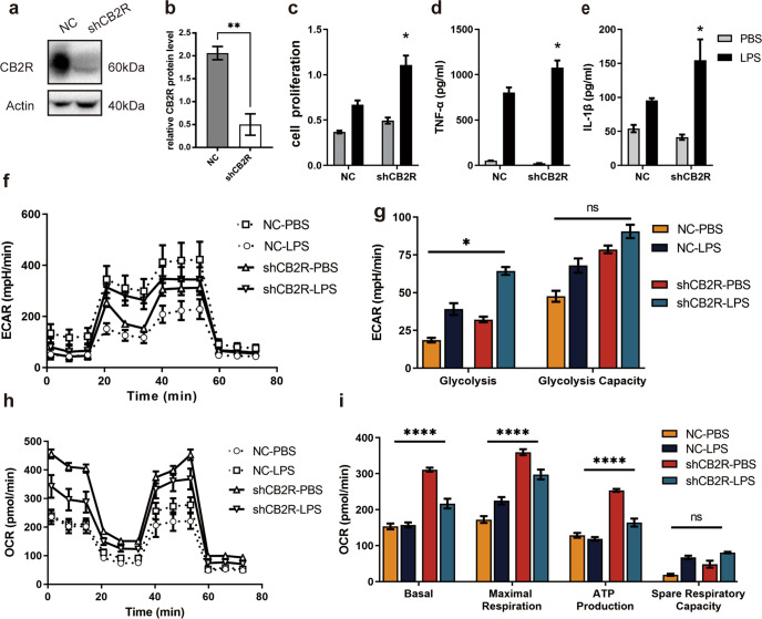
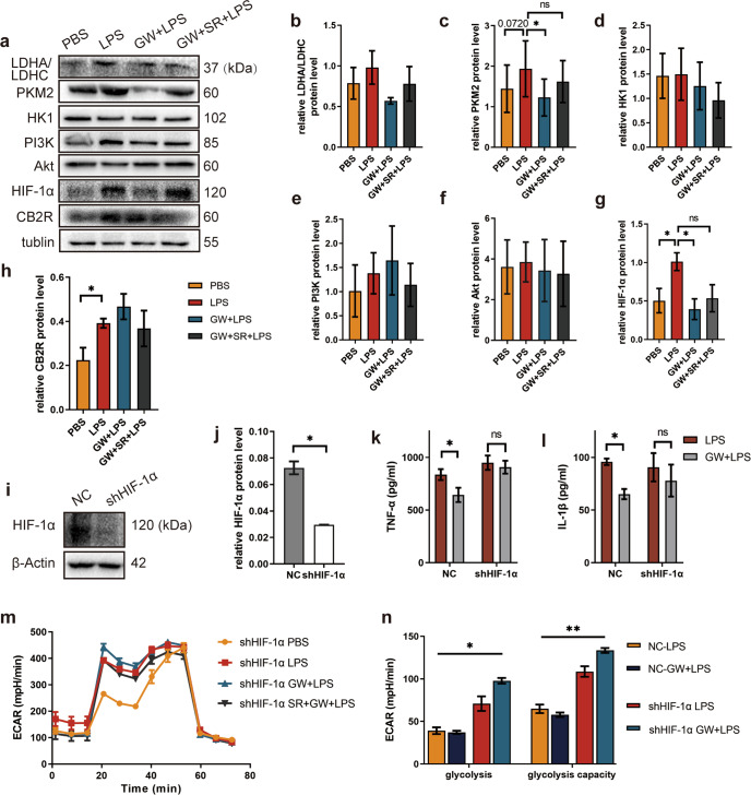
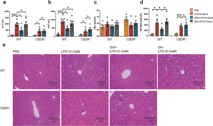
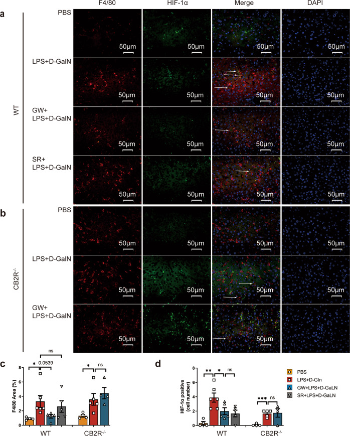
The inflammatory responses involving infiltration and activation of liver macrophages play a vital role in acute liver failure (ALF). In the liver of ALF mice, cannabinoid receptor 2 (CB2R) is significantly upregulated on macrophages, while CB2R agonist GW405833 (GW) could protect against cell death in acute liver damage. In this study, we investigated the molecular mechanisms underlying the protective effects of GW against ALF in vivo and in vitro from a perspective of macrophage glycometabolism. Mice were pretreated with GW (10 mg/kg, i.p.), then were injected with D-GalN (750 mg/kg, i.p.) and LPS (10 mg/kg, i.p.) to induce ALF. We verified the protective effects of GW pretreatment in ALF mice. Furthermore, GW pretreatment significantly reduced liver macrophage infiltration and M1 polarization, and inhibited the release of inflammatory factors TNF-α and IL-1β in ALF mice. These protective effects were eliminated by CB2R antagonist SR144528 or in CB2R-/- ALF mice. We used LPS-stimulated RAW264.7 cells as an in vitro M1 macrophage-centered model of inflammatory response, and demonstrated that pretreatment with GW (10 μM) significantly reduced glucose metabolism by inhibiting glycolysis, which inhibited LPS-induced macrophage proliferation and inflammatory cytokines release. We verified these results in a stable CB2R-/- RAW264.7 cell line. Moreover, we found that GW significantly inhibited the expression of hypoxia inducible factor 1α (HIF-1α). Using a stable HIF-1α-/- RAW264.7 cell line, we confirmed that GW reduced the release of inflammatory cytokines from macrophages and inhibited glycolysis by downregulating HIF-1α expression. In conclusion, activation of CB2Rs inhibits the proliferation of hepatic macrophages and release of inflammatory factors in ALF mice through downregulating HIF-1α to inhibit glycolysis.

Keywords: GW405833; RAW264.7 cells; SR144528; acute liver failure; cannabinoid receptor 2; glycolysis; hypoxia-inducible factor 1α; macrophages.


Conflict of interest statement

The authors declare no competing interests.

Figures

Fig. 1
Fig. 1. Survival analysis of grouped mice.
Kaplan-Meier survival curves of BALB/C mice (n = 8, 13,10, 10 for group with PBS, D-GalN +LPS, GW + D-GalN +LPS, and GW + SR + D-GalN +LPS treatment respectively). (ns, P > 0.05; **P < 0.01; ***P < 0.001)
Fig. 2
Fig. 2. Protective effect of GW in ALF.
a ALT (n = 16, 8, 12, 11 respectively), b AST (n = 16, 11, 11, 8 respectively), c TBIL (n = 16, 15, 15, 13 respectively) in serum of mice was detected 8 h after treatment with PBS, D-GalN +LPS, GW + D-GalN +LPS, or GW + SR + D-GalN +LPS. d HE staining of liver sections of mice at time point 8 h after PBS, D-GalN +LPS, GW + D-GalN +LPS, or GW + SR + D-GalN +LPS treatment. e Statistical analysis of red area in liver section HE staining (n = 4). f TNF-α (n = 13, 7, 14, 14 respectively), g IL-1β (n = 11, 8, 13, 14 respectively) in mice serum was detected 8 h after treatment with PBS, D-GalN +LPS, GW + D-GalN +LPS, or GW + SR + D-GalN +LPS. (ns, P > 0.05; *P < 0.05; **P < 0.01; ****P < 0.0001)
Fig. 3
Fig. 3. Effects of CB2R activation on macrophage activation, inflammation, and apoptosis of liver cells in ALF.
a TUNEL staining of liver sections of mice at 6 h after PBS, D-GalN +LPS, GW + D-GalN +LPS, GW + SR + D-GalN +LPS treatment. b Statistical analysis of TUNEL staining in liver section (n = 8, 6, 4, 9 respectively). c Immunohistochemically labeled with CB2R of mice liver sections 8 h after PBS and D-GalN +LPS treatment. d Statistical analysis of CB2R immunohistochemical staining in liver section (n = 3, 6 respectively). Immunohistochemically labeled with e F4/80+, f CD86, g CD206 of mice liver sections 8 h after PBS, D-GalN +LPS, GW + D-GalN +LPS, or GW + SR + D-GalN +LPS treatment. Statistical analysis of h F4/80+ (n = 7, 6, 9, 6 respectively), i CD86 (n = 3), j CD206 (n = 4, 6, 6, 4, respectively) immunohistochemical staining in liver section. (ns, P > 0.05; *P < 0.05; **P < 0.01; ***P < 0.001; ****P < 0.0001)
Fig. 4
Fig. 4. Effect of CB2R activation on macrophage proliferation and release of inflammatory cytokines.
a Different proliferation of RAW264.7 cells after grouped treatment (n = 6). b TNF-α (n = 5), c IL-1β (n = 6) concentrations in cell supernatant after grouping treatment. (ns, P > 0.05; *P < 0.05; **P < 0.01; ****P < 0.0001)
Fig. 5
Fig. 5. Effect of CB2R activation on glucose metabolism in LPS-activated macrophages.
a ECAR at each point, b Glycolysis parameters of glycolysis stress test (n = 6 for each group). c Lactate, d Pyruvate concentration was detected in grouped treatment cell supernatant (n = 5 for each group). e LDH (n = 4), and f PK (n = 5) activity of grouped treatment cells were detected. g ATP production of grouped treatment cells (n = 3). h OCR at each point, i Aerobic respiration parameters of mitochondrial stress test of grouped treatment RAW264.7 cell (n = 6 for each group). (ns, P > 0.05; *P < 0.05; **P < 0.01; ***P < 0.001; ****P < 0.0001)
Fig. 6
Fig. 6. Effects of CB2R knockdown for LPS-activated macrophages on cell proliferation, release of inflammatory factors and glucose metabolism.
a CB2R knockdown efficiency of RAW264.7 cells were detected by Western blotting. (b) Semi-quantity statistical analysis of CB2R knockdown efficiency Western blotting (n = 3). c Time point 24 h cell proliferation test (n = 6 for each group). d TNF-α, e IL-1β concentration in cell supernatant after the indicated treatment (n = 5 for each group). f ECAR at each point, g Glycolysis parameters of glycolysis stress test of cells after the indicated treatment (n = 6 for each group). h OCR at each point, i Aerobic respiration parameter of mitochondrial stress test (n = 6 for each group). (ns, P > 0.05; *P < 0.05; **P < 0.01; ****P < 0.0001)
Fig. 7
Fig. 7. Effect of HIF-1α knockdown on GW activated CB2R to alter LPS-stimulated macrophages.
a Glycolysis related enzymes and signal proteins detected by Western Blotting. Semi-quantity statistical analysis of b LDHA/LDHC, c PKM2, d HK1, e PI3K, f Akt, g HIF-1α, h CB2R expression detected by Western blotting (n = 3). i HIF-1α knockdown efficiency of RAW264.7 cells were detected by Western blotting. j Semi-quantity statistical analysis of HIF-1α knockdown efficiency detected by Western blotting (n = 3). k TNF-α, l IL-1β concentration in cell supernatant after the indicated treatment (n = 5 for each group). m ECAR at each point, n Glycolysis parameters of glycolysis stress test of cells after the the indicated treatment (n = 6 for each group). (ns, P > 0.05; *P < 0.05; **P < 0.01)
Fig. 8
Fig. 8. Differential liver damage in WT and CB2R−/− mice.
a ALT, b AST, c TBIL (n = 6, 7, 6, 6, 4, 5, 4, respectively) in serum of mice, and d statistical analysis of red area in liver section HE staining (n = 3, 5, 6, 6, 3, 5, 3, respectively) detected 6 h after the indicated treatment with PBS, D-GalN +LPS, GW + D-GalN +LPS, or SR + D-GalN +LPS in WT mice, and PBS, D-GalN +LPS, or GW + D-GalN +LPS in CB2R−/− mice; e Pathological changes of mouse liver tissue after indicated treatment were examined by HE staining. (ns, P > 0.05; *P < 0.05; ***P < 0.001; ****P < 0.0001)
Fig. 9
Fig. 9. Differential expression of HIF-1α in liver macrophages of ALF in WT and CB2R−/− mice.
Liver sections of mice were treated with F4/80+ and HIF-1α immunofluorescence co-staining 6 h after indicated treatment (×400) in a WT mice and b CB2R−/− mice. c Quantitative analysis of F4/80+ expression by Image J. d Analysis of liver macrophage with HIF-1α staining positive. (n = 4, 6, 5, 5, 4, 5, 4, respectively). (ns, P > 0.05; *P < 0.05; **P < 0.01; ***P < 0.001)

Similar articles

Cited by

References

    1. Louvet A, Teixeira-Clerc F, Chobert MN, Deveaux V, Pavoine C, Zimmer A, et al. Cannabinoid CB2 receptors protect against alcoholic liver disease by regulating Kupffer cell polarization in mice. Hepatology. 2011;54:1217–26. doi: 10.1002/hep.24524. - DOI - PubMed
    1. Chen J, Huang ZB, Li H, Zheng X, Chen JJ, Wang XB, et al. Early diagnostic biomarkers of sepsis for patients with acute-on-chronic liver failure: a multicenter study. Infect Dis Ther. 2021;10:281–90. doi: 10.1007/s40121-020-00362-x. - DOI - PMC - PubMed
    1. Chen J, Huang ZB, Fan XG, Hu XW, Qi M, Liao CJ, et al. Potential predictors for prognosis and postpartum recovery time of acute fatty liver of pregnancy. BMC Pregnancy Childbirth. 2020;20:601. doi: 10.1186/s12884-020-03287-y. - DOI - PMC - PubMed
    1. Olah A, Szekanecz Z, Biro T. Targeting cannabinoid signaling in the immune system: “High”-ly exciting questions, possibilities, and challenges. Front Immunol. 2017;8:1487. doi: 10.3389/fimmu.2017.01487. - DOI - PMC - PubMed
    1. Krenkel O, Tacke F. Liver macrophages in tissue homeostasis and disease. Nat Rev Immunol. 2017;17:306–21. doi: 10.1038/nri.2017.11. - DOI - PubMed

MeSH terms

Substances