MATR3 P154S knock-in mice do not exhibit motor, muscle or neuropathologic features of ALS
- PMID: 36689813
- PMCID: PMC10046992
- DOI: 10.1016/j.bbrc.2023.01.032
MATR3 P154S knock-in mice do not exhibit motor, muscle or neuropathologic features of ALS
Abstract
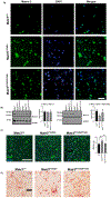
Matrin 3 is a nuclear matrix protein that has many roles in RNA processing including splicing and transport of mRNA. Many missense mutations in the Matrin 3 gene (MATR3) have been linked to familial forms of amyotrophic lateral sclerosis (ALS) and distal myopathy. However, the exact role of MATR3 mutations in ALS and myopathy pathogenesis is not understood. To demonstrate a role of MATR3 mutations in vivo, we generated a novel CRISPR/Cas9 mediated knock-in mouse model harboring the MATR3 P154S mutation expressed under the control of the endogenous promoter. The P154S variant of the MATR3 gene has been linked to familial forms of ALS. Heterozygous and homozygous MATR3 P154S knock-in mice did not develop progressive motor deficits compared to wild-type mice. In addition, ALS-like pathology did not develop in nervous or muscle tissue in either heterozygous or homozygous mice. Our results suggest that the MATR3 P154S variant is not sufficient to produce ALS-like pathology in vivo.
Keywords: ALS; MATR3 P154S mutation; Matrin 3; Mouse model; Neuropathology.
Copyright © 2023 Elsevier Inc. All rights reserved.
Conflict of interest statement
Declaration of competing interest The authors declare that they have no known competing financial interests or personal relationships that could have appeared to influence the work reported in this paper.
Figures


Similar articles
-
MATR3 F115C knock-in mice do not exhibit motor defects or neuropathological features of ALS.Biochem Biophys Res Commun. 2021 Sep 3;568:48-54. doi: 10.1016/j.bbrc.2021.06.052. Epub 2021 Jun 26. Biochem Biophys Res Commun. 2021. PMID: 34182213
-
N-terminal sequences in matrin 3 mediate phase separation into droplet-like structures that recruit TDP43 variants lacking RNA binding elements.Lab Invest. 2019 Jul;99(7):1030-1040. doi: 10.1038/s41374-019-0260-7. Epub 2019 Apr 24. Lab Invest. 2019. PMID: 31019288 Free PMC article.
-
Selective neuronal degeneration in MATR3 S85C knock-in mouse model of early-stage ALS.Nat Commun. 2020 Oct 20;11(1):5304. doi: 10.1038/s41467-020-18949-w. Nat Commun. 2020. PMID: 33082323 Free PMC article.
-
MATR3's Role beyond the Nuclear Matrix: From Gene Regulation to Its Implications in Amyotrophic Lateral Sclerosis and Other Diseases.Cells. 2024 Jun 5;13(11):980. doi: 10.3390/cells13110980. Cells. 2024. PMID: 38891112 Free PMC article. Review.
-
Matrin 3 in neuromuscular disease: physiology and pathophysiology.JCI Insight. 2021 Jan 11;6(1):e143948. doi: 10.1172/jci.insight.143948. JCI Insight. 2021. PMID: 33427209 Free PMC article. Review.
Cited by
-
The role of Matrin-3 in physiology and its dysregulation in disease.Biochem Soc Trans. 2024 Jun 26;52(3):961-972. doi: 10.1042/BST20220585. Biochem Soc Trans. 2024. PMID: 38813817 Free PMC article. Review.
References
-
- Paganoni S, Macklin EA, Hendrix S, Berry JD, Elliott MA, Maiser S, Karam C, Caress JB, Owegi MA, Quick A, Wymer J, Goutman SA, Heitzman D, Heiman-Patterson T, Jackson CE, Quinn C, Rothstein JD, Kasarskis EJ, Katz J, Jenkins L, Ladha S, Miller TM, Scelsa SN, Vu TH, Fournier CN, Glass JD, Johnson KM, Swenson A, Goyal NA, Pattee GL, Andres PL, Babu S, Chase M, Dagostino D, Dickson SP, Ellison N, Hall M, Hendrix K, Kittle G, McGovern M, Ostrow J, Pothier L, Randall R, Shefner JM, Sherman AV, Tustison E, Vigneswaran P, Walker J, Yu H, Chan J, Wittes J, Cohen J, Klee J, Leslie K, Tanzi RE, Gilbert W, Yeramian PD, Schoenfeld D, Cudkowicz ME, Trial of Sodium Phenylbutyrate–Taurursodiol for Amyotrophic Lateral Sclerosis, N. Engl. J. Med 383 (2020) 919–930. 10.1056/NEJMoa1916945. - DOI - PMC - PubMed
-
- Feit H, Silbergleit A, Schneider LB, Gutierrez JA, Fitoussi RP, Réyès C, Rouleau GA, Brais B, Jackson CE, Beckmann JS, Seboun E, Vocal cord and pharyngeal weakness with autosomal dominant distal myopathy: clinical description and gene localization to 5q31, Am. J. Hum. Genet 63 (1998) 1732–1742. 10.1086/302166. - DOI - PMC - PubMed
-
- Senderek J, Garvey SM, Krieger M, Guergueltcheva V, Urtizberea A, Roos A, Elbracht M, Stendel C, Tournev I, Mihailova V, Feit H, Tramonte J, Hedera P, Crooks K, Bergmann C, Rudnik-Schöneborn S, Zerres K, Lochmüller H, Seboun E, Weis J, Beckmann JS, Hauser MA, Jackson CE, Autosomal-Dominant Distal Myopathy Associated with a Recurrent Missense Mutation in the Gene Encoding the Nuclear Matrix Protein, Matrin 3, Am. J. Hum. Genet 84 (2009) 511–518. 10.1016/j.ajhg.2009.03.006. - DOI - PMC - PubMed
Publication types
MeSH terms
Substances
Grants and funding
LinkOut - more resources
Full Text Sources
Medical
Molecular Biology Databases
Research Materials
Miscellaneous

