. 2023 Jan 23;21(1):e3001693.
doi: 10.1371/journal.pbio.3001693. eCollection 2023 Jan.

The enterovirus genome can be translated in an IRES-independent manner that requires the initiation factors eIF2A/eIF2D

Affiliations

The enterovirus genome can be translated in an IRES-independent manner that requires the initiation factors eIF2A/eIF2D

Hyejeong Kim et al. PLoS Biol. .

Abstract

RNA recombination in positive-strand RNA viruses is a molecular-genetic process, which permits the greatest evolution of the genome and may be essential to stabilizing the genome from the deleterious consequences of accumulated mutations. Enteroviruses represent a useful system to elucidate the details of this process. On the biochemical level, it is known that RNA recombination is catalyzed by the viral RNA-dependent RNA polymerase using a template-switching mechanism. For this mechanism to function in cells, the recombining genomes must be located in the same subcellular compartment. How a viral genome is trafficked to the site of genome replication and recombination, which is membrane associated and isolated from the cytoplasm, is not known. We hypothesized that genome translation was essential for colocalization of genomes for recombination. We show that complete inactivation of internal ribosome entry site (IRES)-mediated translation of a donor enteroviral genome enhanced recombination instead of impairing it. Recombination did not occur by a nonreplicative mechanism. Rather, sufficient translation of the nonstructural region of the genome occurred to support subsequent steps required for recombination. The noncanonical translation initiation factors, eIF2A and eIF2D, were required for IRES-independent translation. Our results support an eIF2A/eIF2D-dependent mechanism under conditions in which the eIF2-dependent mechanism is inactive. Detection of an IRES-independent mechanism for translation of the enterovirus genome provides an explanation for a variety of debated observations, including nonreplicative recombination and persistence of enteroviral RNA lacking an IRES. The existence of an eIF2A/eIF2D-dependent mechanism in enteroviruses predicts the existence of similar mechanisms in other viruses.


Conflict of interest statement

The authors have declared that no competing interests exist.

Figures

Fig 1
Fig 1. Hypothesis: Polyprotein determinants presented during translation direct the enteroviral genome to the site of replication.
For recombination to occur between two genetically distinct PV genomes, these genomes must be in the same replication organelle. (A, B) Viral RNAs, shown in blue and red, are targeted to the replication organelle while being translated and facilitate RNA recombination between the two colocalized genomes. Specific viral polyprotein determinants, indicated by a black arrow, mediate this trafficking while still a part of the polyprotein and in association with the ribosome. (C, D) In a case where one genome with impaired polyprotein synthesis, shown in red, is also infected by a second genome, shown in blue, the genome with impaired polyprotein synthesis cannot be trafficked to the replication organelle, so recombination between the two genomes cannot occur. This figure was created by Efraín E. Rivera-Serrano using BioRender. PV, poliovirus; RO, replication organelle.
Fig 2
Fig 2. RNA virus recombinants are recovered after deletion of the PV IRES in a donor subgenomic replicon RNA.
(A) Schematic of the cell-based assay for PV recombination [13,14,75]. Two RNAs are used in the assay, a replication-competent PV subgenomic RNA (Donor, red) in which capsid-coding sequence is replaced with luciferase coding-sequence and a replication-incompetent full-length PV genomic RNA (Acceptor, blue) with a defective cis-acting replication element (indicated by red *). Cotransfection of these RNAs in a L929 mouse fibroblast cell line produces infectious virus if recombination occurs resulting in a replication-competent, full-length PV genomic RNA. Infectious virus produced by recombination can be quantified by plaque assay using HeLa cells. (B) Comparison of infectious virus produced between donor RNAs with either an intact IRES (①) or when the entire IRES and majority of luciferase coding sequence (nt 41–2,393, ΔIRES) was deleted (②) [13]. (C) Recombination between the donor with the deleted IRES (ΔIRES) and acceptor RNA (② x ③) produces 5-fold more recombinant virus compared to the corresponding control (① x ③). Results show titer of recombinant virus (pfu/mL ± SEM; n = 3). Statistical analyses were performed using unpaired, two-tailed t test (* indicates p < 0.05). Numerical data provided as Supporting information (S1 Data). IRES, internal ribosome entry site; PV, poliovirus.
Fig 3
Fig 3. RNA virus recombinants are consistent with the donor RNA as the source of the PV RdRp, 3Dpol, and not the acceptor RNA.
(A) A mutation (GDD to GAA) producing an inactive PV RdRp, shown by a black diamond (◊), was introduced into either the donor or acceptor RNAs (② and ④). Insertion of two STOP codons (UAGUAA) after the 3B-coding sequence (3B STOP), indicated by a red rectangle, was introduced into the acceptor RNAs (③ and ④). (B) The indicated donor and acceptor RNAs were cotransfected into L929 cells. Yields of recombinant virus following transfection are shown (pfu/mL ± SEM; n = 3).—indicates that plaques were not detected (limit of detection: 2 pfu/mL). Statistical analyses were performed using unpaired, two-tailed t test (n.s. indicates not significant). Viral recombinants were recovered only when the donor had an intact 3D gene encoding an active RdRp (① x ③, ① x ④); viral recombinants were not recovered when the donor RNA encoded an inactive RdRp (② x ③, ② x ④). There was no impact on viral recombinants produced when the acceptor RNA had stop codons upstream of the 3D gene encoding an active or inactive RdRp (① x ③, ① x ④). Numerical data provided as Supporting information (S1 Data). PV, poliovirus; RdRp, RNA-dependent RNA polymerase.
Fig 4
Fig 4. A 10-nt deletion in the IRES that is known to prevent translation recapitulates phenotypes observed with a deleted IRES.
(A) The donor RNA was engineered to contain a 10-nucleotide deletion (nt 185–189, nt 198–202) known to disrupt the IRES (referred to as ΔSLII-3) [40] with an active or an inactive RdRp. (B) Comparison of infectious virus produced between the indicated donor and acceptor RNAs with the specific modifications. Results show titer of recombinant virus (pfu/mL ± SEM; n = 3).indicates that plaques were not detected (limit of detection: 2 pfu/mL). Statistical analyses were performed using unpaired, two-tailed t test (n.s. indicates not significant). Viral recombinants were recovered using a donor RNA containing the ΔSLII-3 and an active RdRp with the indicated acceptor RNAs (① x ③, ① x ④); viral recombinants were not recovered when the donor RNA contained ΔSLII-3 and encoded an inactive RdRp (② x ③). Numerical data provided as Supporting information (S1 Data). IRES, internal ribosome entry site; RdRp, RNA-dependent RNA polymerase.
Fig 5
Fig 5. Evidence for IRES-independent translation of the enterovirus A71 genome.
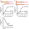
(A) Subgenomic replicon luciferase activity using an EV-A71 C2 subgenomic replicon with the entire IRES (nt 38–767) deleted (ΔIRES) [15]. As a control, the WT subgenomic replicon RNA was transfected in the presence of GuHCl, a replication inhibitor. Luciferase activity is reported in RLUs as a function of time posttransfection. Numerical data provided as Supporting information (S1 Data). (B, C) Comparison of infectious virus generated between the indicated donor and acceptor RNAs with the specific modifications. The indicated set of donor and acceptor RNAs was cotransfected into RD cells. Results show titer of recombinant virus (pfu/mL ± SEM; n = 3).—indicates that plaques were not detected (limit of detection: 2 pfu/mL). Statistical analyses were performed using unpaired, two-tailed t test (n.s. indicates not significant). Viral recombinants were recovered only when the donor had an intact 3D gene encoding an active RdRp (① x ③, ① x ④); viral recombinants were not recovered when the donor RNA encoded an inactive RdRp (② x ③). Numerical data provided as Supporting information (S1 Data). GuHCl, guanidine hydrochloride; IRES, internal ribosome entry site; RD, rhabdomyosarcoma; RdRp, RNA-dependent RNA polymerase; RLU, relative light unit; WT, wild type.
Fig 6
Fig 6. IRES-independent translation may initiate downstream of luciferase-coding sequence within the nonstructural protein-coding sequence.
(A, B) Subgenomic replicon luciferase assay comparing the depicted RNAs: WT vs. ΔIRES (panel A) and WT vs. ΔSLII-3 RNAs (panel B). As a control, the WT RNA was transfected in the presence of GuHCl. Luciferase activity is reported in RLUs as a function of time posttransfection. Luciferase units for ΔIRES and ΔSLII-3 were not detected above 101. Numerical data provided as Supporting information (S1 Data). (C) Luciferase activity observed for a serial dilution of purified recombinant luciferase enzyme. The initial amount of luciferase in the reaction was 12.6 μg. The limit of detection was reached at a 1 × 10⁷ fold dilution (1.26 pg luciferase), indicated by the red arrow. This corresponds to 1.2 × 10⁷ molecules of luciferase and 1,200 molecules of luciferase per cell from RNAs containing either a deleted IRES or ΔSLII-3. Numerical data provided as Supporting information (S1 Data). GuHCl, guanidine hydrochloride; IRES, internal ribosome entry site; RLU, relative light unit, wild-type.
Fig 7
Fig 7. Induction and redistribution of PI4P serve an indirect method to detect production of 3CD by the IRES-independent translation mechanism.
Immunofluorescence of cells after transfection. HeLa cells were transfected with in vitro transcribed subgenomic replicon RNAs: WT; ΔIRES, and a full-length genomic RNA with two STOP codons after the 3B-coding sequence: 3B STOP. WT and ΔIRES transfected cells were also treated with 3 mM GuHCl. Six hours posttransfection, cells were immunostained for the presence of PI4P (red) and 3D (green). Nucleus was stained with DAPI (blue). Mock represents cells that were taken through the transfection protocol in the absence of RNA. PI4P was induced and redistributed in cells transfected with WT and ΔIRES, both in the absence and presence of GuHCl, but not 3B STOP. 3D was detected in cells transfected with WT in the absence of GuHCl only. Scale bars are equivalent to 10 μm. GuHCl, guanidine hydrochloride; IRES, internal ribosome entry site; PI4P, phosphatidylinositol-4-phosphate; WT, wild-type.
Fig 8
Fig 8. Activation of PKR and phosphorylation of eIF2α phosphorylation in response to transfection of PV subgenomic replicon RNA.
Western blot analysis of p-PKR (T446) (panel A), PKR (panel B), p-eIF2α (S51) (panel C), and eIF2α (panel D) in HeLa cell lysates. Cells were transfected individually with PV subgenomic replicon RNAs: ΔIRES, 3B STOP, and WT or with WT in the presence of 3 mM GuHCl (WT_GuHCl). Six hours posttransfection, cells were processed for western blot analysis and probed using anti-p-PKR (T446), PKR, p-eIF2α (S51), and eIF2α antibodies; α/β tubulin was used as a loading control. Blots provided in Supporting information (S1 Raw Images). GuHCl, guanidine hydrochloride; PKR, RNA-activated protein kinase; PV, poliovirus; T446, threonine-446; WT, wild-type.
Fig 9
Fig 9. Deletion of a conserved AUG in the 2A-coding sequence reduces translation of the donor RNA leading to a reduction in viral RNA recombinants.
(A) Primary amino acid sequence alignment of a portion of 2A sequence from PV, EV-A71, CVB3, and EV-D68. Numbers refer to 2A protein sequence. The conserved methionine is shown in red. The sites for the insertion of two STOP codons are shown in red; the codons AAU and UAC were changed to UAG and UAA, respectively. (B) Comparison of infectious virus produced by the indicated donor RNAs with the specified modifications: ①: ΔIRES; ②: Insertion of two STOP codons after the 2A-coding sequence (2A STOP); ③: AUG to UUG; ④: AUG to UUU; ⑤: ΔAUG. Sites for the modifications are depicted. In all cases, the acceptor RNA contained two STOP codons after 3B-coding sequence (3B STOP) and the mutation that inactivates the RdRp (GDD to GAA). Indicated are the relative viral titers with the average viral titer from recombination using ΔIRES donor (①) and acceptor set as 100% (7,500 pfu/mL, mean ± SEM; n = 3). Statistical analyses were performed using unpaired, two-tailed t test (* indicates p < 0.05, *** indicates p < 0.001, n.s. indicates not significant). The 2A STOP and ΔAUG reduced viral recombinants, while the AUG to UUG and AUG to UUU did not. Numerical data provided as Supporting information (S1 Data). IRES, internal ribosome entry site; PV, poliovirus; RdRp, RNA-dependent RNA polymerase.
Fig 10
Fig 10. eIF2A and eIF2D initiation factors contribute to IRES-independent translation.
(A) Start codons utilized by eIF2, eIF2A, and eIF2D initiation factors [32]. Amino acids for each codon is in parentheses. Initiation factor-specific start codons are indicated in red. (B) Comparison of infectious virus produced by the indicated donor RNAs with modifications that alter the conserved AUG to initiation factor specific start codons: ①: ΔIRES; ②: ΔAUG; ③: AUG to UUG; ④: AUG to UUU; ⑤: AUG to CUG; ⑥: AUG to CUA; ⑦: AUG to CUC. Codons UUG, UUU, and CUG can be utilized by initiation factors, but CUA and CUC cannot. In all cases, the acceptor RNA contained two STOP codons after 3B-coding sequence (3B STOP) and the mutation that inactivates the RdRp (GDD to GAA). The indicated set of donor and acceptor RNAs in a 1:5 molar ratio (total 0.3 μg) was cotransfected into HAP1 WT cells. Indicated are the relative viral titers with the average viral titer from recombination using ΔIRES donor (①) and acceptor set as 100% (647 pfu/mL, mean ± SEM; n = 3). Statistical analyses were performed using unpaired, two-tailed t test (*significance level p = < 0.05, ** p = < 0.01, n.s. indicates not significant). The ΔAUG, CUA, and CUC reduced viral recombinants significantly, while UUG, UUU, and CUG did not, consistent with eIF2A and eIF2D contributing to IRES-independent translation. Numerical data provided as Supporting information (S1 Data). (C, D) Viral recombinants are reduced in HAP1 cells deficient for eIF2A and eIF2D expression. The indicated set of donor and acceptor RNAs was cotransfected into HAP1 WT or eIF2A-KO or eIF2D-KO cells. Donor RNA: ΔIRES (panel C); RLuc-WT (panel D). Relative viral titers with the average viral titer from HAP1 WT set as 100% were shown (panel C, 7,433 pfu/mL; panel D, 243 pfu/mL; mean ± SEM). Statistical analyses were performed using unpaired, two-tailed t test (**** indicates p < 0.0001). Numerical data provided as Supporting information (S1 Data). IRES, internal ribosome entry site; KO, knockout; RdRp, RNA-dependent RNA polymerase; WT, wild-type.
Fig 11
Fig 11. Identification of an RNA sequence upstream of the AUG codon required for eIF2A/2D-dependent translation.
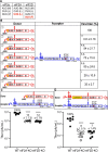
(A) Nucleotide substitutions at each position across the EV-A71 genome have been defined by deep RNA sequencing. The average substitution frequency is 38. Some regions exhibit a lower average; these regions of the genome may encode cis-acting RNA elements. Numerical data provided as Supporting information (S1 Data). The entire data set is available at the GEO repository under accession number GSE183959. (B) The region from 3,602–3,679 of the EV-A71 genome exhibits a below-average frequency of nucleotide substitutions. This region corresponds to a sequence 78-nt upstream of the AUG used for eIF2A/2D-dependent translation. Numerical data provided as Supporting information (S1 Data). (C) Alignment of the corresponding nucleotide sequence and amino acid sequence reveals moderate to high sequence identity among enterovirus (PV, EV-A71, CVB3, and EV-D68). Black asterisks indicate conservation across all enteroviruses; red asterisks indicate conservation in three of the four enteroviruses. (D) RNA secondary structure is predicted in this region for all enteroviruses. The RNAfold algorithm was used. The details of the fold varied across the enteroviruses more substantially than sequence might predict. (E) Comparison of infectious PV produced using donor RNAs harboring a deletion of the 78-nt sequence (Δ78 nt) or containing the corresponding sequences from EV-A71 or EV-D68. The acceptor RNA used does not support translation of 3CD or an active polymerase. Virus produced (pfu/mL ± SEM; n = 3) from each cotransfection is shown. Statistical analyses were performed using unpaired, two-tailed t test (* indicates p < 0.05, ** indicates p < 0.01; n.s. indicates nonsignificant). The sequence contributes to production of virus. Numerical data provided as Supporting information (S1 Data). (F) Contribution of the 78-nt sequence to IRES-independent translation indirectly by monitoring PI4P levels and localization as described in the legend to Fig 7. Donor RNAs of panel E were used, in addition to the ΔAUG donor RNA used in Fig 9 that has a strong defect to virus production. Quantitation of PI4P staining in approximately 60 cells selected randomly from three separate fields expressed as fluorescence intensity is shown. A significant reduction of PI4P/translation of the genome (p < 0.0001) was observed for Δ78 and ΔAUG only. Scale bars are equivalent to 10 μm. Numerical data provided as Supporting information (S1 Data). GEO, Gene Expression Omnibus; IRES, internal ribosome entry site; PI4P, phosphatidylinositol-4-phosphate; PV, poliovirus.
Fig 12
Fig 12. Contribution of eIF2A and eIF2D to translation of enteroviral RNAs.
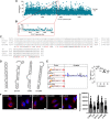
(A) NanoLuc activity in HAP1 WT cells transfected with a full-length PV genome with the nanoLuc-coding sequence embedded between 2C- and 3A-coding regions (2C/3A-Nluc). As a control, the PV 2C/3A-Nluc RNA was transfected in the presence of GuHCl. Numerical data provided as Supporting information (S1 Data). (B) Comparison of nanoLuc activity at six hours posttransfection using PV 2C/3A-Nluc RNA in HAP1 WT, eIF2A-KO, and eIF2D-KO cells. Data from one of two biological replicates with similar results, each with two technical replicates. Numerical data provided as Supporting information (S1 Data). (C) Comparison of nanoLuc activity at six hours postinfection using an MOI of 0.1 or 1 in HAP1 WT, eIF2A-KO, and eIF2D-KO cells. Data from one of two biological replicates with similar results, each with two technical replicates. Numerical data provided as Supporting information (S1 Data). (D) Comparison of nanoLuc activity at six hours posttransfection using PV 2C/3A-Nluc RNA (Transfection), and at six hours postinfection using an MOI of 0.1 or 1 (Infection) in HAP1 WT, and eIF2A-KO/eIF2D-KO cells. Data from one of two biological replicates with similar results, each with two or three technical replicates. Statistical analyses were performed using unpaired, two-tailed t test (** indicates p < 0.01, n.s. indicates not significant). Numerical data provided as Supporting information (S1 Data). (E-G) Western blot analysis of eIF2A (panel E), eIF2D (panel F), and eIF2α (panel G) in HAP1 WT, eIF2A-KO, eIF2D-KO, and eIF2A-KO/eIF2D-KO cells. Cells were processed for western blot analysis and probed using anti-eIF2A, eIF2D, and eIF2α antibodies. GAPDH and tubulin were used as a loading control for western blot. Blots provided in Supporting information (S1 Raw Images). GuHCl, guanidine hydrochloride; KO, knockout; PV, poliovirus; RLU, relative light unit; WT, wild-type.
Fig 13
Fig 13. Single-cell analysis suggests a role for eIF2A and eIF2D during infection.
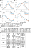
Single-cell analysis [50,51] using an MOI of 5 of PV-eGFPPV in HAP1 WT, eIF2A-KO, and eIF2D-KO cells. Comparison of the distributions of each parameter: start point (panel A); maximum (panel B); slope (panel C); and infection time (panel D); is shown (E) Percentage of PV-infected cells using HAP1 WT, eIF2A-KO, and eIF2D-KO cells (mean ± SD, n = 3) (Left). Adjusted P values of the t tests (Right). (F, G) Quantitative analysis from the data presented in panels A-D. Shown are the mean and standard deviation for each of the indicated parameters (panel F). Adjusted P values of the t tests (panel G). Numerical data provided as Supporting information (S1 Data). hpi, hours postinfection; KO, knockout; NFI, normalized fluorescence intensity; PV, poliovirus; WT, wild-type.
Fig 14
Fig 14. Models for initiation of translation on the enterovirus genome.
(A) Under normal conditions, eIF4G and eIF4A bind to the primary IRES and recruit the 43S preinitiation complex composed of the 40S ribosomal subunit (yellow), eIF2/GTP/Met-tRNAiMet (green), and eIF3, among other factors. Initiation may also be facilitated by interactions of the poly(rA)-binding protein, PABP, bound to the 3′-poly(rA) tail. This interaction may use eIF4G or viral/cellular factors interacting with the cloverleaf. Translation begins at the AUG start site in an eIF2-directed manner. (B) Multiple distinct eIF2-independent mechanisms exist. Both eIF5B and MCT-1•DENR (brown) can substitute for eIF2 to promote recruitment of initiator tRNA and translation initiation at the AUG start site. These factors may also initiate at non-AUG codons. (C) A canonical IRES-independent, eIF2A/eIF2D-dependent mechanism. Activation of intrinsic antiviral defense mechanisms, for example, as a result of the presence of the 5′-OH as a component of structured RNA in our biosynthetic enteroviral genomes, will lead to inactivation of eIF2 and perhaps even sequestration of eIF3. The noncanonical translation initiation factors eIF2A and eIF2D (black) direct translation initiation from a region of RNA (red) within 2A-coding sequence in a manner that is not dependent on the presence of an AUG codon but may require cis-acting RNA element upstream of the site of translation initiation as suggested by the studies reported herein. Other than the 40S ribosomal subunit, factors leading to formation of the translation–initiation complex are not known.

Comment in

Similar articles

Cited by

References

    1. Muller HJ. The Relation of Recombination to Mutational Advance. Mutat Res. 1964;106:2–9. doi: 10.1016/0027-5107(64)90047-8 - DOI - PubMed
    1. Felsenstein J. The evolutionary advantage of recombination. Genetics. 1974;78(2):737–756. doi: 10.1093/genetics/78.2.737 - DOI - PMC - PubMed
    1. Simon-Loriere E, Holmes EC. Why do RNA viruses recombine? Nat Rev Microbiol. 2011;9(8):617–626. doi: 10.1038/nrmicro2614 - DOI - PMC - PubMed
    1. Cuervo NS, Guillot S, Romanenkova N, Combiescu M, Aubert-Combiescu A, Seghier M, et al.. Genomic features of intertypic recombinant sabin poliovirus strains excreted by primary vaccinees. J Virol. 2001;75(13):5740–5751. doi: 10.1128/JVI.75.13.5740-5751.2001 - DOI - PMC - PubMed
    1. Hahn CS, Lustig S, Strauss EG, Strauss JH. Western equine encephalitis virus is a recombinant virus. Proc Natl Acad Sci U S A. 1988;85(16):5997–6001. doi: 10.1073/pnas.85.16.5997 - DOI - PMC - PubMed

Publication types