

Micropattern Silk Fibroin Film Facilitates Tendon Repair In Vivo and Promotes Tenogenic Differentiation of Tendon Stem/Progenitor Cells through the α 2 β 1/FAK/PI3K/AKT Signaling Pathway In Vitro
- PMID: 36684388
- PMCID: PMC9859702
- DOI: 10.1155/2023/2915826
Micropattern Silk Fibroin Film Facilitates Tendon Repair In Vivo and Promotes Tenogenic Differentiation of Tendon Stem/Progenitor Cells through the α 2 β 1/FAK/PI3K/AKT Signaling Pathway In Vitro
Abstract
Background: Tendon injuries are common clinical disorders. Due to the limited regeneration ability of tendons, tissue engineering technology is often used as an adjuvant treatment. This study explored the molecular pathways underlying micropattern SF film-regulated TSPC propensity and their repairing effects to highlight the application value of micropattern SF films.
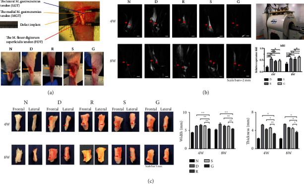
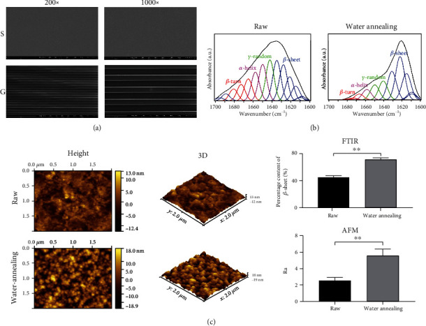
Methods: First, we characterized the physical properties of the micropattern SF films and explored their repairing effects on the injured tendons in vivo. Then, we seeded TSPCs on SF films in vitro and determined the micropattern SF film-induced gene expression and activation of signaling pathways in TSPCs through high-throughput RNA sequencing and proteomics assays.
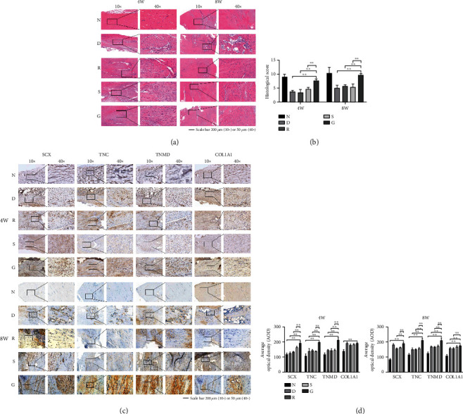
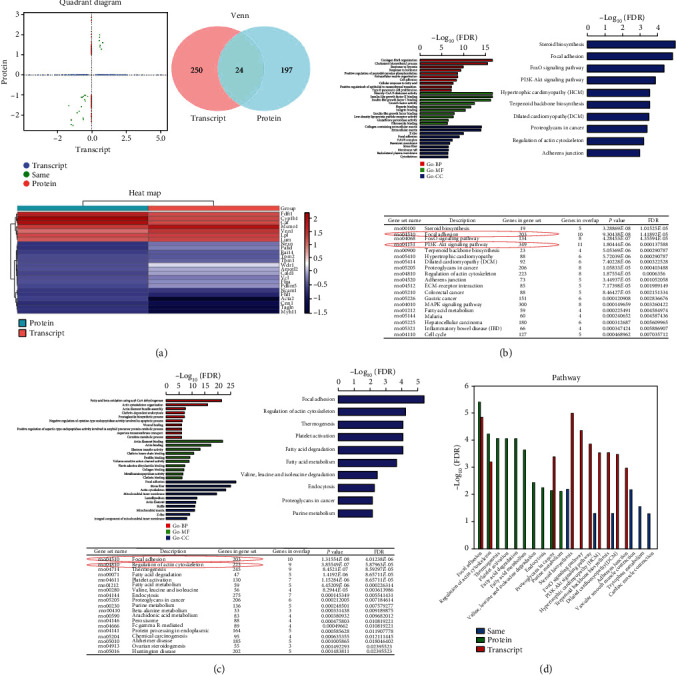
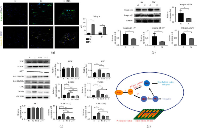
Results: The results of in vivo studies suggested that micropattern SF films can promote remodeling of the injured tendon. In addition, immunohistochemistry (IHC) results showed that tendon marker genes were significantly increased in the micropattern SF film repair group. Transcriptomic and proteomic analyses demonstrated that micropattern SF film-induced genes and proteins in TSPCs were mainly enriched in the focal adhesion kinase (FAK)/actin and phosphoinositide 3-kinase (PI3K)/AKT pathways. Western blot analysis showed that the expression of integrins α2β1, tenascin-C (TNC), and tenomodulin (TNMD) and the phosphorylation of AKT were significantly increased in the micropattern SF film group, which could be abrogated by applying PI3K/AKT inhibitors.
Conclusion: Micropattern SF films modified by water annealing can promote remodeling of the injured tendon in vivo and regulate the tendon differentiation of TSPCs through the α2β1/FAK/PI3K/AKT signaling pathway in vitro. Therefore, they have great medical value in tendon repair.
Copyright © 2023 Kang Lu et al.
Conflict of interest statement
The authors declare no conflicts of interest.
Figures
Similar articles
-
Bionic Silk Fibroin Film Promotes Tenogenic Differentiation of Tendon Stem/Progenitor Cells by Activating Focal Adhesion Kinase.Stem Cells Int. 2020 Nov 4;2020:8857380. doi: 10.1155/2020/8857380. eCollection 2020. Stem Cells Int. 2020. PMID: 33204279 Free PMC article.
-
Bionic Silk Fibroin Film Induces Morphological Changes and Differentiation of Tendon Stem/Progenitor Cells.Appl Bionics Biomech. 2020 Dec 1;2020:8865841. doi: 10.1155/2020/8865841. eCollection 2020. Appl Bionics Biomech. 2020. PMID: 33343699 Free PMC article.
-
N-Acetyl-L-cysteine facilitates tendon repair and promotes the tenogenic differentiation of tendon stem/progenitor cells by enhancing the integrin α5/β1/PI3K/AKT signaling.BMC Mol Cell Biol. 2023 Jan 5;24(1):1. doi: 10.1186/s12860-022-00463-0. BMC Mol Cell Biol. 2023. PMID: 36604630 Free PMC article.
-
Targeting Senescent Tendon Stem/Progenitor Cells to Prevent or Treat Age-Related Tendon Disorders.Stem Cell Rev Rep. 2023 Apr;19(3):680-693. doi: 10.1007/s12015-022-10488-9. Epub 2022 Dec 15. Stem Cell Rev Rep. 2023. PMID: 36520409 Review.
-
Mesenchymal stem cells in tendon repair and regeneration: basic understanding and translational challenges.Ann N Y Acad Sci. 2016 Nov;1383(1):88-96. doi: 10.1111/nyas.13262. Epub 2016 Oct 5. Ann N Y Acad Sci. 2016. PMID: 27706825 Review.
Cited by
-
Current and potential future biological uses of Saussurea costus (Falc.) Lipsch: A comprehensive review.Heliyon. 2024 Sep 11;10(18):e37790. doi: 10.1016/j.heliyon.2024.e37790. eCollection 2024 Sep 30. Heliyon. 2024. PMID: 39323795 Free PMC article. Review.
References
-
- Rudisill S. S., Kucharik M. P., Varady N. H., Martin S. D. Evidence-based management and factors associated with return to play after acute hamstring injury in athletes: a systematic review. Orthopaedic Journal of Sports Medicine . 2021;9(11):p. 232596712110538. doi: 10.1177/23259671211053833. - DOI - PMC - PubMed
-
- Müller S. A., Todorov A., Heisterbach P. E., Martin I., Majewski M. Tendon healing: an overview of physiology, biology, and pathology of tendon healing and systematic review of state of the art in tendon bioengineering. Knee Surgery, Sports Traumatology, Arthroscopy . 2015;23(7):2097–2105. doi: 10.1007/s00167-013-2680-z. - DOI - PubMed
LinkOut - more resources
Full Text Sources
Miscellaneous
