. 2023 Jan 4;15(1):157.
doi: 10.3390/v15010157.

Movement Protein Mediates Systemic Necrosis in Tomato Plants with Infection of Tomato Mosaic Virus

Affiliations

Movement Protein Mediates Systemic Necrosis in Tomato Plants with Infection of Tomato Mosaic Virus

Qiansheng Liao et al. Viruses. .

Abstract

The necrogenic strain N5 of tomato mosaic virus (ToMV-N5) causes systemic necrosis in tomato cultivar Hezuo903. In this work, we mapped the viral determinant responsible for the induction of systemic necrosis. By exchanging viral genes between N5 and a non-necrogenic strain S1, we found that movement protein (MP) was the determinant for the differential symptoms caused by both strains. Compared with S1 MP, N5 MP had an additional ability to increase virus accumulation, which was not due to its functions in viral cell-to-cell movement. Actually, N5 MP, but not S1 MP, was a weak RNA silencing suppressor, which assisted viral accumulation. Sequence alignment showed that both MPs differed by only three amino acid residues. Experiments with viruses having mutated MPs indicated that the residue isoleucine at position 170 in MP was the key site for MP to increase virus accumulation, but also was required for MP to induce systemic necrosis in virus-infected tomato plants. Collectively, the lethal necrosis caused by N5 is dependent on its MP protein that enhances virus accumulation via its RNA silencing suppressor activity, probably leading to systemic necrosis responses in tomato plants.

Keywords: RNA silencing suppressor; Solanum lycopersicum; movement protein; systemic necrosis; tomato mosaic virus.


Conflict of interest statement

The authors declare no conflict of interest.

Figures

Figure 1
Figure 1
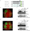
Genetic mapping of the viral protein responsible for the induction of systemic necrosis in tomato plants with infection of ToMV-N5. (a) Schematic diagrams of the 35S promoter-based infectious clones of wildtype N5 and S1 strains of ToMV, and their derivatives. The DNA fragments of viral genomes were inserted between the 35S promoter (2 × 35S) and HDV cis-cleaving ribozyme sequence (Rz) in the binary vector pCB301. Viral sequences of N5 and S1 are colored gray and light blue, respectively. Six chimeric viruses expressing heterologous 183 KD protein, MP, or CP were shown. For instance, pCB301-N5S1-183KD is the construct where the 183 KD in N5 was replaced with that of S1, and pCB301-S1N5-183KD is the construct where the 183 KD in S1 was replaced with that of N5. (b) Viral symptoms on the tomato plants inoculated with N5, S1, or their chimeric viruses, as illustrated in (a). Plants were photographed at 7 days post-infiltration (dpi). (c) Northern blotting analyses of viral RNAs in the upper systematic leaves of the infected plants at 7 dpi. Viral RNAs were detected with a digoxigenin (DIG)-labeled DNA oligonucleotide complementary to the sequence of ToMV genomic 3′ untranslated region (3′ UTR). Genomic RNAs and subgenomic RNAs of these viruses were indicated with gRNA and sgRNA, respectively, to the right. Relative accumulation levels of gRNA were shown below. Ethidium bromide (EB)-stained ribosomal RNAs (rRNAs) were used to indicate the relative amounts of RNA samples loaded.
Figure 2
Figure 2
N5 MP increased viral accumulations in Nicotiana benthamiana. (a) Schematic diagrams of ToMV constructs harboring the coding sequence of green fluorescent protein (GFP). (b) Appearance of green fluorescence on the plants inoculated with N5-gfp, S1-gfp, or their MP recombinants under a long wavelength UV illuminator at 8 dpi. (c) Northern blot and immune blot analyses of viral RNAs and GFP proteins in upper systemic leaves, respectively. Genomic RNAs and subgenomic RNAs of these viruses were indicated with gRNA and sgRNA, respectively, to the right. Relative accumulation levels of gRNA and GFP protein are shown below. Ethidium bromide-stained ribosomal RNAs (rRNAs) were used as a loading indicator. The large subunit of Rubisco stained by Ponceau S was used to assess the relative amounts of protein samples loaded.
Figure 3
Figure 3
The difference in both N5 and S1 MPs-mediated viral accumulations was restored in the presence of the RNA silencing suppressor p19. (a) Schematic diagrams of N5-Δcp-gfp, S1-Δcp-gfp, and their MP exchangers, where the CP gene was deleted. (b,d) Green fluorescence appearance on the leaves of N. benthamiana inoculated with N5-Δcp-gfp, S1-Δcp-gfp, and their MP exchangers in the absence of p19, or in the presence of p19, respectively. GFP fluorescence was photographed at 4 dpi. (c,e) RNA blotting analyses of GFP-expressing subgenomic RNA (sgRNA) and Western blotting detection of GFP protein from the leaf samples as shown in (b,d), respectively. Relative accumulation levels of sgRNA and GFP protein are shown below. Ethidium bromide-stained ribosomal RNAs (rRNAs) and Ponceau S-stained Rubisco were used as loading controls for Northern blot and Western blot, respectively.
Figure 4
Figure 4
Comparison of both MPs in facilitating viral cell-to-cell movement. (a) Schematic diagram of agroinfiltration to inoculate N5-Δcp-gfp (left half) and N5S1-MP-Δcp-gfp (right half) on the same leaf of N. benthamaina plants. As shown in Figure 4a, N5-Δcp-gfp was constructed by deleting the CP gene in N5-gfp, and N5S1-MP-Δcp-gfp was constructed by replacing MP in N5-Δcp-gfp with S1-MP. (b) Distribution patterns of GFP fluorescence foci produced from N5-Δcp-gfp and N5S1-MP-Δcp-gfp at 5, 6, and 7 dpi. The concentration of Agrobacterium cells harboring N5-Δcp-gfp or N5S1-MP-Δcp-gfp was 0.0001 at OD595. (c) Statistic results of GFP foci measured from N5-Δcp-gfp and N5S1-MP-Δcp-gfp at 5, 6, and 7 dpi. All GFP foci visualized at 5 dpi were measured again by Image J at 6 and 7 dpi. The collected data were analyzed with GraphPad Prism. The dark lines indicate the mean values of GFP foci size at a certain time point.
Figure 5
Figure 5
Comparison of N5 and S1 MPs in suppressing local GFP post-transcriptional gene silencing. (a) Observation of GFP fluorescence under UV light at 3 dpi. The leaves of N. benthamiana were co-infiltrated with Agrobacterium cells containing 35S:GFP and 35S:dsFP (GFP-derived inverted repeat RNA), together with the cells carrying a vector control, 35S:N5-MP, 35:S1-MP or 35S:p19. (b) Analyses of GFP protein, GFP transcripts and GFP-derived small-interfering RNAs (siRNAs) using Western blot and Northern blot, respectively. All these RNAs were detected by DIG-labeled DNA oligonucleotides. Ethidium bromide-stained ribosomal RNAs (rRNAs) were used as RNA loading control for analysis of GFP transcripts. U6 RNA was detected as a loading control. Rubisco protein stained by Ponceau S was used as a loading control. Relative accumulation levels of GFP transcript, GFP-derived siRNA, and GFP protein are shown below.
Figure 6
Figure 6
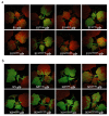
Differential green fluorescence intensities produced by S1-gfp and its MP mutants (a), or N5-gfp and its MP mutants (b) in Nicotiana benthamiana. The fifth true leaves were inoculated with viruses via agroinfiltration. Plants were photographed under UV light at 8 dpi.
Figure 7
Figure 7
The S1 mutants harboring isoleucine at position 170 (170I) in its MP induced systemic necrosis in tomato plants. (a) Disease symptoms in tomato plants infected with S1 or its MP mutants, as well as S1N5-mp, at 7 dpi. (b) Steady-stage levels of viral RNAs in the upper systemic leaves were analyzed using Northern blot using a DIG-labeled DNA oligonucleotide. The bands corresponding to genomic RNA and subgenomic RNAs of these viruses were indicated with gRNA and sgRNA, respectively. Ethidium bromide-stained ribosomal RNAs (rRNAs) were used to assess relative loading amounts of RNA samples. Relative accumulation levels of gRNA are shown below.

Similar articles

Cited by

References

    1. Rivarez M.P.S., Vučurović A., Mehle N., Ravnikar M., Kutnjak D. Global Advances in Tomato Virome Research: Current Status and the Impact of High-Throughput Sequencing. Front. Microbiol. 2021;12:671925. doi: 10.3389/fmicb.2021.671925. - DOI - PMC - PubMed
    1. Alfaro-Fernández A., Castillo P., Sanahuja E., Rodríguez-Salido M.D.C., Font M.I. First report of Tomato brown rugose fruit virus in tomato in Spain. Plant Dis. 2020;105:515-515. doi: 10.1094/PDIS-06-20-1251-PDN. - DOI - PubMed
    1. Creager A.N., Scholthof K.B., Citovsky V., Scholthof H.B. Tobacco mosaic virus. Pioneering research for a century. Plant Cell. 1999;11:301–308. doi: 10.1105/tpc.11.3.301. - DOI - PMC - PubMed
    1. Hak H., Spiegelman Z. The Tomato Brown Rugose Fruit Virus Movement Protein Overcomes Tm-2(2) Resistance in Tomato While Attenuating Viral Transport. Mol. Plant-Microbe Interact. 2021;34:1024–1032. doi: 10.1094/MPMI-01-21-0023-R. - DOI - PubMed
    1. Nishikiori M., Dohi K., Mori M., Meshi T., Naito S., Ishikawa M. Membrane-bound tomato mosaic virus replication proteins participate in RNA synthesis and are associated with host proteins in a pattern distinct from those that are not membrane bound. J. Virol. 2006;80:8459–8468. doi: 10.1128/JVI.00545-06. - DOI - PMC - PubMed

Publication types

Substances

Supplementary concepts

Grants and funding

This research was funded by the National Natural Science Foundation of China (32070154, 31870144) and the project of Genetically Modified Rrganisms Breeding Major Projects (2018ZX08001-03B).

LinkOut - more resources