. 2023 Jan 20;9(3):eadc9830.
doi: 10.1126/sciadv.adc9830. Epub 2023 Jan 20.

HPV is a cargo for the COPI sorting complex during virus entry

Affiliations

HPV is a cargo for the COPI sorting complex during virus entry

Mara C Harwood et al. Sci Adv. .

Abstract

During entry, human papillomavirus (HPV) traffics from the cell surface to the endosome and then to the trans-Golgi network (TGN) and Golgi apparatus. HPV must transit across the TGN/Golgi and exit these compartments to reach the nucleus to cause infection, although how these steps are accomplished is unclear. Combining cellular fractionation, unbiased proteomics, and gene knockdown strategies, we identified the coat protein complex I (COPI), a highly conserved protein complex that facilitates retrograde trafficking of cellular cargos, as a host factor required for HPV infection. Upon TGN/Golgi arrival, the cytoplasmic segment of HPV L2 binds directly to COPI. COPI depletion causes the accumulation of HPV in the TGN/Golgi, resembling the fate of a COPI binding-defective L2 mutant. We propose that the L2-COPI interaction drives HPV trafficking through the TGN and Golgi stacks during virus entry. This shows that an incoming virus is a cargo of the COPI complex.


Figures

Fig. 1.
Fig. 1.. Cell fractionation identifies COPI as an HPV-interacting host factor at the TGN/Golgi.
(A) Sequence alignment of the L2 protein of various HPV types. Two conserved arginine residues (magenta) are mutated to alanine (red) in R302/5A HPV16 L2. Asterisks indicate fully conserved residues. Colons indicate conservation between groups of strongly similar properties. Multiple sequence alignment of full-length L2 was performed with Clustal Omega, and the fragments containing the di-arginine motif are shown. (B) Overview of cellular fractionation to isolate material for mass spectrometry analysis. See the main text for details. Figure created with BioRender.com. (C) Representative blots from fractionated extract obtained after step 3. Extracts were subjected to SDS-PAGE followed by immunoblotting with the indicated antibodies. Fractions 3 to 5 containing TGN46 and GM130 were pooled before FLAG-immunoprecipitation (step 4) and mass spectrometry (step 5). (D) Total number of peptides corresponding to subunits of the COPI complex identified by mass spectrometry (step 5). WT, material obtained from cells infected with WT HPV16.L2F PsV; R302/5A, material obtained from cells infected with R302/5A HPV16.L2F PsV; control, material obtained from cells that were uninfected but mixed with WT HPV16.L2F PsV between steps 3 and 4. See table S1 for a complete list of peptides identified by mass spectrometry. (E) Simplified diagram of the COPI complex. Blue and green represent the two subcomplexes of COPI (32). Figure created with BioRender.com.
Fig. 2.
Fig. 2.. The di-arginine motif in the L2 protein mediates binding between HPV16 and COPI upon TGN/Golgi arrival.
(A) Whole-cell extracts of HeLa cells uninfected or infected with WT or R302/5A HPV16.L2F for 22 hours were subjected to immunoprecipitation with an anti–β-COP antibody or a rabbit normal IgG as a negative control. Immunoprecipitated samples were analyzed with anti-FLAG and anti–β-COP antibodies. (B) As in (A), except an anti-β′-COP antibody was used. (C) As in (A), except an anti–γ-COP antibody was used. (D) Whole-cell extracts of uninfected HeLa cells were incubated with biotin-tagged peptides containing residues T299 to L312 of the WT HPV16 L2 (lane 3) or the corresponding R302/5A mutant (lane 4). Samples precipitated with streptavidin-beads were analyzed with antibodies recognizing α-COP, γ-COP, or retromer subunit VPS35. (E) WT HPV16.L2F–infected HeLa cells were lysed at different hpi for immunoprecipitation experiments as in (C). (F) HeLa cells were infected with WT HPV16.L2F for 20 hours in the presence of dimethyl sulfoxide (DMSO) or 2 μM XXI and subjected to immunoprecipitation experiments as in (C). (G) HeLa-S3 cells were uninfected or infected with WT HPV16.L2F in the presence of DMSO or 2 μM XXI. At 24 hpi, PLA (signals shown in green) was performed with antibodies recognizing HPV16 L1 and γ-COP. Nuclei were stained with 4′,6-diamidino-2-phenylindole (DAPI; blue). Similar results were obtained in two independent experiments. (H) PLA fluorescence intensity per cell in multiple images as in (G) was measured, and the individual cell fluorescence intensity values, means, and SDs of 30 cells are shown. A two-tailed, unequal variance t test was used to determine statistical significance. ****P < 0.0001.
Fig. 3.
Fig. 3.. COPI directly binds to HPV16 L2 in vitro.
(A) Coomassie stain (left to right): purified rat liver COPI with subunits marked left of the gel, WT HPV16.L2F PsV purified from HEK 293T cells, and purified C-terminal FLAG-tagged EGFP. (B) The rat COPI complex was incubated with WT HPV16.L2F PsV or EGFP-FLAG. Samples from immunoprecipitation using anti-FLAG beads were analyzed by immunoblotting with antibodies recognizing FLAG or human γ-COP. (C) Coomassie stain of purified proteins. FLAG-γ1-COP, N-terminal FLAG-tagged human γ1-COP; HA-(Δ1–67) L2-3xFLAG, HPV16 L2 (amino acids 1 to 67 deleted) tagged with N-terminal HA and C-terminal 3xFLAG; HA-(Δ1–67) L2-R302/5A-3xFLAG, HA-(Δ1–67) L2-3xFLAG with R302/5A mutation. (D) FLAG-γ1-COP was incubated with EGFP-FLAG or HA-(Δ1–67) L2-3xFLAG followed by immunoprecipitation using anti–γ-COP antibodies. Coimmunoprecipitated proteins were analyzed by immunoblotting for HA-(Δ1–67) L2-3xFLAG and EGFP-FLAG (anti-FLAG antibody) or FLAG-γ1-COP (anti–γ-COP antibody). (E) Same experiments as in (D) except that HA-(Δ1–67) L2-R302/5A-3xFLAG was analyzed for the comparison with HA-(Δ1–67) L2-3xFLAG. (F) Quantification of the coimmunoprecipitated proteins shown in top of (E). The protein band intensity of HA-(Δ1–67) L2-3xFLAG (WT) or HA-(Δ1–67) L2-R302/5A-3xFLAG (R302/5A) was normalized to that of FLAG-γ1-COP in the same precipitated sample. Data represents means normalized to WT and SDs (n = 4). A two-tailed, unequal variance t test was used to determine statistical significance. **P < 0.01. (G) Biotin peptide pulldown as in (Fig. 2D), except biotinylated peptides were incubated with the purified FLAG-γ1-COP. Precipitated samples were immunoblotted with an antibody recognizing γ-COP.
Fig. 4.
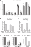
Fig. 4.. COPI promotes HPV infection.
(A) HeLa cells transfected with the indicated siRNA for 24 hours were infected with WT HPV16.L2F PsV containing a GFP reporter plasmid. At 48 hpi, flow cytometry was used to determine the fraction of GFP-expressing cells. The results were normalized to the infected fraction of cells treated with Scr siRNA. The means and SDs are shown (n = 3). A two-tailed, unequal variance t test was used to determine statistical significance compared to Scr siRNA–treated cells. NS, not significant; *P < 0.05, **P < 0.01, ***P < 0.001, and ****P < 0.0001. (B) HeLa cells were transfected with Scr or COPG1 siRNAs for 9 hours, followed by another transfection with DNA constructs for 15 hours for the expression of HA-γ1-COP-mCherry or the control HA-mCherry. The transfected cells were then infected with WT HPV16.L2F PsV containing a GFP reporter plasmid. At 48 hpi, flow cytometry was used to measure GFP and mCherry fluorescence. The fraction of cells expressing GFP in the mCherry-positive population is graphed. The results were normalized to the infected fraction of cells cotransfected with Scr siRNA and HA-mCherry–expressing plasmid. Data from three independent experiments were analyzed and presented as in (A). (C) As in (B), except the fraction of cells expressing GFP in the mCherry-negative population is graphed. (D) As in (A), except WT HPV5.L2F PsV was used. (E) As in (A), except WT HPV18.L2F PsV was used. (F) As in (A), except HaCaT cells were transfected with pooled Scr or COPG1 siRNAs for 48 hours before infection with WT HPV16.L2F PsV.
Fig. 5.
Fig. 5.. Arf1-dependent COPI recruitment to the TGN/Golgi is critical for HPV infection.
(A) Overview of COPI recruitment to the Golgi membrane by the small GTPase Arf1. The chemical inhibitor NAV disrupts Arf1 nucleotide exchange. Figure created with BioRender.com. (B) HeLa cells treated with DMSO or 25 μM of NAV for 48 hours were subjected to immunofluorescence staining with antibodies recognizing GM130 (green, first column) or γ-COP (green, second column). Nuclei were stained with DAPI (blue). Representative images of a single, medial Z plane taken by confocal microscopy are shown. (C) HeLa cells treated with DMSO or 25 μM NAV for 48 hours were lysed, and the resulting whole-cell extract was analyzed by immunoblotting with the indicated antibodies. β-Actin, loading control. (D) HeLa cells were infected with WT HPV16.L2F for 22 hours in the presence of DMSO or 20 μM NAV. Immunoprecipitation experiments followed by immunoblotting were carried out as in (Fig. 2C). (E) HeLa cells treated with the indicated siRNA for 72 hours were lysed, and the resulting whole-cell extract was analyzed by immunoblotting with antibodies recognizing Arf1 or Hsp90 as a loading control. (F) Infectivity analysis as in (Fig. 4A) except cells were treated with Scr or ARF1 siRNA before infection (n = 3). A two-tailed, unequal variance t test was used to determine statistical significance compared to Scr siRNA–treated cells. *P < 0.05.
Fig. 6.
Fig. 6.. The COPI complex drives TGN/Golgi transit of HPV.
(A) HeLa-S3 cells treated with 2 nM Scr or a mixture of 1 nM COPA and 1 nM COPG1 (COPA/G1) siRNA for 18 hours were uninfected or infected with WT HPV16.L2F PsV. At 30 hpi, PLA was performed with antibodies recognizing HPV16 L1 and TGN46 (PLA signals shown in green). Nuclei were stained with DAPI (blue). (B) Multiple images as in (A) were analyzed and presented as in (Fig. 2H). ****P < 0.0001. Similar results were obtained in three independent experiments. (C) As in (A), except PLA was performed with antibodies recognizing HPV16 L1 and GM130. (D) As in (B), except images as in (C) were analyzed. Similar results were obtained in three independent experiments. (E) HeLa cells transfected with the indicated siRNA for 48 hours were incubated with Hoechst 33342 and analyzed by flow cytometry to measure relative Hoechst 33342 fluorescence (DNA content). The percentage of cells in each phase is indicated. The representative histograms of three independent experiments are shown. (F) Summary of cell cycle phase distributions as in (E). Statistical significance for each phase was determined by a two-tailed, paired t test. NS, not significant (P > 0.05).

Similar articles

Cited by

References

    1. C. de Martel, M. Plummer, J. Vignat, S. Franceschi, Worldwide burden of cancer attributable to HPV by site, country and HPV type. Int. J. Cancer 141, 664–670 (2017). - PMC - PubMed
    1. CDC, “Cancers Associated with Human Papillomavirus, United States—2014–2018,” USCS Data Brief, no. 26 (Centers for Disease Control and Prevention, US Department of Health and Human Services, Atlanta, GA, 2021).
    1. J. C. Cardoso, E. Calonje, Cutaneous manifestations of human papillomaviruses: A review. Acta Dermatovenerol. Alp. Pannonica Adriat. 20, 145–154 (2011). - PubMed
    1. C. B. Buck, N. Cheng, C. D. Thompson, D. R. Lowy, A. C. Steven, J. T. Schiller, B. L. Trus, Arrangement of L2 within the papillomavirus capsid. J. Virol. 82, 5190–5197 (2008). - PMC - PubMed
    1. D. M. Belnap, N. H. Olson, N. M. Cladel, W. W. Newcomb, J. C. Brown, J. W. Kreider, N. D. Christensen, T. S. Baker, Conserved features in papillomavirus and polyomavirus capsids. J. Mol. Biol. 259, 249–263 (1996). - PMC - PubMed

MeSH terms

Substances