. 2023 Feb 1;16(2):dmm049594.
doi: 10.1242/dmm.049594. Epub 2023 Feb 1.

Patient-specific variants of NFU1/NFU-1 disrupt cholinergic signaling in a model of multiple mitochondrial dysfunctions syndrome 1

Affiliations

Patient-specific variants of NFU1/NFU-1 disrupt cholinergic signaling in a model of multiple mitochondrial dysfunctions syndrome 1

Peter A Kropp et al. Dis Model Mech. .

Abstract

Neuromuscular dysfunction is a common feature of mitochondrial diseases and frequently presents as ataxia, spasticity and/or dystonia, all of which can severely impact individuals with mitochondrial diseases. Dystonia is one of the most common symptoms of multiple mitochondrial dysfunctions syndrome 1 (MMDS1), a disease associated with mutations in the causative gene (NFU1) that impair iron-sulfur cluster biogenesis. We have generated Caenorhabditis elegans strains that recreated patient-specific point variants in the C. elegans ortholog (nfu-1) that result in allele-specific dysfunction. Each of these mutants, Gly147Arg and Gly166Cys, have altered acetylcholine signaling at neuromuscular junctions, but opposite effects on activity and motility. We found that the Gly147Arg variant was hypersensitive to acetylcholine and that knockdown of acetylcholine release rescued nearly all neuromuscular phenotypes of this variant. In contrast, we found that the Gly166Cys variant caused predominantly postsynaptic acetylcholine hypersensitivity due to an unclear mechanism. These results are important for understanding the neuromuscular conditions of MMDS1 patients and potential avenues for therapeutic intervention.

Keywords: C. elegans; Acetylcholine; GABA; MMDS1; Mitochondria; Motility.


Conflict of interest statement

Competing interests The authors declare no competing or financial interests.

Figures

Fig. 1.
Fig. 1.
Crawling defects of nfu-1 variants on solid medium. (A) Speed of crawling. (B) Wavelength of sinusoidal wave. (C) Amplitude of sinusoidal wave. (D) Schematic of wavelength and amplitude, and representative images of animals analyzed. (E) Number of turns in 1 min. (F) Number of reversals in 1 min. (G) Percentage of time reversing. Each data point is an individual animal (n=36-40). **P≤0.01; ***P≤0.001; ****P≤0.0001 by one-way ANOVA with Tukey correction for multiple comparisons. Scale bars: 500 µm. WT, wild type.
Fig. 2.
Fig. 2.
Swimming defect of nfu-1 variants in liquid. (A) Wave rate. (B) Swimming speed. (C) Activity index. (D) Representative traces of animal center point in 1 min. (E) Stretch: a measure of bend in the body during a wave where stretch is measured as the inverse of distance (i.e. more stretch brings the anterior and posterior of the animal together giving a larger value). (F) Percentage of time curling. (G) Percentage of time reversing. (H) Representative curvature maps of individual animals during measurement. Color represents the body's curvature during a wave, with deeper red indicating a deeper clockwise curve and deeper blue indicating a deeper counterclockwise curve. In each frame (x-axis point), curvature was measured across the length of each animal in 17 slices (y-axis point). Each data point is an individual animal (n=28-35). *P≤0.05; **P≤0.01; ****P≤0.0001 by one-way ANOVA with Tukey correction for multiple comparisons.
Fig. 3.
Fig. 3.
nfu-1 variants experience different sensitivities to neuromuscular signaling pathways. (A) Schematic of a simplified neuromuscular junction. Cholinergic neurons stimulate body wall muscle contraction by secreting acetylcholine (ACh; red dots), which binds to the ACh receptor (AChR; shown as yellow/orange receptor). ACh is degraded in the synapse by acetylcholinesterase (AChE). GABAergic neurons inhibit body wall muscle contraction by secreting GABA (blue dots), which binds to the GABA receptor (GABAR; blue receptor). Drugs used: levamisole, an AChR agonist; aldicarb, an AChE antagonist; and piperazine, a GABAR agonist. (B) Aldicarb paralysis curve (n=75-105). (C) Levamisole paralysis curve (n=45-65). (D) Piperazine paralysis curve (n=89-95). Data plotted as Kaplan–Meier survival curves. *P≤0.05; ***P≤0.001; ****P≤0.0001 by log-rank analysis with Bonferroni correction for multiple comparisons.
Fig. 4.
Fig. 4.
Muscle-specific knockout of nfu-1 results in more severe neuromuscular defects than neuron-specific knockout of nfu-1. (A) Schematic of FLP/FRT system used for tissue-specific knockouts of nfu-1. All but the first exon of nfu-1 was removed. Transgenic reporter construct shown in Fig. S3. Chr, chromosome; FLP, flippase; FRT, flippase recognition target. (B) Representative maximum-intensity projections of confocal images of nfu-1Δmuscle and nfu-1Δneurons. Cells in which recombination successfully occurred express GFP (green) and all other cells express mCherry (magenta). Scale bars: 250 µm. DIC, differential interference contrast. (C,E) Levamisole paralysis curve for muscle-specific (C) and neuron-specific (E) knockout of nfu-1 (n=45). (D,F) Piperazine paralysis curve for muscle-specific (D) and neuron-specific (F) knockout of nfu-1 (n=42-44). Data plotted as Kaplan–Meier survival curves. (G) Wave rate. (H) Swimming speed. (I) Activity index. (J) Stretch. (K) Percentage of time curling. (L) Percentage of time reversing. Each data point represents an individual animal (n=23-27). For C, ***P≤0.001 by log-rank analysis. For G-L, **P≤0.01; ***P≤0.001; ****P≤0.0001 by parallel two-tailed Student's t-tests.
Fig. 5.
Fig. 5.
Tissue-specific re-expression of nfu-1 alone and in patient-specific variants. (A) Schematic representation of the SKI LODGE approach for single-copy transgene insertion. (B) Wave rate. (C) Swimming speed. (D) Activity index. (E) Stretch. (F) Percentage of time curling. (G) Percentage of time reversing. Each data point represents an individual animal (n=25-37). For B-G, *P≤0.05; **P≤0.01; ***P≤0.001; ****P≤0.0001 from WT by one-way ANOVA with Dunnett correction for multiple comparisons.
Fig. 6.
Fig. 6.
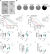
Knockdown of ACh signaling rescues Gly147Arg motility phenotypes. (A) Representative images of shrinker analysis. Black arrowheads indicate head position; white arrowheads indicate tail position at time of nose touch (top row). Bottom images represent 1 s after nose touch. Arrowheads are in the same position in both images. Shrinker animals [unc-47(n2409) as positive control] contract from the anterior body without any posterior movement. WT animals reverse. Scale bars: 500 μm. (B) Shrinker phenotype as represented as percentage of total population (n=101-138). (C) Levamisole paralysis curve. Empty vector (EV)-treated samples in solid lines and unc-17 RNAi interference (RNAi)-treated samples in dotted lines. (n=40-41). (D) Piperazine paralysis curve. Solid lines, EV-treated samples; dotted lines, unc-17 RNAi-treated samples (n=40-54). Data plotted as Kaplan–Meier survival curves. (E-J) Swimming phenotypes of EV- or unc-17 RNAi-treated samples in filled or open circles, respectively (n=25-33). (E) Wave rate. (F) Swimming speed. (G) Activity index. (H) Stretch. (I) Percentage of time curling. (J) Percentage of time reversing. Each data point represents an individual animal (n=25-33). *P≤0.05; **P≤0.01; ****P≤0.0001 by parallel two-tailed Student's t-tests.
Fig. 7.
Fig. 7.
Summary of findings. In WT animals, NFU-1 functions normally within neuronal mitochondria, and there are normal levels of ACh packaged into vesicles and secreted in the synapse. Normal mitochondrial function is indirectly necessary for vesicular packaging, indicated by dashed line arrows in neurons. ACh binding to AChRs thereby stimulates contraction. Normal NFU-1 function in muscle mitochondria allows for contraction to occur normally. In Gly147Arg animals, NFU-1 dimerizes around iron–sulfur clusters (ISCs) less efficiently, thereby altering mitochondrial function. By an unknown mechanism, this mitochondrial dysfunction in neurons causes hypersecretion of ACh either through increased packaging of ACh into synaptic vesicles, increased vesicular release or both. Increased ACh signaling increases contractions irrespective of muscular mitochondrial dysfunction. In Gly166Cys animals, NFU-1 fails to release ISCs, thus inhibiting mitochondrial function. Neuronal ACh secretion is unaffected, but muscular contraction is altered by an unclear mechanism (dashed line arrow in muscle). Animals' hypersensitivity to aldicarb and levamisole suggest that contraction is potentiated or increased, consequently causing reduced motility. The nature of contractile defect and mechanism are unknown, indicated by dashed line arrow in muscle and question marks.

Similar articles

References

    1. Abdel-Wahab, W. M. and Moussa, F. I. (2019). Neuroprotective effect of N-acetylcysteine against cisplatin-induced toxicity in rat brain by modulation of oxidative stress and inflammation. Drug. Des. Dev. Ther. 13, 1155-1162. 10.2147/DDDT.S191240 - DOI - PMC - PubMed
    1. Ahting, U., Mayr, J. A., Vanlander, A. V., Hardy, S. A., Santra, S., Makowski, C., Alston, C. L., Zimmermann, F. A., Abela, L., Plecko, B.et al. . (2015). Clinical, biochemical, and genetic spectrum of seven patients with NFU1 deficiency. Front. Genet. 6, 123. 10.3389/fgene.2015.00123 - DOI - PMC - PubMed
    1. Alfonso, A., Grundahl, K., Duerr, J. S., Han, H.-P. and Rand, J. B. (1993). The Caenorhabditis elegans unc-17 gene: a putative vesicular acetylcholine transporter. Science 261, 617-619. 10.1126/science.8342028 - DOI - PubMed
    1. Ames, E. G., Neville, K. L., McNamara, N. A., Keegan, C. E. and Elsea, S. H. (2020). Clinical Reasoning: A 12-month-old child with hypotonia and developmental delays. Neurology 95, 184-187. 10.1212/WNL.0000000000009912 - DOI - PubMed
    1. Babu, K., Hu, Z., Chien, S.-C., Garriga, G. and Kaplan, J. M. (2011). The immunoglobulin super family protein RIG-3 prevents synaptic potentiation and regulates Wnt signaling. Neuron 71, 103-116. 10.1016/j.neuron.2011.05.034 - DOI - PMC - PubMed

Publication types

Supplementary concepts