

Estrogen fluctuations during the menopausal transition are a risk factor for depressive disorders
- PMID: 36639604
- PMCID: PMC9889489
- DOI: 10.1007/s43440-022-00444-2
Estrogen fluctuations during the menopausal transition are a risk factor for depressive disorders
Abstract
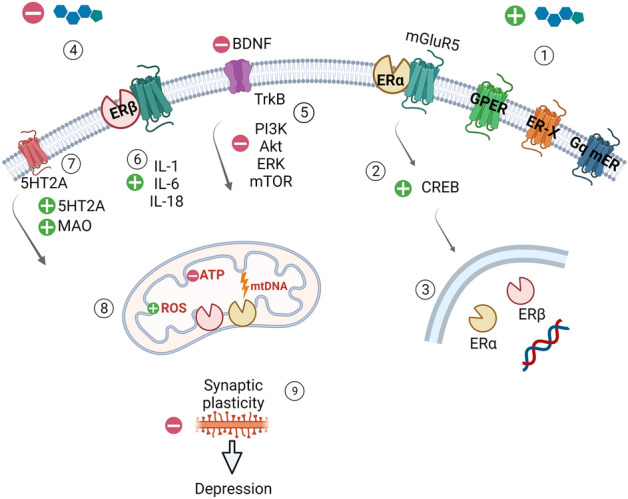
Women are significantly more likely to develop depression than men. Fluctuations in the ovarian estrogen hormone levels are closely linked with women's well-being. This narrative review discusses the available knowledge on the role of estrogen in modulating brain function and the correlation between changes in estrogen levels and the development of depression. Equally discussed are the possible mechanisms underlying these effects, including the role of estrogen in modulating brain-derived neurotrophic factor activity, serotonin neurotransmission, as well as the induction of inflammatory response and changes in metabolic activity, are discussed.
Keywords: Depression; Estrogen receptors; Menopause.
© 2023. The Author(s).
Conflict of interest statement
The authors declare that they have no competing interests to disclose.
Figures

Similar articles
-
Estrogen receptor β deficiency impairs BDNF-5-HT2A signaling in the hippocampus of female brain: A possible mechanism for menopausal depression.Psychoneuroendocrinology. 2017 Aug;82:107-116. doi: 10.1016/j.psyneuen.2017.05.016. Epub 2017 May 18. Psychoneuroendocrinology. 2017. PMID: 28544903 Free PMC article.
-
AMERICAN ASSOCIATION OF CLINICAL ENDOCRINOLOGISTS AND AMERICAN COLLEGE OF ENDOCRINOLOGY POSITION STATEMENT ON MENOPAUSE-2017 UPDATE.Endocr Pract. 2017 Jul;23(7):869-880. doi: 10.4158/EP171828.PS. Endocr Pract. 2017. PMID: 28703650
-
Depression, menopause and estrogens: is there a correlation?Maturitas. 2002 Apr 15;41 Suppl 1:S3-8. doi: 10.1016/s0378-5122(02)00009-9. Maturitas. 2002. PMID: 11955789 Review.
-
[The effects of hormone replacement therapy on mind and brain].Nervenarzt. 2013 Jan;84(1):14-9. doi: 10.1007/s00115-011-3456-7. Nervenarzt. 2013. PMID: 22318360 Review. German.
-
About the menopausal depression.Neuropsychopharmacol Hung. 2005 Dec;7(4):208-14. Neuropsychopharmacol Hung. 2005. PMID: 16496486 Clinical Trial.
Cited by
-
New Role of the Serotonin as a Biomarker of Gut-Brain Interaction.Life (Basel). 2024 Oct 9;14(10):1280. doi: 10.3390/life14101280. Life (Basel). 2024. PMID: 39459580 Free PMC article. Review.
-
Understanding the Biological Relationship between Migraine and Depression.Biomolecules. 2024 Jan 30;14(2):163. doi: 10.3390/biom14020163. Biomolecules. 2024. PMID: 38397400 Free PMC article. Review.
-
The impact of 17β-estradiol on the estrogen-deficient female brain: from mechanisms to therapy with hot flushes as target symptoms.Front Endocrinol (Lausanne). 2024 Jan 8;14:1310432. doi: 10.3389/fendo.2023.1310432. eCollection 2023. Front Endocrinol (Lausanne). 2024. PMID: 38260155 Free PMC article. Review.
-
Dorsal brain activity reflects the severity of menopausal symptoms.Menopause. 2024 May 1;31(5):399-407. doi: 10.1097/GME.0000000000002347. Epub 2024 Apr 15. Menopause. 2024. PMID: 38626372 Free PMC article.
-
The role of estrogen in Alzheimer's disease pathogenesis and therapeutic potential in women.Mol Cell Biochem. 2024 Aug 1. doi: 10.1007/s11010-024-05071-4. Online ahead of print. Mol Cell Biochem. 2024. PMID: 39088186 Review.
References
-
- Cyranowski JM, Frank E, Young E, Shear MK. Adolescent onset of the gender difference in lifetime rates of major depression: a theoretical model. Arch Gen Psychiatry. 2000;57:21–27. - PubMed
-
- Wesselhoeft R, Pedersen CB, Mortensen PB, Mors O, Bilenberg N. Gender-age interaction in incidence rates of childhood emotional disorders. Psychol Med. 2015;45:829–839. - PubMed
-
- Hindmarch I. Expanding the horizons of depression: beyond the monoamine hypothesis. Hum Psychopharmacol. 2001;16:203–218. - PubMed
Publication types
MeSH terms
Substances
LinkOut - more resources
Full Text Sources
Medical