. 2023 Mar;1866(1):194908.
doi: 10.1016/j.bbagrm.2023.194908. Epub 2023 Jan 10.

The cellular pathways that maintain the quality control and transport of diverse potassium channels

Affiliations

The cellular pathways that maintain the quality control and transport of diverse potassium channels

Nga H Nguyen et al. Biochim Biophys Acta Gene Regul Mech. 2023 Mar.

Abstract

Potassium channels are multi-subunit transmembrane proteins that permit the selective passage of potassium and play fundamental roles in physiological processes, such as action potentials in the nervous system and organismal salt and water homeostasis, which is mediated by the kidney. Like all ion channels, newly translated potassium channels enter the endoplasmic reticulum (ER) and undergo the error-prone process of acquiring post-translational modifications, folding into their native conformations, assembling with other subunits, and trafficking through the secretory pathway to reach their final destinations, most commonly the plasma membrane. Disruptions in these processes can result in detrimental consequences, including various human diseases. Thus, multiple quality control checkpoints evolved to guide potassium channels through the secretory pathway and clear potentially toxic, aggregation-prone misfolded species. We will summarize current knowledge on the mechanisms underlying potassium channel quality control in the secretory pathway, highlight diseases associated with channel misfolding, and suggest potential therapeutic routes.

Keywords: Endocytosis; Endoplasmic reticulum associated degradation (ERAD); Endosomal sorting; Molecular chaperones; Plasma membrane quality control; Protein quality control.


Conflict of interest statement

Declaration of competing interest The authors declare no conflicts of interest.

Figures
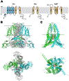
Fig. 1.
Fig. 1.
Classes and general structures of potassium channels A) Cartoon depictions of the three main classes of potassium channels. All three classes share a common structure of the pore domain (P, in red) selective for potassium ions. Note the differences in the number of transmembrane domains (TMDs), which contribute to their respective oligomeric states (Kv: 4-mer, Kir: 4-mer, K2P: 2-mer). The S4 domain of Kv channels is lined with positively charged residues for voltage sensing, and the T1 domain in Kv1-4 channels plays a role in channel assembly and function. Meanwhile, the unique cap structure in K2P channels is important for channel conductance. B) Structure of the human tetrameric Kv11.1 (hERG) channel depicted from the side (top) and from the extracellular pore (bottom), with the membrane marked with light gray lines. Two opposite monomers are highlighted in green and blue, while the remaining monomers are in gray. The cryo-EM structure of Kv11.1 (PDB: 5VA2) is shown and covers residues 3–863 out of the total 1159 amino acids in the channel. C) Structure of the human dimeric K2P2.1 (TREK-1) channel depicted from the side (top) and from the extracellular pore (bottom), with four potassium ions in the pore domain. The membrane is marked with light gray lines. Note the cap helixes on the extracellular side. Shown is a SWISS-MODEL structure built based on the crystal structure of mouse K2P2.1 (PDB: 6CQ6), which covers residues 50–336 out of the total 426 amino acids in the channel. Both figures in B and C were rendered in PyMOL, ver. 2.6.0a0.
Fig. 2.
Fig. 2.
Disease-causing missense mutations in a representative channel, Kir1.1 (ROMK) Twenty seven selected mutations in Kir1.1 associated with Bartter syndrome type 2 from the NIH ClinVar database are highlighted (neon blue sticks). While the channel is a tetramer, only two opposite monomers are shown in (A) and are displayed in dark and light gray, with four potassium ions in the pore domain. Mutations in the β sheet-rich C-terminal domain (zoomed in for clarity) are highlighted in (B). In ClinVar, there are 57 missense Kir1.1 mutations (see Table 2). Omitting 9 that fail to meet the database’s minimum standard of having at least one assertion criteria, the remaining 48 are divided into two groups: Bartter-associated (the 27 highlighted with neon blue sticks) and uncertain significance (21 in dark blue). For a complete list of the 57 mutations, see Table 2. Shown is a Kir1.1 structure using a SWISS-MODEL homology model that was built based on the crystal structure of Kir2.2 (PDB: 3SPG) that covers residues 38–364 out of the total 391 amino acids. For full coverage of all 391 residues, an AlphaFold structure was superimposed onto the left monomer using PyMOL, ver. 2.6.0a0.
Fig. 3.
Fig. 3.
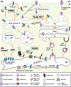
Cellular processing of potassium channels in the secretory pathway Upon being translated by the ribosome and translocated into the lumen of the endoplasmic reticulum (ER) through a translocon, a peptide that encodes a potassium channel monomer undergoes multiple folding steps to achieve its final conformation. At this point, monomeric subunits assemble to form a channel protein (a tetramer, in this example) for export from the ER to the Golgi via COPII (coatomer complex II) vesicles. The channel then transits through the rest of the secretory pathway, from the cis-, medial-, to trans-Golgi, the trans-Golgi network (TGN), and finally to the plasma membrane (PM). In contrast, if recognized as misfolded by molecular chaperones in the ER, the protein is retained, polyubiquitinated, retrotranslocated from the ER, and then degraded by the proteasome. This process is termed ER-associated degradation (ERAD). Accumulation of some misfolded proteins in the ER can lead to protein aggregation and ER stress, which could trigger ER-phagy and the unfolded protein response (UPR). Both ER-phagy and the UPR (in some cases) support degradation of misfolded proteins by the lysosome. Another quality control (QC) checkpoint is channel retrieval from the Golgi back to the ER, which is mediated by coatomer complex I (COPI). While at the Golgi, a protein can also be transported to early endosomes for lysosomal degradation, though this process of Golgi QC is poorly defined for potassium channels. Once at the PM, a channel protein can be monoubiquitinated or possibly polyubiquitinated, which leads to its retrieval by clathrin- or non-clathrin-mediated endocytosis (CME and non-CME, respectively). Internalized vesicles containing cargo proteins from the PM eventually fuse with early endosomes, where endosomal sorting takes place to either recycle the protein back to the cell surface, to the Golgi (via retromer), or on to the lysosome for degradation. Protein recycling to the PM from early endosomes may rely on cargo fusion with the recycling endosome (slow recycling). On the other hand, channel proteins destined for degradation are recruited by components of the ESCRT (endosomal complexes required for transport) complex into the multivesicular body (MVB) degradation pathway in late endosomes. Late endosomes eventually fuse with the lysosome for protein degradation. Please see text for further details and note that the three main quality control pathways are highlighted in boxes.

Similar articles

Cited by

References

    1. Kuo MM, Haynes WJ, Loukin SH, Kung C, Saimi Y, Prokaryotic K(+) channels: from crystal structures to diversity, FEMS Microbiol Rev 29 (2005) 961–985. 10.1016/j.femsre.2005.03.003. - DOI - PubMed
    1. Lawrence JH, Tomaselli GF, Marban E, Ion channels: structure and function, Heart Dis Stroke 2 (1993) 75–80. - PubMed
    1. Hodgkin AL, Keynes RD, Active transport of cations in giant axons from Sepia and Loligo, J Physiol 128 (1955) 28–60. 10.1113/jphysiol.1955.sp005290. - DOI - PMC - PubMed
    1. Elliott TL, Braun M, Electrolytes: Potassium Disorders, FP Essent 459 (2017) 21–28. - PubMed
    1. Boyd-Shiwarski CR, Subramanya AR, The renal response to potassium stress: integrating past with present, Curr Opin Nephrol Hypertens 26 (2017) 411–418. 10.1097/MNH.0000000000000352. - DOI - PMC - PubMed

Publication types

MeSH terms

LinkOut - more resources