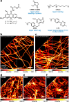
Photoactivatable Large Stokes Shift Fluorophores for Multicolor Nanoscopy
- PMID: 36626161
- PMCID: PMC9880998
- DOI: 10.1021/jacs.2c12567
Photoactivatable Large Stokes Shift Fluorophores for Multicolor Nanoscopy
Abstract
We designed caging-group-free photoactivatable live-cell permeant dyes with red fluorescence emission and ∼100 nm Stokes shifts based on a 1-vinyl-10-silaxanthone imine core structure. The proposed fluorophores undergo byproduct-free one- and two-photon activation, are suitable for multicolor fluorescence microscopy in fixed and living cells, and are compatible with super-resolution techniques such as STED (stimulated emission depletion) and PALM (photoactivated localization microscopy). Use of photoactivatable labels for strain-promoted tetrazine ligation and self-labeling protein tags (HaloTag, SNAP-tag), and duplexing of an imaging channel with another large Stokes shift dye have been demonstrated.
Conflict of interest statement
The authors declare the following competing financial interest(s): R.L., M.L.B., and A.N.B. are co-inventors of a patent application covering the photoactivatable dyes of this work, filed by the Max Planck Society. S.W.H. owns shares of Abberior GmbH and Abberior Instruments GmbH, whose dyes and STED microscope, respectively, have been used in this study. The remaining authors declare no competing interests.
Figures


Similar articles
-
Cell-Permeant Large Stokes Shift Dyes for Transfection-Free Multicolor Nanoscopy.J Am Chem Soc. 2017 Sep 13;139(36):12378-12381. doi: 10.1021/jacs.7b06412. Epub 2017 Aug 30. J Am Chem Soc. 2017. PMID: 28845665
-
A general design of caging-group-free photoactivatable fluorophores for live-cell nanoscopy.Nat Chem. 2022 Sep;14(9):1013-1020. doi: 10.1038/s41557-022-00995-0. Epub 2022 Jul 21. Nat Chem. 2022. PMID: 35864152 Free PMC article.
-
Labeling Strategies Matter for Super-Resolution Microscopy: A Comparison between HaloTags and SNAP-tags.Cell Chem Biol. 2019 Apr 18;26(4):584-592.e6. doi: 10.1016/j.chembiol.2019.01.003. Epub 2019 Feb 7. Cell Chem Biol. 2019. PMID: 30745239 Free PMC article.
-
Strategies to maximize performance in STimulated Emission Depletion (STED) nanoscopy of biological specimens.Methods. 2020 Mar 1;174:27-41. doi: 10.1016/j.ymeth.2019.07.019. Epub 2019 Jul 22. Methods. 2020. PMID: 31344404 Free PMC article. Review.
-
Photoactivatable BODIPYs for Live-Cell PALM.Molecules. 2023 Mar 7;28(6):2447. doi: 10.3390/molecules28062447. Molecules. 2023. PMID: 36985424 Free PMC article. Review.
Cited by
-
PyrAtes: Modular Organic Salts with Large Stokes Shifts for Fluo-rescence Microscopy.Angew Chem Int Ed Engl. 2024 May 6;63(19):e202318127. doi: 10.1002/anie.202318127. Epub 2024 Apr 3. Angew Chem Int Ed Engl. 2024. PMID: 38570814 Free PMC article.
-
Bioorthogonal Caging-Group-Free Photoactivatable Probes for Minimal-Linkage-Error Nanoscopy.ACS Cent Sci. 2023 Jul 26;9(8):1581-1590. doi: 10.1021/acscentsci.3c00746. eCollection 2023 Aug 23. ACS Cent Sci. 2023. PMID: 37637742 Free PMC article.
-
Unprecedented perspectives on the application of CinNapht fluorophores provided by a "late-stage" functionalization strategy.Chem Sci. 2023 May 3;14(22):6000-6010. doi: 10.1039/d3sc01365k. eCollection 2023 Jun 7. Chem Sci. 2023. PMID: 37293654 Free PMC article.
References
-
- Chozinski T. J.; Gagnon L. A.; Vaughan J. C. Twinkle, twinkle little star: Photoswitchable fluorophores for super-resolution imaging. FEBS Lett. 2014, 588, 3603–3612. 10.1016/j.febslet.2014.06.043. - DOI - PubMed
- Zhang Y.; Raymo F. M. Photoactivatable fluorophores for single-molecule localization microscopy of live cells. Methods Appl. Fluoresc. 2020, 8, 032002.10.1088/2050-6120/ab8c5c. - DOI - PubMed
Publication types
MeSH terms
Substances
LinkOut - more resources
Full Text Sources

