. 2022 Dec 23:13:1069499.
doi: 10.3389/fimmu.2022.1069499. eCollection 2022.

Cdc42 regulates cytokine expression and trafficking in bronchial epithelial cells

Affiliations

Cdc42 regulates cytokine expression and trafficking in bronchial epithelial cells

Rowayna Shouib et al. Front Immunol. .

Abstract

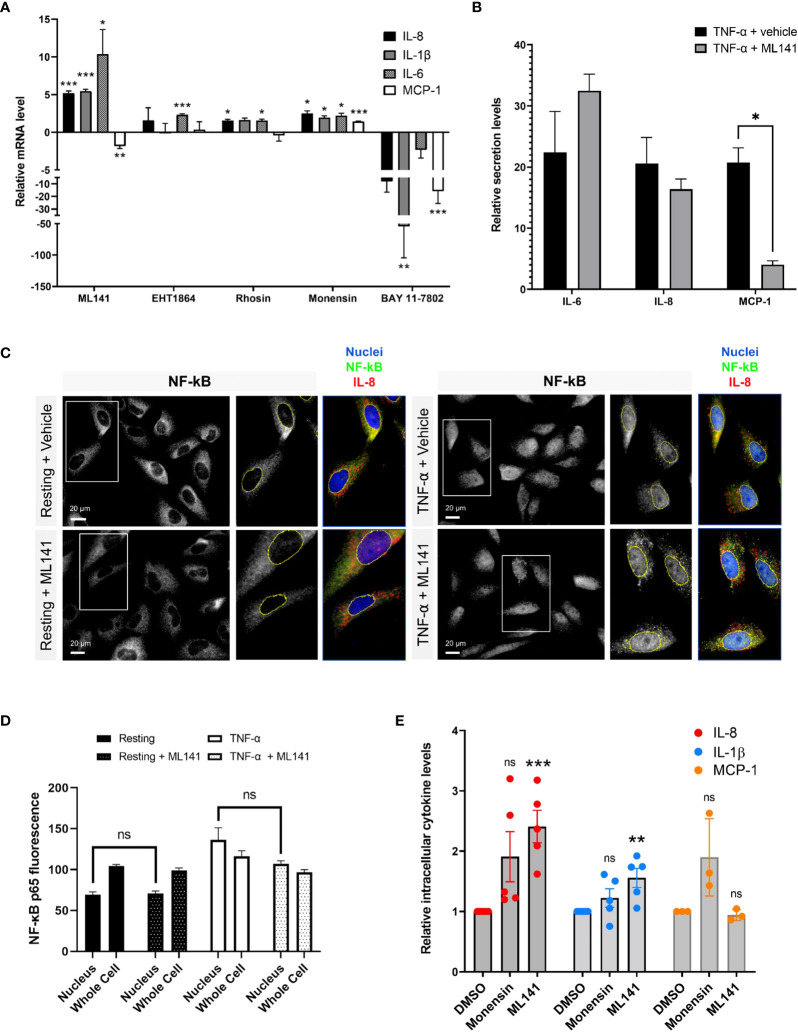
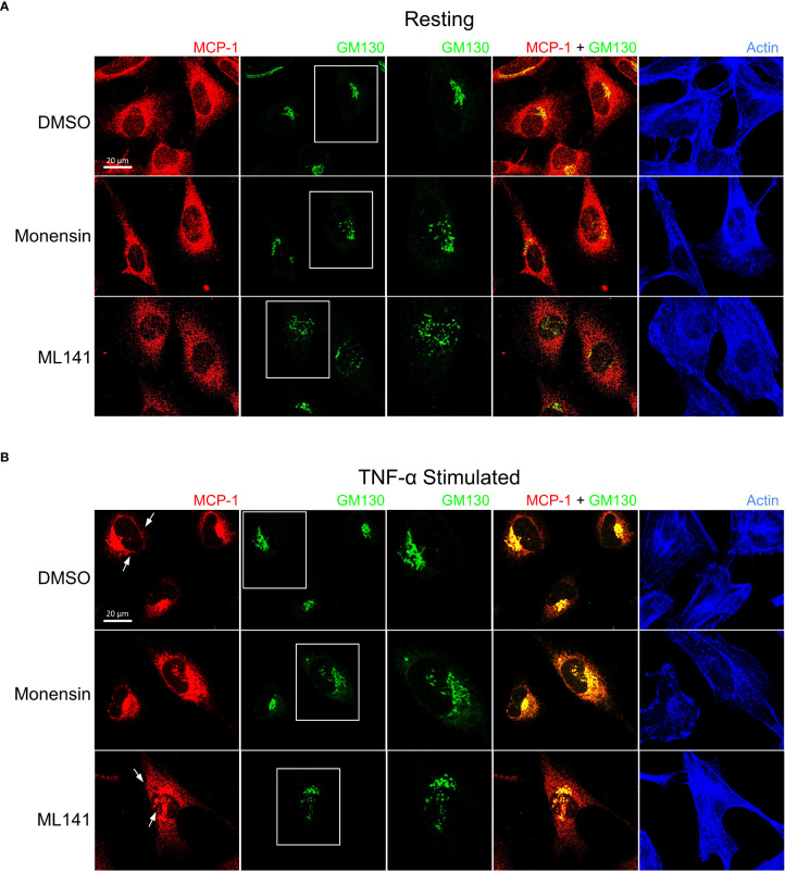
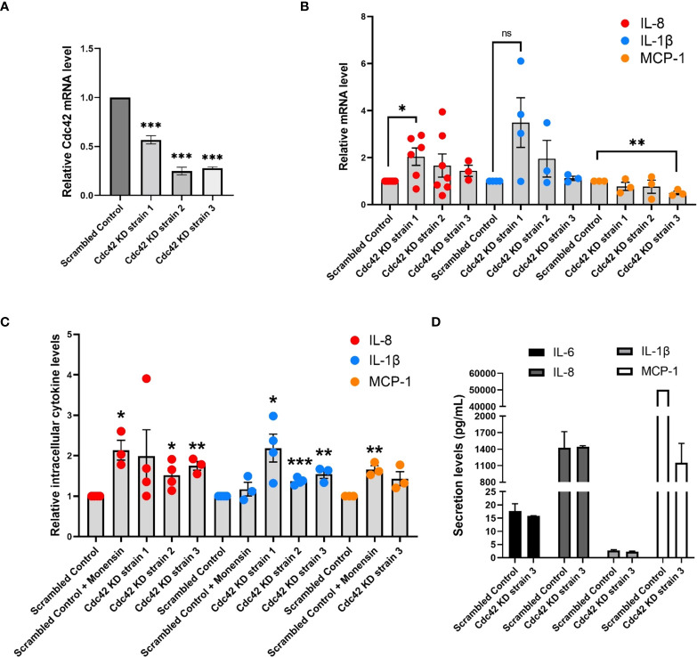
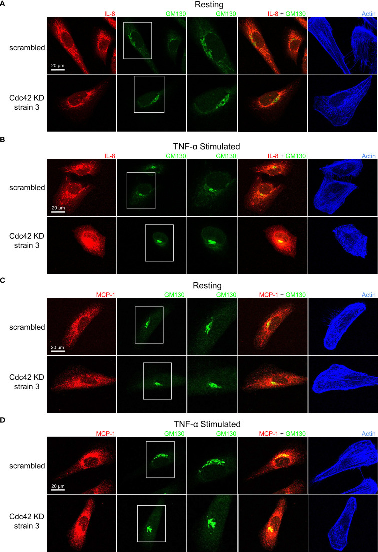
Airway epithelial cells can respond to incoming pathogens, allergens and stimulants through the secretion of cytokines and chemokines. These pro-inflammatory mediators activate inflammatory signaling cascades that allow a robust immune response to be mounted. However, uncontrolled production and release of cytokines and chemokines can result in chronic inflammation and appears to be an underlying mechanism for the pathogenesis of pulmonary disorders such as asthma and COPD. The Rho GTPase, Cdc42, is an important signaling molecule that we hypothesize can regulate cytokine production and release from epithelial cells. We treated BEAS-2B lung epithelial cells with a set of stimulants to activate inflammatory pathways and cytokine release. The production, trafficking and secretion of cytokines were assessed when Cdc42 was pharmacologically inhibited with ML141 drug or silenced with lentiviral-mediated shRNA knockdown. We found that Cdc42 inhibition with ML141 differentially affected gene expression of a subset of cytokines; transcription of IL-6 and IL-8 were increased while MCP-1 was decreased. However, Cdc42 inhibition or depletion disrupted IL-8 trafficking and reduced its secretion even though transcription was increased. Cytokines transiting through the Golgi were particularly affected by Cdc42 disruption. Our results define a role for Cdc42 in the regulation of cytokine production and release in airway epithelial cells. This underscores the role of Cdc42 in coupling receptor activation to downstream gene expression and also as a regulator of cytokine secretory pathways.

Keywords: Rho GTPase; cytokine; epithelial cells; golgi; lung inflammation; secretion.


Conflict of interest statement

The authors declare that the research was conducted in the absence of any commercial or financial relationships that could be construed as a potential conflict of interest.

Figures

Figure 1
Figure 1
BEAS-2B cells respond to pro-inflammatory stimuli with upregulated cytokine production and release. BEAS-2B cells were stimulated with 10 µg/ml poly(I:C), 20 µg/ml cockroach extract and 10 ng/ml TNF-α. (A) Relative levels of IL-8, IL-1β, IL-6 and MCP-1 in cell culture media after 8 h of stimulation as detected by multiplex assay. Values represent secretion levels normalized to unstimulated samples. (B) Fold change in mRNA levels of cytokine genes after 4 h of stimulation as detected by qPCR. Values represent ΔΔCt fold changes normalized to unstimulated samples. Bars are the mean ± s.e.m.; n=3; **p <0.01, *p < 0.05 by unpaired two-tailed Student’s t-test.
Figure 2
Figure 2
Immunofluorescence microscopy shows cytokine upregulation in response to treatment with pro-inflammatory stimuli. (A, B) BEAS-2B cells were serum-starved for 16 h, then stimulated with 20 µg/ml cockroach extract, 10 µg/ml poly(I:C) or 10 ng/ml TNF-α. (A) Cells were stimulated for 4 h then fixed and stained with anti-IL8 and anti-β-tubulin antibodies, Alexa546-phalloidin (F-actin), and DAPI (nuclei). (B, C) Quantification of NF-κB levels in the nucleus. Cells were stimulated for 30 min then fixed and stained with anti-NF-κB antibodies and DAPI (nuclei) for immunofluorescence (panel B) and levels of NF-κB staining in the nuclei or whole cells (nuclei and cytosolic regions) was quantified (panel C). Bars are the mean ± s.e.m.; n=4; * p < 0.05 by unpaired two-tailed Student’s t-test. (D) DAPI staining of BEAS-2B cells to assess apoptosis. Cells were treated with the indicated stimuli for 4 h then fixed and stained with DAPI to analyze nuclear fragmentation which indicates apoptosis.
Figure 3
Figure 3
The Cdc42 inhibitor, ML141, affects cytokine production and secretion. (A) Change in cytokine mRNA levels due to incubation with Rho drugs. BEAS-2B cells were serum-starved for 16 h and pretreated with drugs for 1 h, then stimulated with 10 ng/ml TNF-α for 4 h. Values represent ΔΔCt fold changes in mRNA normalized to vehicle-treated control. Drug concentrations: 20 μM ML141, 10 μM EHT1864, 10 μM Rhosin, 2 μM monensin, 10 μM BAY 11-7082. (B) Relative levels of secreted IL-6, IL-8 and MCP-1 in cell culture media of BEAS-2B cells. Conditioned media was collected after 8 h of stimulation with 10 ng/ml TNF-α +/- 20 µM ML141, and cytokines detected by human cytokine multiplex assay (EveTechnologies™). (C, D) Quantification of NF-κB levels in the nucleus. BEAS-2B cells were pre-treated with 20 µM ML141 or vehicle (DMSO) for 1 h, then stimulated for 30 min with 10 ng/ml TNF-α. Cells were fixed and stained with NF-κB antibodies for immunofluorescence (panel C) and levels of NF-κB staining in the nuclei or whole cells (nuclei and cytosolic regions) was quantified (panel D). (E) Intracellular levels of cytokines in stimulated BEAS-2B cells detected by flow cytometry. Cells were pre-treated with 2 μM monensin, 20 μM ML141 or vehicle (DMSO) for 1 h then stimulated with 10 ng/ml TNF-α for 4 h. Cells were fixed and stained with IL-8, IL-1β, and MCP-1 antibodies. Bars are the mean ± s.e.m; n=4 (15 – 20 cells from at least 3 different images); *** p < 0.001, ** p < 0.01, * p < 0.05, ns, not significant, by unpaired two-tailed Student’s t-test.
Figure 4
Figure 4
Cdc42 inhibition disrupts cytokine trafficking. BEAS-2B cells were pretreated with 20 µM ML141 or vehicle (DMSO) for 1 h, then stimulated with 10 ng/ml TNF-α for 4 h. Cells were then fixed and stained with cytokine antibodies. (A) IL-8 staining pattern. (B) IL-1β staining pattern. Zoomed panels show the two cytokines display different characteristic staining patterns upon stimulation. ML141 affects the IL-8 staining pattern but does not affect IL-1β staining.
Figure 5
Figure 5
Cdc42 inhibition induces changes in Golgi structure and in IL-8 trafficking. BEAS-2B cells were pre-treated with 20 μM ML141, 2 μM monensin or vehicle (DMSO) for 1 h. (A) Cells left unstimulated (resting) for 4 h. (B) Cells were stimulated with 10 ng/ml TNF-α for 4 h. Cells were then fixed and stained with IL-8 and GM130 (Golgi marker) antibodies while actin was labeled with phalloidin-iFluor 405 dye. Monensin induced a Golgi-localized staining pattern for IL-8 while ML141 treatment resulted in less Golgi-localized IL-8 and dispersed GM130 staining.
Figure 6
Figure 6
Cdc42 inhibition induces changes in Golgi structure and in MCP-1 trafficking. BEAS-2B cells were pre-treated with 20 μM ML141, 2 μM monensin or vehicle (DMSO) for 1 h. (A) Cells unstimulated (resting) for 4 h. (B) Cells were stimulated with 10 ng/ml TNF-α for 4 h. Cells were then fixed and stained with MCP-1 and GM130 (Golgi marker) antibodies while actin was labeled with phalloidin-iFluor 405 dye. Monensin did not induced an enrichment of Golgi-localized MCP-1 and ML141 treatment resulted in no change in the MCP-1 staining pattern.
Figure 7
Figure 7
Cdc42 knockdown (KD) affects expression and secretion of a subset of cytokines. (A) Cdc42 mRNA levels detected by qPCR for three Cdc42 KD strains in BEAS-2B cells. Values represent ΔΔCt fold changes normalized to scrambled control KD. (B) mRNA levels of various cytokines detected with qPCR in Cdc42 KD strains compared to scrambled control. All cells were serum-starved for 16 h and stimulated with 10 ng/ml TNF-α for 4 h. Values represent ΔΔCt fold changes normalized to scrambled control. (C) Intracellular levels of cytokine proteins detected with flow cytometry. Serum-starved cells were treated with 2 μM monensin to block secretion or vehicle control (DMSO) for 1 h. Cells were then stimulated with 10 ng/ml TNF-α for 4 h. Cells were then trypsinized, fixed for staining and analyzed by flow cytometry. (D) Secretion levels of IL-6, IL-8, IL-1β and MCP-1 cytokines in cell culture media performed by human cytokine multiplex assay (EveTechnologies™). All cells were serum-starved for 16 h then treated with 10 ng/ml TNF-α for 8 h. Data represents three independent experiments. Bars are the mean ± s.e.m; n=3; *** p < 0.001, ** p < 0.01, * p < 0.05 **, ns, not significant, by unpaired two-tailed Student’s t-test. Note, secreted levels of MCP-1 for scrambled control samples were at the upper limit of detection, therefore s.e.m. was not calculated.
Figure 8
Figure 8
Cdc42 knockdown (KD) induces changes in IL-8 and MCP-1 trafficking. Serum-starved Cdc42 KD and scrambled control BEAS-2B cells were either left unstimulated (panels A, C) or stimulated with 10 ng/ml TNF-α for 4 h (panels B, D). Cells were fixed and stained with IL-8 or MCP-1 cytokine antibodies, GM130 antibody to label Golgi and phalloidin-iFluor 405 dye to label F-actin. (A, B) IL-8 staining pattern is tubular in control cells (scrambled) but punctate in Cdc42 KD cells, which indicates a significant disruption in trafficking. (C, D) MCP-1 staining pattern shows Cdc42 KD disrupted MCP-1 trafficking, but had a less significant effect compared to IL-8.

Similar articles

Cited by

References

    1. Gohy ST, Hupin C, Pilette C, Ladjemi MZ. Chronic inflammatory airway diseases: the central role of the epithelium revisited. Clin Exp Allergy (2016) 46(4):529–42. doi: 10.1111/cea.12712 - DOI - PubMed
    1. Frey A, Lunding LP, Ehlers JC, Weckmann M, Zissler UM, Wegmann M. More than just a barrier: The immune functions of the airway epithelium in asthma pathogenesis. Front Immunol (2020) 11:761. doi: 10.3389/fimmu.2020.00761 - DOI - PMC - PubMed
    1. Holgate ST, Lackie P, Wilson S, Roche W, Davies D. Bronchial epithelium as a key regulator of airway allergen sensitization and remodeling in asthma. Am J Respir Crit Care Med (2000) 162(supplement_2):S113–7. doi: 10.1164/ajrccm.162.supplement_2.ras-12 - DOI - PubMed
    1. Nhu QM, Shirey K, Teijaro JR, Farber DL, Netzel-Arnett S, Antalis TM, et al. . Novel signaling interactions between proteinase-activated receptor 2 and toll-like receptors in vitro and in vivo . Mucosal Immunol (2010) 3(1):29–39. doi: 10.1038/mi.2009.120 - DOI - PMC - PubMed
    1. Kawai T, Akira S. Toll-like receptors and their crosstalk with other innate receptors in infection and immunity. Immunity (2011) 34(5):637–50. doi: 10.1016/j.immuni.2011.05.006 - DOI - PubMed

Publication types