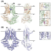
Snapshots of ABCG1 and ABCG5/G8: A Sterol's Journey to Cross the Cellular Membranes
- PMID: 36613930
- PMCID: PMC9820320
- DOI: 10.3390/ijms24010484
Snapshots of ABCG1 and ABCG5/G8: A Sterol's Journey to Cross the Cellular Membranes
Abstract
The subfamily-G ATP-binding cassette (ABCG) transporters play important roles in regulating cholesterol homeostasis. Recent progress in the structural data of ABCG1 and ABCG5/G8 disclose putative sterol binding sites that suggest the possible cholesterol translocation pathway. ABCG1 and ABCG5/G8 share high similarity in the overall molecular architecture, and both transporters appear to use several unique structural motifs to facilitate cholesterol transport along this pathway, including the phenylalanine highway and the hydrophobic valve. Interestingly, ABCG5/G8 is known to transport cholesterol and phytosterols, whereas ABCG1 seems to exclusively transport cholesterol. Ligand docking analysis indeed suggests a difference in recruiting sterol molecules to the known sterol-binding sites. Here, we further discuss how the different and shared structural features are relevant to their physiological functions, and finally provide our perspective on future studies in ABCG cholesterol transporters.
Keywords: ABCG1; ABCG5; ABCG8; RCT; TICE; sterol trasportation; structural motif.
Conflict of interest statement
The authors declare no conflict of interest.
Figures


Similar articles
-
Structural Analysis of Cholesterol Binding and Sterol Selectivity by ABCG5/G8.J Mol Biol. 2022 Oct 30;434(20):167795. doi: 10.1016/j.jmb.2022.167795. Epub 2022 Aug 18. J Mol Biol. 2022. PMID: 35988751
-
Picky ABCG5/G8 and promiscuous ABCG2 - a tale of fatty diets and drug toxicity.FEBS Lett. 2020 Dec;594(23):4035-4058. doi: 10.1002/1873-3468.13938. Epub 2020 Oct 14. FEBS Lett. 2020. PMID: 32978801 Free PMC article. Review.
-
Association of ABCG5 and ABCG8 Transporters with Sitosterolemia.Adv Exp Med Biol. 2024;1440:31-42. doi: 10.1007/978-3-031-43883-7_2. Adv Exp Med Biol. 2024. PMID: 38036873 Review.
-
Transmembrane Polar Relay Drives the Allosteric Regulation for ABCG5/G8 Sterol Transporter.Int J Mol Sci. 2020 Nov 19;21(22):8747. doi: 10.3390/ijms21228747. Int J Mol Sci. 2020. PMID: 33228147 Free PMC article.
-
Metformin and AMP Kinase Activation Increase Expression of the Sterol Transporters ABCG5/8 (ATP-Binding Cassette Transporter G5/G8) With Potential Antiatherogenic Consequences.Arterioscler Thromb Vasc Biol. 2018 Jul;38(7):1493-1503. doi: 10.1161/ATVBAHA.118.311212. Epub 2018 May 31. Arterioscler Thromb Vasc Biol. 2018. PMID: 29853564 Free PMC article.
Cited by
-
Endoplasmic Reticulum Stress and Its Impact on Adipogenesis: Molecular Mechanisms Implicated.Nutrients. 2023 Dec 12;15(24):5082. doi: 10.3390/nu15245082. Nutrients. 2023. PMID: 38140341 Free PMC article. Review.
-
NPC1L1 Plays a Novel Role in Nonalcoholic Fatty Liver Disease.ACS Omega. 2023 Dec 13;8(51):48586-48589. doi: 10.1021/acsomega.3c07337. eCollection 2023 Dec 26. ACS Omega. 2023. PMID: 38162748 Free PMC article. Review.
-
TNFα Activates the Liver X Receptor Signaling Pathway and Promotes Cholesterol Efflux from Human Brain Pericytes Independently of ABCA1.Int J Mol Sci. 2023 Mar 22;24(6):5992. doi: 10.3390/ijms24065992. Int J Mol Sci. 2023. PMID: 36983062 Free PMC article.
-
Alleviation of Lipid Disorder and Liver Damage in High-Fat Diet-Induced Obese Mice by Selenium-Enriched Cardamine violifolia with Cadmium Accumulation.Nutrients. 2024 Sep 22;16(18):3208. doi: 10.3390/nu16183208. Nutrients. 2024. PMID: 39339808 Free PMC article.
-
Advances in the role of GPX3 in ovarian cancer (Review).Int J Oncol. 2024 Mar;64(3):31. doi: 10.3892/ijo.2024.5619. Epub 2024 Feb 1. Int J Oncol. 2024. PMID: 38299269 Free PMC article.
References
Publication types
MeSH terms
Substances
Grants and funding
LinkOut - more resources
Full Text Sources
Medical

