Review
. 2022 Dec 23;15(1):101.
doi: 10.3390/cancers15010101.

CRAC and SK Channels: Their Molecular Mechanisms Associated with Cancer Cell Development

Affiliations
Review

CRAC and SK Channels: Their Molecular Mechanisms Associated with Cancer Cell Development

Adéla Tiffner et al. Cancers (Basel). .

Abstract

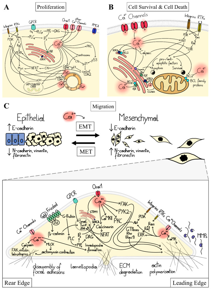
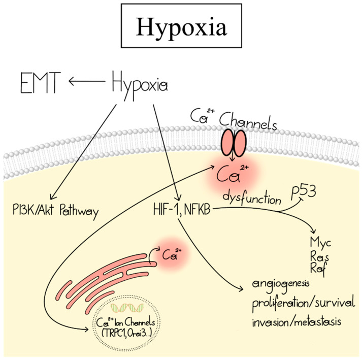
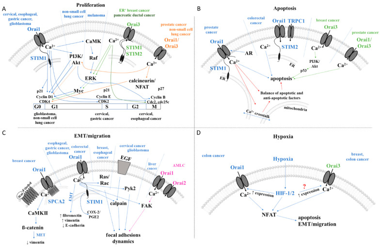
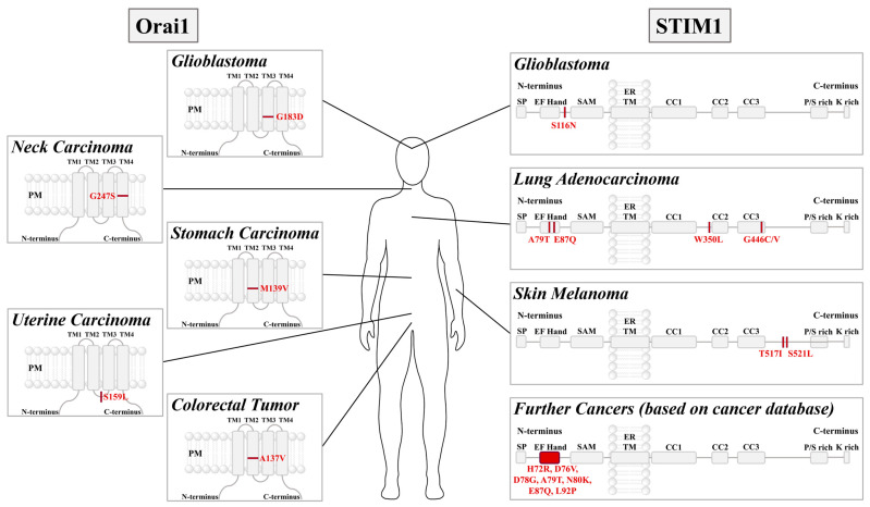
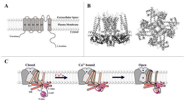
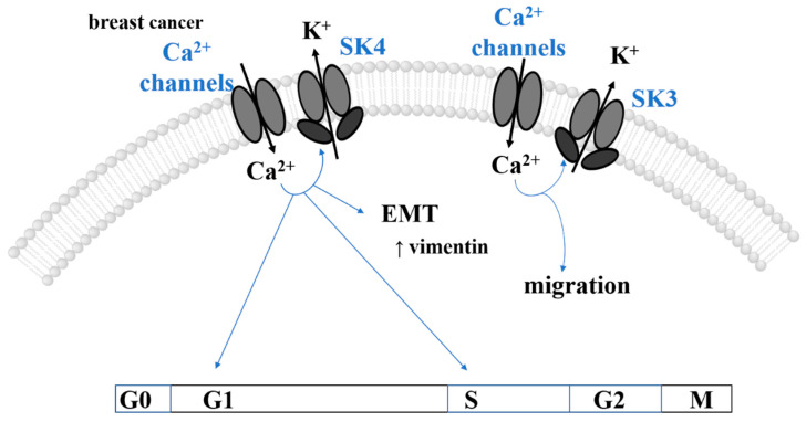
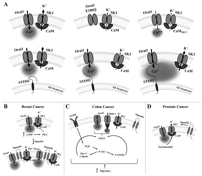
Cancer represents a major health burden worldwide. Several molecular targets have been discovered alongside treatments with positive clinical outcomes. However, the reoccurrence of cancer due to therapy resistance remains the primary cause of mortality. Endeavors in pinpointing new markers as molecular targets in cancer therapy are highly desired. The significance of the co-regulation of Ca2+-permeating and Ca2+-regulated ion channels in cancer cell development, proliferation, and migration make them promising molecular targets in cancer therapy. In particular, the co-regulation of the Orai1 and SK3 channels has been well-studied in breast and colon cancer cells, where it finally leads to an invasion-metastasis cascade. Nevertheless, many questions remain unanswered, such as which key molecular components determine and regulate their interplay. To provide a solid foundation for a better understanding of this ion channel co-regulation in cancer, we first shed light on the physiological role of Ca2+ and how this ion is linked to carcinogenesis. Then, we highlight the structure/function relationship of Orai1 and SK3, both individually and in concert, their role in the development of different types of cancer, and aspects that are not yet known in this context.

Keywords: CRAC channel; SK3 channel; cancer; cancer hallmarks; cancer signaling pathways.


Conflict of interest statement

The authors declare no conflict of interest.

Figures

Figure 1
Figure 1
Ca2+-dependent cancer signaling pathways controlling proliferation, cell survival, and death and migration. (A) The scheme depicts critical pathways for elevation of cytosolic Ca2+ levels (via PM ion channels (here, the focus is on the store-operated Ca2+ ion channel named the CRAC channel, i.e., thepore-forming component Orai1) or upon ER-Ca2+ depletion) that control proliferation at the level of the cell cycle machinery (cyclins, CDKs, CDK inhibitors, centrosome cycle) in a Ca2+-dependent manner, either directly via Ca2+-binding proteins (CaM, CaMK, CN), or indirectly via transcription factors (NFAT, or immediate early gene (JUN, Myc, and FOS)) and the oncogenic pathways, Ras/ERK and PI3K/Akt. (B) Scheme highlights pathways that control cell survival and death factors (e.g., BCL family proteins such as BCL2) in a Ca2+-dependent manner. These include factors modulating ER Ca2+ uptake and Ca2+ crosstalk between ER and mitochondria (SERCA, p53, Akt, IP3R, calpains, caspases). Moreover, the Ca2+-dependent modulation of cell fate via oncogenic pathways is shown. (C) Top: Schematic representation of the transitions between epithelial and mesenchymal states (MET-EMT (mesenchymal-epithelial transition <-> epithelial-mesenchymal transition)) and the characteristic up- and down-regulations of specific gene expression markers (E-/N-cadherin, vimentin, fibronectin), which are controlled by Ca2+. Bottom: Ca2+-dependent pathways controlling highly coordinated migration together with contacts to the extracellular matrix at the rear and the leading edge of the cell. At the leading edge, Ca2+ triggers lamellipodia formation by actin polymerization, the assembly of focal adhesions, the formation of contact with the bottom, and matrix metalloproteinases (involving small GTPase, such as Ras and Rac1, Ca2+-dependent factors, including CaMK and proline-rich tyrosine kinase 2 (PYK2); focal adhesion kinase (FAK)). At the rear edge, the cell disconnects from the bottom, involving focal adhesion disassembly (Ca2+-sensitive protease calpain, which cleaves FAK, vinculin, talin, and integrins) and actomyosin contraction (MLCK). Software used for all figure drawings: Procreate, Chemdraw (RRID:SCR_016768).
Figure 2
Figure 2
Linkage of hypoxia and the Ca2+-signaling machinery in cancer. Hypoxia induces EMT and activates PI3K/Akt, HIF, and NFkB signaling pathways. HIF-1 is involved in angiogenesis, cell proliferation/survival, and invasion/metastasis. It can trigger upregulated expression of Ca2+ ion channels, leading to enhanced Ca2+ levels that promote transcription of oncogenes. Conversely, Ca2+ can modulate HIF-1 signaling. Software used for all figure drawings: Procreate, Chemdraw (RRID:SCR_016768).
Figure 3
Figure 3
The STIM1-Orai1 activation mechanism and STIM and Orai isoforms. (A) Illustration of the activation mechanism of the CRAC channel, starting with inactive STIM1 and Orai1 in the resting state (left), followed by the depiction of the two proteins in the activated state allowing Ca2+ influx indicated by the green arrow (right), with the most important interaction sites highlighted. (B) A schematic illustration of the STIM1 and STIM2 isoforms consisting of the respective canonical EF-hand domain (cEF) and the hidden EF-hand domain (hEF), followed by the sterile alpha motif (SAM) in the ER lumen and subsequently the single TM. The C-terminus located in the cytosol contains three coiled-coil domains (CC1–3), more specifically, the three predicted α-helices of CC1 (α1–3). These coiled-coil regions comprise the Orai-activating small fragment (OASF), which spans all three coiled-coil domains, the CRAC-activating domain (CAD), or the STIM1-Orai-activating region (SOAR), which includes CC2 and CC3. The coiled-coil regions are followed by the inactivation domain (ID), the microtubule end-binding domain (EB), and finally, the polybasic domain (PBD) at the very end of the C-terminus. (C) An overview of all Orai proteins (Orai1, Orai2, Orai3) highlighting the TM domains alongside major positions for CRAC channel gating (Orai1 aa 72-90—ETON region, R91—SCID mutation position, E106—selectivity filter, H134, P245—critical gating checkpoints, L273—critical STIM1 binding site and analogue sites are labeled in Orai2 and Orai3). Software used for all figure drawings: Procreate, Chemdraw (RRID:SCR_016768).
Figure 4
Figure 4
Cancer features and associated CRAC channel-dependent signaling pathways in different cancer types. The schematics summarize the current knowledge of signaling pathways controlling cancer features (proliferation (A), apoptosis (B), EMT/migration (C), and hypoxia (D)) of the mentioned cancer types depending on CRAC channel components, TRP channels, or SPCA2. ? … signaling pathway is unknow. Colors indicate which cancer type correlates (arrow in corresponding color) which signaling pathway. Software used for all figure drawings: Procreate, Chemdraw (RRID:SCR_016768).
Figure 5
Figure 5
STIM1 and Orai1 cancer-related mutations. Schematic representation of the major components of STIM1 and Orai1 highlighting those point mutations associated with cancer cell development, including glioblastoma, cervical cancer, gastric cancer, uterine cancer, colorectal tumor, glioblastoma, lung adenocarcinoma, skin melanoma, and others [144,290]. Software used for all figure drawings: Procreate, Chemdraw (RRID:SCR_016768).
Figure 6
Figure 6
A general overview of SK channels and their activation mechanism. (A) Simplified scheme of the 6 TM domains (S1–S6) of the SK channel, with the pore located between S5 and S6 and both the N- as well as the C-terminus in the cytosol. (B) Side and top view of SK4 with the constitutively bound CaM in dark gray (PDB ID: 6CNM—visualized by ChimeraX [336]). (C) Activation mechanism of the SK channel, starting with an inactive channel and the free CaM N-lobe with no bound Ca2+. The C-lobe of CaM is constitutively bound to the channel (left). The binding of Ca2+, mainly to the CaM N-lobe, triggers a conformational change leading to the interaction between the CaM N-lobe and the S45A helix (middle), further communicating a conformational change to the channel, pulling the S45B away from the pore and rendering the channel open (right) (adapted from Lee et al., 2018 [312]). Software used for all figure drawings: Procreate, Chemdraw (RRID:SCR_016768).
Figure 7
Figure 7
Cancer features and associated SK channel-dependent signaling pathways in different cancer types. The scheme summarizes the current knowledge of signaling pathways controlling cancer features (proliferation and EMT/migration) of the mentioned cancer types depending on SK channels as well as Ca2+ signaling. Software used for all figure drawings: Procreate, Chemdraw (RRID:SCR_016768).
Figure 8
Figure 8
The Orai1/SK3 interplay and methods of its modulation in breast, colon, and prostate cancer. (A) Generally, the co-localization of Orai1 and SK3 channels is sufficient for their interplay, which leads to a positive feedback mechanism. Orai1 E106Q pore mutant does not affect its co-localization with SK3 but is unable to boost SK3 K+ currents. CaMMUT, which typically abolishes SK3 K+ currents, can be rescued by co-localized Orai1. In the presence of STIM1, the SK3–Orai1 interplay remains unaffected in the resting state, and upon store-depletion, STIM1 binds to Orai1 and thus moves SK3 and Orai1 apart from each other. Local enhancements of Ca2+ due to STIM1-mediated Orai1 activation do not boost SK3 K+ currents, whereas global Ca2+ enhancements strongly augment SK3 K+ currents. (B) In breast cancer cells, the interplay of Orai1 and SK3 boosts metastasis, which can be modulated by the cAMP-PKA pathway (adapted from Clarysse et al. (2014) [322]). Additionally, the stress-activated chaperone SigmaR1 promotes the interplay of Orai1 and SK3 in breast cancer cells (adapted from Gueguinou et al. (2017) [321]). (C) In colon cancer cells, migration is promoted by SOCE via a complex of TRPC1, Orai1, and SK3. This involves the activation of the Akt pathway and the phosphorylation of STIM1, leading to a positive feedback loop (adapted from Gueguinou et al. (2016) [323]). (D) SK3, reported to be endogenously expressed in the prostate cancer cell line LNCaP, is involved in the regulation of cell proliferation. The application of Enzalutamide enables the upregulation of SK3 expression in LNCaP cells (adapted from Figiel et al. (2019) [364]). Further involvement of accessory proteins, such as SigmaR1, cannot be ruled out and require further investigation. Throughout the figure, arrows indicate the direction of the ion flux. Software used for all figure drawings: Procreate, Chemdraw (RRID:SCR_016768).

Similar articles

Cited by

References

    1. Sung H., Ferlay J., Siegel R.L., Laversanne M., Soerjomataram I., Jemal A., Bray F. Global Cancer Statistics 2020: GLOBOCAN Estimates of Incidence and Mortality Worldwide for 36 Cancers in 185 Countries. CA Cancer J. Clin. 2021;71:209–249. doi: 10.3322/caac.21660. - DOI - PubMed
    1. Hanahan D., Weinberg R.A. The hallmarks of cancer. Cell. 2000;100:57–70. doi: 10.1016/S0092-8674(00)81683-9. - DOI - PubMed
    1. Prevarskaya N., Ouadid-Ahidouch H., Skryma R., Shuba Y. Remodelling of Ca2+ transport in cancer: How it contributes to cancer hallmarks? Philos. Trans. R. Soc. Lond. B Biol. Sci. 2014;369:20130097. doi: 10.1098/rstb.2013.0097. - DOI - PMC - PubMed
    1. Fouad Y.A., Aanei C. Revisiting the hallmarks of cancer. Am. J. Cancer Res. 2017;7:1016–1036. - PMC - PubMed
    1. Hanahan D., Weinberg R.A. Hallmarks of cancer: The next generation. Cell. 2011;144:646–674. doi: 10.1016/j.cell.2011.02.013. - DOI - PubMed

LinkOut - more resources