

Oxidative Stress and Mitochondrial Dysfunction in Chronic Kidney Disease
- PMID: 36611880
- PMCID: PMC9818928
- DOI: 10.3390/cells12010088
Oxidative Stress and Mitochondrial Dysfunction in Chronic Kidney Disease
Abstract
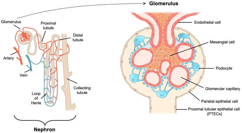
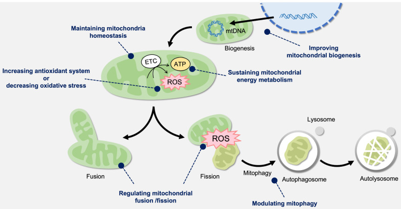
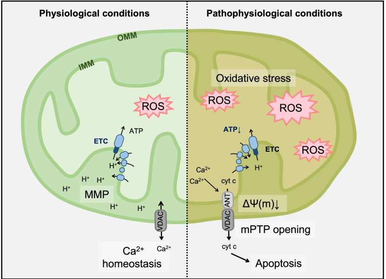
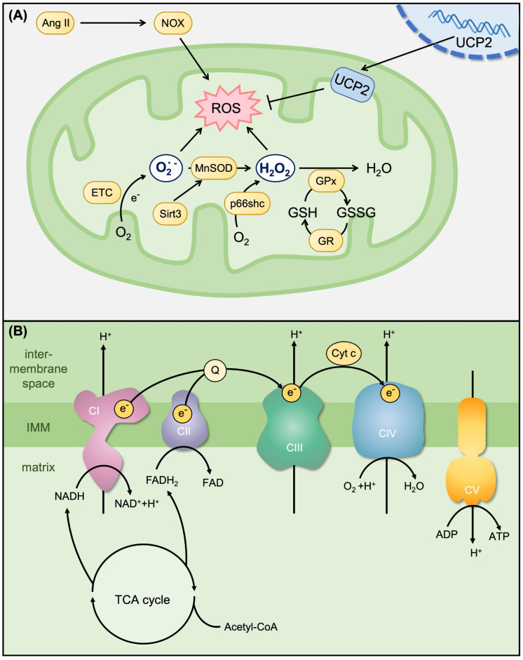
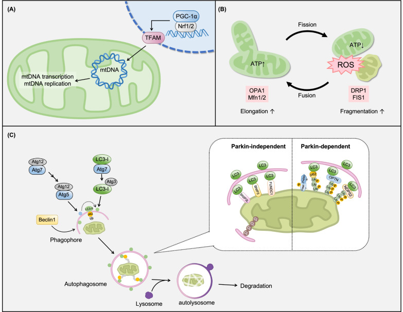
The kidney contains many mitochondria that generate ATP to provide energy for cellular processes. Oxidative stress injury can be caused by impaired mitochondria with excessive levels of reactive oxygen species. Accumulating evidence has indicated a relationship between oxidative stress and kidney diseases, and revealed new insights into mitochondria-targeted therapeutics for renal injury. Improving mitochondrial homeostasis, increasing mitochondrial biogenesis, and balancing mitochondrial turnover has the potential to protect renal function against oxidative stress. Although there are some reviews that addressed this issue, the articles summarizing the relationship between mitochondria-targeted effects and the risk factors of renal failure are still few. In this review, we integrate recent studies on oxidative stress and mitochondrial function in kidney diseases, especially chronic kidney disease. We organized the causes and risk factors of oxidative stress in the kidneys based in their mitochondria-targeted effects. This review also listed the possible candidates for clinical therapeutics of kidney diseases by modulating mitochondrial function.
Keywords: chronic kidney disease; mitochondrial homeostasis; mitochondrial turnover; oxidative stress.
Conflict of interest statement
The authors declare no conflict of interest.
Figures
Similar articles
-
Mitochondrial Redox Signaling and Oxidative Stress in Kidney Diseases.Biomolecules. 2021 Aug 3;11(8):1144. doi: 10.3390/biom11081144. Biomolecules. 2021. PMID: 34439810 Free PMC article. Review.
-
The Role of Mitochondria in Acute Kidney Injury and Chronic Kidney Disease and Its Therapeutic Potential.Int J Mol Sci. 2021 Oct 19;22(20):11253. doi: 10.3390/ijms222011253. Int J Mol Sci. 2021. PMID: 34681922 Free PMC article. Review.
-
Mitochondrial dysfunction in the pathophysiology of renal diseases.Am J Physiol Renal Physiol. 2014 Feb 15;306(4):F367-78. doi: 10.1152/ajprenal.00571.2013. Epub 2013 Dec 4. Am J Physiol Renal Physiol. 2014. PMID: 24305473 Review.
-
Mitochondrial dysfunction and oxidative stress: Role in chronic kidney disease.Life Sci. 2023 Apr 15;319:121432. doi: 10.1016/j.lfs.2023.121432. Epub 2023 Jan 24. Life Sci. 2023. PMID: 36706833 Review.
-
High-fat diet induces an initial adaptation of mitochondrial bioenergetics in the kidney despite evident oxidative stress and mitochondrial ROS production.Am J Physiol Endocrinol Metab. 2011 Jun;300(6):E1047-58. doi: 10.1152/ajpendo.00666.2010. Epub 2011 Mar 8. Am J Physiol Endocrinol Metab. 2011. PMID: 21386058 Free PMC article.
Cited by
-
Transcriptome Analysis of Differentially Expressed Genes and Molecular Pathways Involved in C2C12 Cells Myogenic Differentiation.Mol Biotechnol. 2024 Sep 18. doi: 10.1007/s12033-024-01259-7. Online ahead of print. Mol Biotechnol. 2024. PMID: 39289290
-
Aerobic Exercise Ameliorates Cognitive Disorder and Declined Oxidative Stress via Modulating the Nrf2 Signaling Pathway in D-galactose Induced Aging Mouse Model.Neurochem Res. 2024 Sep;49(9):2408-2422. doi: 10.1007/s11064-024-04164-2. Epub 2024 Jun 6. Neurochem Res. 2024. PMID: 38839706
-
Renal-Protective Roles of Lipoic Acid in Kidney Disease.Nutrients. 2023 Apr 1;15(7):1732. doi: 10.3390/nu15071732. Nutrients. 2023. PMID: 37049574 Free PMC article. Review.
-
Evaluation of Unsaponifiable Fraction of Avocado Oil on Liver and Kidney Mitochondrial Function in Rats Fed a High-Fat and High-Carbohydrate Diet.Metabolites. 2024 Aug 4;14(8):431. doi: 10.3390/metabo14080431. Metabolites. 2024. PMID: 39195527 Free PMC article.
-
Biovalorisation of agro-industrial wastes into astaxanthin by Xanthophyllomyces dendrorhous.Appl Microbiol Biotechnol. 2024 Jul 27;108(1):429. doi: 10.1007/s00253-024-13257-5. Appl Microbiol Biotechnol. 2024. PMID: 39066896 Free PMC article.
References
-
- Wang Z.M., Yang Z.L., Bosy-westphal A., Zhang J.Y., Schautz B., Later W., Heymsfield S.B., Muller M.J. Specific meta-bolic rates of major organs and tissues across adulthood: Evaluation by mechanistic model of resting energy expenditure. Am. J. Clin. Nutr. 2010;92:1369–1377. doi: 10.3945/ajcn.2010.29885. - DOI - PMC - PubMed
-
- Fredericks W.J., Yin H., Lal P., Puthiyaveettil R., Malkowicz S.B., Fredericks N.J., Tomaszewski J., Rauscher F.J., 3rd, Malkowicz S.B. Ectopic expression of the TERE1 (UBIAD1) protein inhibits growth of renal clear cell carcinoma cells: Altered metabolic phenotype associated with reactive oxygen species, nitric oxide and SXR target genes involved in cholesterol and lipid metabolism. Int. J. Oncol. 2013;43:638–652. doi: 10.3892/ijo.2013.1985. - DOI - PubMed
Publication types
MeSH terms
Substances
Grants and funding
LinkOut - more resources
Full Text Sources
Medical
