. 2023 Jan 3;12(1):e028215.
doi: 10.1161/JAHA.122.028215. Epub 2022 Dec 24.

Proteolytic Degradation Is a Major Contributor to Bioprosthetic Heart Valve Failure

Affiliations

Proteolytic Degradation Is a Major Contributor to Bioprosthetic Heart Valve Failure

Alexander E Kostyunin et al. J Am Heart Assoc. .

Abstract

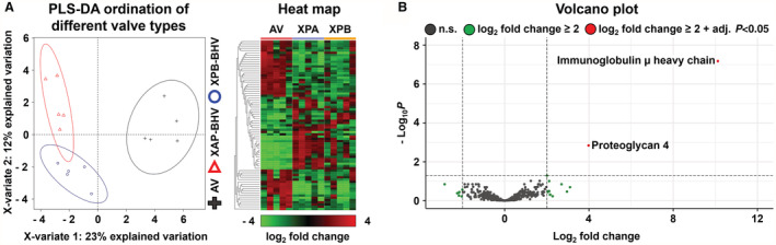
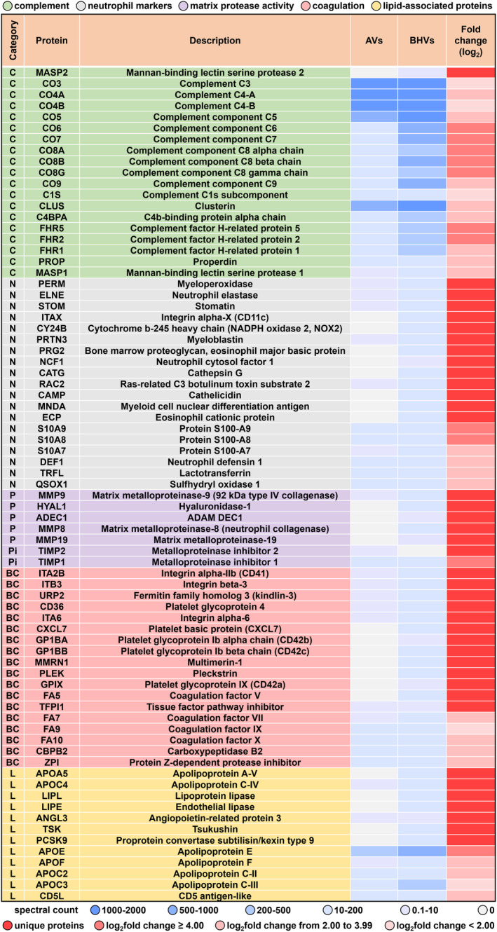
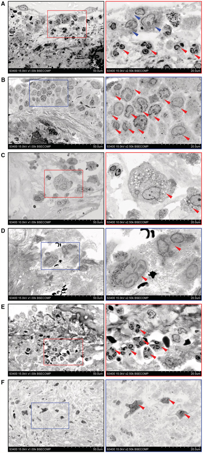
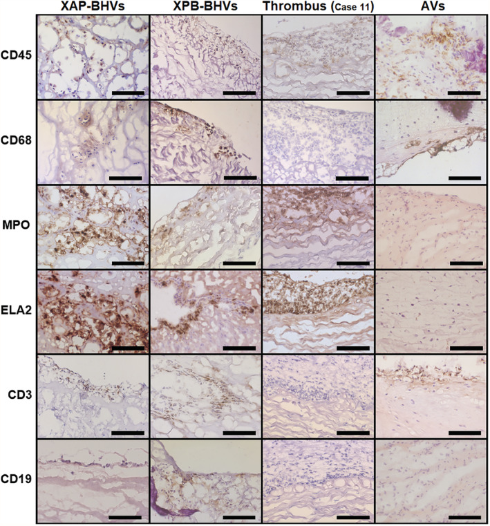
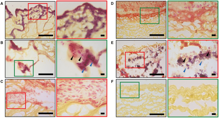
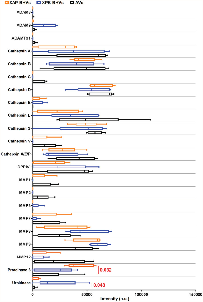
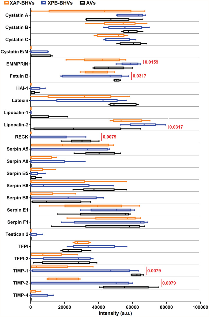
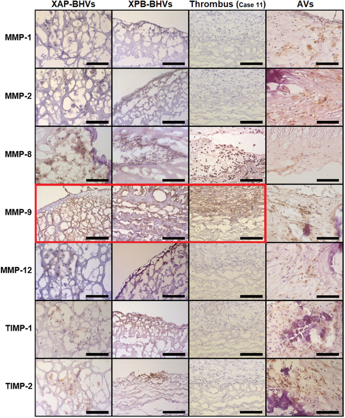
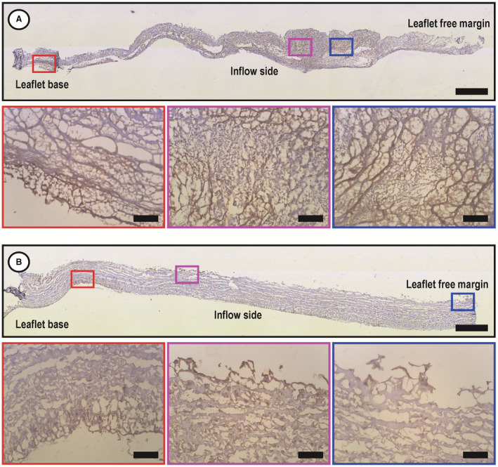
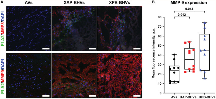
Background Whereas the risk factors for structural valve degeneration (SVD) of glutaraldehyde-treated bioprosthetic heart valves (BHVs) are well studied, those responsible for the failure of BHVs fixed with alternative next-generation chemicals remain largely unknown. This study aimed to investigate the reasons behind the development of SVD in ethylene glycol diglycidyl ether-treated BHVs. Methods and Results Ten ethylene glycol diglycidyl ether-treated BHVs excised because of SVD, and 5 calcified aortic valves (AVs) replaced with BHVs because of calcific AV disease were collected and their proteomic profile was deciphered. Then, BHVs and AVs were interrogated for immune cell infiltration, microbial contamination, distribution of matrix-degrading enzymes and their tissue inhibitors, lipid deposition, and calcification. In contrast with dysfunctional AVs, failing BHVs suffered from complement-driven neutrophil invasion, excessive proteolysis, unwanted coagulation, and lipid deposition. Neutrophil infiltration was triggered by an asymptomatic bacterial colonization of the prosthetic tissue. Neutrophil elastase, myeloblastin/proteinase 3, cathepsin G, and matrix metalloproteinases (MMPs; neutrophil-derived MMP-8 and plasma-derived MMP-9), were significantly overexpressed, while tissue inhibitors of metalloproteinases 1/2 were downregulated in the BHVs as compared with AVs, together indicative of unbalanced proteolysis in the failing BHVs. As opposed to other proteases, MMP-9 was mostly expressed in the disorganized prosthetic extracellular matrix, suggesting plasma-derived proteases as the primary culprit of SVD in ethylene glycol diglycidyl ether-treated BHVs. Hence, hemodynamic stress and progressive accumulation of proteases led to the extracellular matrix degeneration and dystrophic calcification, ultimately resulting in SVD. Conclusions Neutrophil- and plasma-derived proteases are responsible for the loss of BHV mechanical competence and need to be thwarted to prevent SVD.

Keywords: bacterial invasion; bioprosthetic heart valves; matrix metalloproteinases; neutrophil infiltration; structural valve degeneration.


Figures

Figure 1
Figure 1. Label‐free proteomic profiling of calcified AVs, XAP‐BHVs, and XPB‐BHVs.
A, PLS‐DA and heat map demonstrated considerable differences between the AVs and BHVs, while XAP‐BHVs and XPB‐BHVs have not been significantly different from each other with regards to the proteomic composition. B, Volcano plot illustrates the absence of statistically significant differences between XAP‐BHVs and XPB‐BHVs, showing only 2 different proteins. AVs indicates aortic valves; PLS‐DA, partial least squares discriminant analysis; XAP‐BHVs, xenoaortic porcine bioprosthetic heart valves; and XPB‐BHVs, xenopericardial bovine bioprosthetic heart valves.
Figure 2
Figure 2. Annotation of unique or upregulated proteins in XAP‐BHVs and XPB‐BHVs.
The list of 75 selected proteins that have been unique or differentially expressed between the AVs and BHVs (XAP‐BHVs and XPB‐BHVs were pooled for this analysis as their proteomic profiles were similar on PLS‐DA and volcano plot). AVs indicates aortic valves; PLS‐DA, partial least squares discriminant analysis; XAP‐BHVs, xenoaortic porcine bioprosthetic heart valves; and XPB‐BHVs, xenopericardial bovine bioprosthetic heart valves.
Figure 3
Figure 3. Immune cell populations inhabitating the BHVs and calcified AVs.
A, Both macrophages (indicated by blue arrows) and neutrophils (indicated by red arrows) were among the frequent findings in the BHVs, often being colocalized with each other. B, Canonical macrophages (indicated by red arrows) infiltrating the BHV. C, Foam cells (indicated by red arrows) formed because of excessive lipid load in the BHVs. D, Multinucleated giant cells (indicated by red arrows) reflecting the ECM degradation have been observed exclusively in the BHVs. E, Multiple neutrophils (indicated by red arrows) also were commonly found in the BHVs. F, Among all immune cells, calcified AVs contained only canonical macrophages (shown in the figure, indicated by red arrows) and foam cells, while multinucleated giant cells and neutrophils have not been detected. Images to the right are the close‐ups of those to the left (demarcated by red or blue contour). EM‐BSEM, representative images, magnification: ×1000 (left) and ×2500 (right), scale bars: 50 μm (left) and 20 μm (right). Accelerating voltage: 10 kV (C) or 15 kV (A, B, DF). AVs indicates aortic valves; BHVs, bioprosthetic heart valves; ECM, extracellular matrix; and EM‐BSEM, embedding and backscattered scanning electron microscopy.
Figure 4
Figure 4. Macrophages and neutrophils are prevailing cell populations in BHVs while calcified AVs are devoid of neutrophils.
Cells infiltrating XAP‐BHVs and XPB‐BHVs were positively stained (brown color) for pan‐leukocyte marker CD45, macrophage marker CD68 and neutrophil markers MPO and NE/ELA2, with rare T (CD3+) and no B (CD19+) cells. Prosthetic thrombus served as a positive control for CD45, MPO, and NE/ELA2 stainings. AVs contained only CD45+, CD68+, and CD3+‐positive cells. Immunohistochemical staining, representative images, magnification: ×400, scale bar: 100 μm. AVs indicates aortic valves; BHVs, bioprosthetic heart valves; CD, cluster of differentiation; MPO, myeloperoxidase; NE/ELA2, neutrophil elastase; XAP‐BHVs, xenoaortic porcine bioprosthetic heart valves; XPB‐BHVs, xenopericardial bovine bioprosthetic heart valves.
Figure 5
Figure 5. Bacterial colonization of XAP‐BHVs and XPB‐BHVs.
A, Moderate amounts of spherical and rodlike bacteria beneath the BHV surface. B, Colocalization of immune cells (indicated by black arrows) and bacteria (indicated by blue arrows) has been rarely observed. C, Immune cells were mostly located in bacteria‐free areas. D, BHV‐associated thrombotic masses have been heavily infiltrated with neutrophils but were devoid of bacteria. E, Vegetations in the BHV suffering from the prosthetic endocarditis caused by Staphylococcus aureus (positive control). F, Nonimplanted bovine pericardium used as a negative control to exclude an accidental contamination. Note the large bacterial colonies (indicated by blue arrows) and aggressive infiltration by immune cells (indicated by black arrows). Images to the right are the close‐ups of those to the left (demarcated by red or green contour). Gram staining, magnification: ×400 (left) and ×1000 (right), scale bars: 100 μm (left) and 10 μm (right). XAP‐BHVs indicates xenoaortic porcine bioprosthetic heart valves; and XPB‐BHVs, xenopericardial bovine bioprosthetic heart valves.
Figure 6
Figure 6. Densitometry analysis (ImageJ) of protease levels in XAP‐BHVs, XPB‐BHVs, and calcified AVs.
Box‐and‐whisker plot, center lines indicate median, boxes bounds indicate 25th–75th percentiles, whiskers indicate range. Kruskal–Wallis test with post hoc FDR adjustment for multiple comparisons by 2‐stage linear step‐up procedure of Benjamini, Krieger, and Yekutieli. Statistically significant, FDR‐corrected q values are provided above boxes. AVs indicates aortic valves; FDR, false discovery rate; XAP‐BHVs, xenoaortic porcine bioprosthetic heart valves; and XPB‐BHVs, xenopericardial bovine bioprosthetic heart valves.
Figure 7
Figure 7. Densitometry analysis (ImageJ) of protease inhibitor levels in XAP‐BHVs, XPB‐BHVs, and calcified AVs.
Box‐and‐whisker plot, center lines indicate median, boxes bounds indicate 25th to 75th percentiles, whiskers indicate range. Kruskal–Wallis test with post hoc FDR adjustment for multiple comparisons by 2‐stage linear step‐up procedure of Benjamini, Krieger, and Yekutieli. Statistically significant, FDR‐corrected q values are provided above boxes. ADAM indicates a disintegrin and metalloproteinase; ADAMTS, a disintegrin and metalloproteinase with thrombospondin motifs; AVs, aortic valves; DPPIV, dipeptidyl peptidase 4; EMMPRIN, extracellular matrix metalloproteinase inducer; FDR, false discovery rate; HAI‐1, hepatocyte growth factor activator inhibitor 1; MMP, matrix metalloproteinase; RECK, reversion‐inducing‐cysteine‐rich protein with Kazal motifs; TFPI, tissue factor pathway inhibitor; TIMP, tissue inhibitor of metalloproteinases; XAP‐BHVs, xenoaortic porcine bioprosthetic heart valves; and XPB‐BHVs, xenopericardial bovine bioprosthetic heart valves.
Figure 8
Figure 8. Expression patterns of MMPs/TIMPs within failing BHVs and calcified AVs.
All proteases and protease inhibitors (brown color) were colocalized with cells except for the MMP‐9 within the BHVs, which has been colocalized with the degraded extracellular matrix. Note that MMP‐8 and MMP‐9 are abundant within the clot in the BHV diagnosed with immunothrombosis and excised as early as 2 days postimplantation, indicating neutrophils as the principal source of MMP‐8 (colocalized with the cells) and plasma as the primary source of MMP‐9 (diffuse staining pattern). Immunohistochemical staining, magnification: ×400, scale bar: 100 μm. AVs indicates aortic valves; BHVs, bioprosthetic heart valves; MMP, matrix metalloproteinase; and TIMPs, tissue inhibitors of metalloproteinases.
Figure 9
Figure 9. MMP‐9 is colocalized with the spongiosa and degraded extracellular matrix of failing BHVs.
A, XAP‐BHVs, note the highest MMP‐9 expression (brown color) in the spongiosa. B, XPB‐BHVs, note the highest MMP‐9 expression in the degraded extracellular matrix beneath the surface. For A and B: images below are the close‐ups of the image above (demarcated by red, purple, or blue contour). Immunohistochemical staining, magnification: ×50 (overview, merged image), scale bar: 1000 μm (overview) and magnification: ×200, scale bar: 100 μm (close‐ups). MMP‐9 indicates matrix metalloproteinase 9; XAP‐BHVs, xenoaortic porcine bioprosthetic heart valves; and XPB‐BHVs, xenopericardial bovine bioprosthetic heart valves.
Figure 10
Figure 10. Immunofluorescence analysis of MMP‐8 and MMP‐9 expression in calcified AVs, XAP‐BHVs, and XPB‐BHVs.
A, Combined immunofluorescence staining for the neutrophil marker NE/ELA2 (green color) either with neutrophil‐secreted MMP‐8 or with plasma‐derived MMP‐9 (red color). Nuclei are counterstained with DAPI (blue color). Representative images. Note the colocalization of MMP‐8 with NE/ELA2 and ubiquitous staining pattern of MMP‐9. Magnification: ×200, scale bar: 50 μm. B, Semiquantitative analysis of mean fluorescence intensity (MMP‐9, red channel) in confocal images (n=10 per group). Box‐and‐whisker plot combined with univariate scatterplot, center lines indicate median, boxes bounds indicate 25th–75th percentiles, whiskers indicate range. Each dot reflects a measurement from 1 image of a prosthetic or native valve. Kruskal–Wallis test with post hoc false discovery rate (FDR) adjustment for multiple comparisons by 2‐stage linear step‐up procedure of Benjamini, Krieger, and Yekutieli. Statistically significant, FDR‐corrected q values are provided above boxes. AVs indicates aortic valves; DAPI, 4′,6‐diamidino‐2‐phenylindole; FDR, false discovery rate; MMP, matrix metalloproteinase; NE/ELA2, neutrophil elastase; XAP‐BHVs, xenoaortic porcine bioprosthetic heart valves; and XPB‐BHVs, xenopericardial bovine bioprosthetic heart valves.

Similar articles

Cited by

References

    1. Otto CM, Nishimura RA, Bonow RO, Carabello BA, Erwin JP III, Gentile F, Jneid H, Krieger EV, Mack M, McLeod C, et al. 2020 ACC/AHA guideline for the management of patients with valvular heart disease: a report of the American College of Cardiology/American Heart Association joint committee on clinical practice guidelines. Circulation. 2021;143:e72–e227. doi: 10.1161/CIR.0000000000000923 - DOI - PubMed
    1. Lindman BR, Clavel MA, Mathieu P, Iung B, Lancellotti P, Otto CM, Pibarot P. Calcific aortic stenosis. Nat Rev Dis Primers. 2016;2:16006. doi: 10.1038/nrdp.2016.6 - DOI - PMC - PubMed
    1. Bax JJ, Delgado V. Bioprosthetic heart valves, thrombosis, anticoagulation, and imaging surveillance. JACC Cardiovasc Interv. 2017;10:388–390. doi: 10.1016/j.jcin.2017.01.017 - DOI - PubMed
    1. Kostyunin AE, Yuzhalin AE, Ovcharenko EA, Kutikhin AG. Development of calcific aortic valve disease: do we know enough for new clinical trials? J Mol Cell Cardiol. 2019;132:189–209. doi: 10.1016/j.yjmcc.2019.05.016 - DOI - PubMed
    1. Head SJ, Çelik M, Kappetein AP. Mechanical versus bioprosthetic aortic valve replacement. Eur Heart J. 2017;38:2183–2191. doi: 10.1093/eurheartj/ehx141 - DOI - PubMed

Publication types