. 2022 Dec 9;23(24):15630.
doi: 10.3390/ijms232415630.

A Study on the Protective Effect of sRAGE-MSCs in a Rodent Reperfusion Model of Myocardial Infarction

Affiliations

A Study on the Protective Effect of sRAGE-MSCs in a Rodent Reperfusion Model of Myocardial Infarction

Delger Bayarsaikhan et al. Int J Mol Sci. .

Abstract

Acute myocardial infarction (AMI) is one of the major leading causes of death in humans globally. Recently, increased levels of recruited macrophages and AGE-albumin were observed in the hearts of humans and animals with acute myocardial infarction. Thus, the purposes of this study were to investigate whether the elevated levels of AGE-albumin from activated macrophage cells are implicated in ischemia-induced cardiomyocyte death and to develop therapeutic strategies for AMI based on its underlying molecular mechanisms with respect to AGEs. The present study demonstrated that activated macrophages and AGE-albumin were observed in heart tissues obtained from humans and rats with AMI incidences. In the cellular model of AMI, it was found that increased expression of AGE-albumin was shown to be co-localized with macrophages, and the presence of AGE-albumin led to increased expression of RAGE through the mitogen-activated protein kinase pathway. After revealing cardiomyocyte apoptosis induced by toxicity of the AGE-RAGE system, sRAGE-secreting MSCs were generated using the CRISPR/Cas9 platform to investigate the therapeutic effects of sRAGE-MSCs in an AMI rat model. Gene-edited sRAGE-MSCs showed greater therapeutic effects against AMI pathogenesis in rat models compared to mock MSCs, and promising results of the functional improvement of stem cells could result in significant improvements in the clinical management of cardiovascular diseases.

Keywords: AGE-albumin; MSCs; acute myocardial ischemia; cardiomyocyte death; gene editing; macrophage; soluble RAGE.


Conflict of interest statement

The authors declare no conflict of interest.

Figures

Figure 1
Figure 1
Co-localization of AGE-albumin and activated macrophage cells in the ischemic reperfusion injured heart of human and rat. (A) Triple-labeled immunostaining of AGE (blue), albumin (green), and Iba1 (red, activated microglial cell marker) in the left ventricle of human (A) and rat (C). Merged image shows that AGE (blue), albumin (green), and Iba1 (red, activated microglial cell marker) were co-localized mostly in infarcted area of heart. Fluorescence expression level (B,D) and co-localization coefficient analyzed by densitometry analysis software using Zen software (Zeiss). Scale bar = 50 μm; ***, p < 0.001.
Figure 2
Figure 2
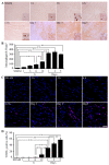
Time-dependent distribution of activated macrophages and apoptotic cardiomyocytes in the left ventricle of the sham and ischemic reperfusion injured heart of rats. (A) Iba-1-positive macrophage cells were measured by immunohistochemical staining in control or ischemic reperfusion injured rat hearts at different time periods. (B) A number of Iba-1-positive cells in the heart of control and ischemic reperfusion injured rats. (C) TUNEL staining used to detect apoptotic cardiomyocytes in control or ischemic reperfusion injured rat hearts at different time periods. (D) A percent of TUNEL-positive cells in the heart of control and ischemic reperfusion injured rats. Scale bar = 50 µm. In the statistical analysis, a one-way ANOVA was performed, and Tukey’s test was used to compare means: *, p < 0.05; **, p < 0.01.
Figure 3
Figure 3
Increased synthesis and secretion of AGE-albumin in activated macrophage cells. (A) Triple-labeled confocal microscopic image analyses were used to study the distribution and relative levels of AGE (blue), albumin (green), and Iba-1 (red) in rat macrophage cells after treated with hypoxia-exposed cardiomyocyte conditioned medium. Scale bar = 50 μm. (B) The dose-dependent increases in AGE-albumin in total lysates of RAW cells treated with 0, 1, 6, 12, or 24 h hypoxia-exposed cardiomyocyte conditioned medium for 48 h were determined by co-immunoprecipitation. (C) A graph illustrating the dose-dependent changes in intracellular (cell lysates) and extracellular (culture supernatant) levels of AGE-albumin in RAW cells treated with 0, 1, 6, 12, or 24 h hypoxia-exposed cardiomyocyte conditioned medium for 48 h, as determined by ELISA.
Figure 4
Figure 4
The relative level of RAGE and MAPKs in ischemic reperfusion injured rat heart. (A) RAGE expression is shown in double-labeled confocal images: RAGE (green) and DAPI (blue) in the left ventricle of sham and ischemic reperfusion injured hearts of rats at day 3, 5, 7, 10, and 14 in the border area. (B) Immunoblot analysis was performed to determine the expressed levels of RAGE, ERK1/2, p38, SAPK/JNK, pERK1/2, pp38, pSAPK/JNK, and β-actin, used as internal controls for equal protein loading of each lane. (CF) Densitometry analyses of MAPK proteins were evaluated using the Image-J software. Scale bar = 50 µm; **, p < 0.01; ***, p < 0.001.
Figure 5
Figure 5
Protective effect of sRAGE on AGE-albumin-induced cardiomyocyte death by decreasing RAGE level. (A) RAGE expression is shown in double-labeled confocal images: RAGE (red) and DAPI (blue) using H9C2 cell before and after exposing AGE-albumin or co-treated with AGE-albumin and sRAGE. Cardiomyocyte cell death was evaluated by double staining with TUNEL (red) and DAPI (blue). (B) Immunoblot analysis of cardiomyocytes lysates after AGE-albumin or AGE-albumin with sRAGE co-treatment. RAGE expression was increased after AGE-albumin treatment but decreased after co-treatment with sRAGE. In MAPK analysis, pp38 and pSAPK/JNK were increased after AGE-albumin treatment but decreased after co-treatment. (CF) Densitometry analyses of MAPK proteins were evaluated using the Image-J software. Scale bar = 50 µm; **, p < 0.01; ***, p < 0.001.
Figure 6
Figure 6
sRAGE-secreting MSC line characterization. (A) The illustration picture represents the gene information of pZDonor-AAVS1 puromycin vector. Each arrow describes a certain gene. (B) The illustration of sRAGE insertion coding sequence. (C) Genome integration was confirmed with genomic DNAs of MSCs which were transfected with mock, GFP, and sRAGE-containing pZDonor-AAVS1 plasmids. (D) Western blot analysis of supernatant and extract from MSCs transfected with GFP (lane 1) and FLAG-tagged sRAGE in pZDonor-AAVS1 vector (lane 2). β-actin loaded as a positive control. (E) The secretion of human sRAGE levels was confirmed with ELISA after puromycin selection; and statistical analysis was performed using Student’s t-test; ***, p < 0.001.
Figure 7
Figure 7
Soluble RAGE-secreting MSC protection of ischemia-mediated cardiomyocyte death by decreasing RAGE level. (A) Representative Masson’s trichrome staining revealed left ventricular fibrosis 4 weeks after AMI (magnification: 10X). Red color indicates viable myocardium; blue color indicates fibrosis due to infarction damage. (B) Infarct size was expressed as a percentage of ischemic area (C) and as the length of wall thickness; *, p < 0.05; **, p < 0.01; ***, p < 0.001.

Similar articles

References

    1. Bayarsaikhan D., Bayarsaikhan G., Lee B. Recent advances in stem cells and gene editing: Drug discovery and therapeutics. Prog. Mol. Biol. Transl. Sci. 2021;181:231–269. - PubMed
    1. Mc Namara K., Alzubaidi H., Jackson J.K. Cardiovascular disease as a leading cause of death: How are pharmacists getting involved? Integr. Pharm. Res. Pract. 2019;8:1–11. doi: 10.2147/IPRP.S133088. - DOI - PMC - PubMed
    1. Buja L.M. Myocardial ischemia and reperfusion injury. Cardiovasc Pathol. 2005;14:170–175. doi: 10.1016/j.carpath.2005.03.006. - DOI - PubMed
    1. Frank A., Bonney M., Bonney S., Weitzel L., Koeppen M., Eckle T. Myocardial ischemia reperfusion injury: From basic science to clinical bedside. Semin. Cardiothorac. Vasc. Anesth. 2012;16:123–132. doi: 10.1177/1089253211436350. - DOI - PMC - PubMed
    1. Hausenloy D.J., Yellon D.M. Myocardial ischemia-reperfusion injury: A neglected therapeutic target. J. Clin. Investig. 2013;123:92–100. doi: 10.1172/JCI62874. - DOI - PMC - PubMed