Detection of De Novo Dividing Stem Cells In Situ through Double Nucleotide Analogue Labeling
- PMID: 36552766
- PMCID: PMC9777310
- DOI: 10.3390/cells11244001
Detection of De Novo Dividing Stem Cells In Situ through Double Nucleotide Analogue Labeling
Abstract
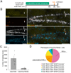
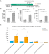
Tissue-specific somatic stem cells are characterized by their ability to reside in a state of prolonged reversible cell cycle arrest, referred to as quiescence. Maintenance of a balance between cell quiescence and division is critical for tissue homeostasis at the cellular level and is dynamically regulated by numerous extrinsic and intrinsic factors. Analysis of the activation of quiescent stem cells has been challenging because of a lack of methods for direct detection of de novo dividing cells. Here, we present and experimentally verify a novel method based on double labeling with thymidine analogues to detect de novo dividing stem cells in situ. In a proof of concept for the method, we show that memantine, a drug widely used for Alzheimer's disease therapy and a known strong inducer of adult hippocampal neurogenesis, increases the recruitment into the division cycle of quiescent radial glia-like stem cells-primary precursors of the adult-born neurons in the hippocampus. Our method could be applied to assess the effects of aging, pathology, or drug treatments on the quiescent stem cells in stem cell compartments in developing and adult tissues.
Keywords: adult hippocampal neurogenesis; memantine; proliferation; radial glia-like stem cells; stem cell quiescence; thymidine analogues.
Conflict of interest statement
The authors declare no conflict of interest.
Figures


Similar articles
-
Aging Modulates the Ability of Quiescent Radial Glia-Like Stem Cells in the Hippocampal Dentate Gyrus to be Recruited into Division by Pro-neurogenic Stimuli.Mol Neurobiol. 2024 Jun;61(6):3461-3476. doi: 10.1007/s12035-023-03746-5. Epub 2023 Nov 23. Mol Neurobiol. 2024. PMID: 37995077
-
Identification of De Novo Dividing Stem Cells.Methods Mol Biol. 2024 Jul 6. doi: 10.1007/7651_2024_560. Online ahead of print. Methods Mol Biol. 2024. PMID: 38967913
-
The Alzheimer's disease drug memantine increases the number of radial glia-like progenitor cells in adult hippocampus.Glia. 2009 Aug 1;57(10):1082-90. doi: 10.1002/glia.20831. Glia. 2009. PMID: 19115386
-
Quiescence Entry, Maintenance, and Exit in Adult Stem Cells.Int J Mol Sci. 2019 May 1;20(9):2158. doi: 10.3390/ijms20092158. Int J Mol Sci. 2019. PMID: 31052375 Free PMC article. Review.
-
Quiescence of Adult Mammalian Neural Stem Cells: A Highly Regulated Rest.Neuron. 2019 Dec 4;104(5):834-848. doi: 10.1016/j.neuron.2019.09.026. Neuron. 2019. PMID: 31805262 Review.
Cited by
-
Aging Modulates the Ability of Quiescent Radial Glia-Like Stem Cells in the Hippocampal Dentate Gyrus to be Recruited into Division by Pro-neurogenic Stimuli.Mol Neurobiol. 2024 Jun;61(6):3461-3476. doi: 10.1007/s12035-023-03746-5. Epub 2023 Nov 23. Mol Neurobiol. 2024. PMID: 37995077
-
Methods for Inferring Cell Cycle Parameters Using Thymidine Analogues.Biology (Basel). 2023 Jun 20;12(6):885. doi: 10.3390/biology12060885. Biology (Basel). 2023. PMID: 37372169 Free PMC article. Review.
References
Publication types
MeSH terms
Substances
Grants and funding
LinkOut - more resources
Full Text Sources
Medical

