

The enigma of ultraviolet radiation stress granules: Research challenges and new perspectives
- PMID: 36533077
- PMCID: PMC9751325
- DOI: 10.3389/fmolb.2022.1066650
The enigma of ultraviolet radiation stress granules: Research challenges and new perspectives
Abstract
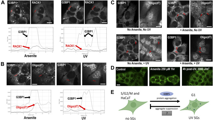
Stress granules (SGs) are non-membrane bound cytoplasmic condensates that form in response to a variety of different stressors. Canonical SGs are thought to have a cytoprotective role, reallocating cellular resources during stress by activation of the integrated stress response (ISR) to inhibit translation and avoid apoptosis. However, different stresses result in compositionally distinct, non-canonical SG formation that is likely pro-apoptotic, though the exact function(s) of both SGs subtypes remain unclear. A unique non-canonical SG subtype is triggered upon exposure to ultraviolet (UV) radiation. While it is generally agreed that UV SGs are bona fide SGs due to their dependence upon the core SG nucleating protein Ras GTPase-activating protein-binding protein 1 (G3BP1), the localization of other key components of UV SGs are unknown or under debate. Further, the dynamics of UV SGs are not known, though unique properties such as cell cycle dependence have been observed. This Perspective compiles the available information on SG subtypes and on UV SGs in particular in an attempt to understand the formation, dynamics, and function of these mysterious stress-specific complexes. We identify key gaps in knowledge related to UV SGs, and examine the unique aspects of their formation. We propose that more thorough knowledge of the distinct properties of UV SGs will lead to new avenues of understanding of the function of SGs, as well as their roles in disease.
Keywords: biomolecular condensation; cell cycle; neurodegeneration; poly(A)+ RNA; stress granules; ultraviolet radiation (UV).
Copyright © 2022 Cabral, Costello and Farny.
Conflict of interest statement
The authors declare that the research was conducted in the absence of any commercial or financial relationships that could be construed as a potential conflict of interest.
Figures
Similar articles
-
Newcastle disease virus induces stable formation of bona fide stress granules to facilitate viral replication through manipulating host protein translation.FASEB J. 2017 Apr;31(4):1337-1353. doi: 10.1096/fj.201600980R. Epub 2016 Dec 23. FASEB J. 2017. PMID: 28011649
-
Stress granule components G3BP1 and G3BP2 play a proviral role early in Chikungunya virus replication.J Virol. 2015 Apr;89(8):4457-69. doi: 10.1128/JVI.03612-14. Epub 2015 Feb 4. J Virol. 2015. PMID: 25653451 Free PMC article.
-
Mammalian Orthoreovirus Factories Modulate Stress Granule Protein Localization by Interaction with G3BP1.J Virol. 2017 Oct 13;91(21):e01298-17. doi: 10.1128/JVI.01298-17. Print 2017 Nov 1. J Virol. 2017. PMID: 28794026 Free PMC article.
-
Stress Granules as Causes and Consequences of Translation Suppression.Antioxid Redox Signal. 2023 Aug;39(4-6):390-409. doi: 10.1089/ars.2022.0164. Epub 2023 Jun 28. Antioxid Redox Signal. 2023. PMID: 37183403 Free PMC article. Review.
-
Plant Stress Granules: Trends and Beyond.Front Plant Sci. 2021 Aug 9;12:722643. doi: 10.3389/fpls.2021.722643. eCollection 2021. Front Plant Sci. 2021. PMID: 34434210 Free PMC article. Review.
Cited by
-
Connecting the dots: Neuronal senescence, stress granules, and neurodegeneration.Gene. 2023 Jun 30;871:147437. doi: 10.1016/j.gene.2023.147437. Epub 2023 Apr 20. Gene. 2023. PMID: 37084987 Free PMC article. Review.
-
Nascent mRNA damage: depot and disposal.Signal Transduct Target Ther. 2024 Aug 9;9(1):198. doi: 10.1038/s41392-024-01900-6. Signal Transduct Target Ther. 2024. PMID: 39117645 Free PMC article. No abstract available.
-
TRIM25 predominately associates with anti-viral stress granules.Nat Commun. 2024 May 15;15(1):4127. doi: 10.1038/s41467-024-48596-4. Nat Commun. 2024. PMID: 38750080 Free PMC article.
References
-
- An H., Litscher G., Watanabe N., Wei W., Hashimoto T., Iwatsubo T., et al. (2022). ALS-linked cytoplasmic FUS assemblies are compositionally different from physiological stress granules and sequester hnRNPA3, a novel modifier of FUS toxicity. Neurobiol. Dis. 162, 105585. 10.1016/j.nbd.2021.105585 - DOI - PMC - PubMed
LinkOut - more resources
Full Text Sources
Research Materials
Miscellaneous