. 2022 Nov 23:12:1069875.
doi: 10.3389/fonc.2022.1069875. eCollection 2022.

Hydroxyacyl-CoA dehydrogenase trifunctional multienzyme complex subunit beta gene as a tumour suppressor in stomach adenocarcinoma

Affiliations

Hydroxyacyl-CoA dehydrogenase trifunctional multienzyme complex subunit beta gene as a tumour suppressor in stomach adenocarcinoma

Yun Li et al. Front Oncol. .

Abstract

Background: Stomach adenocarcinoma (STAD) is the most common type of gastric cancer. In this study, the functions and potential mechanisms of hydroxyacyl-CoA dehydrogenase trifunctional multienzyme complex subunit beta (HADHB) in STAD were explored.

Methods: Different bioinformatics analyses were performed to confirm HADHB expression in STAD. HADHB expression in STAD tissues and cells was also evaluated using western blot, qRT-PCR, and immunohistochemistry. Further, the viability, proliferation, colony formation, cell cycle determination, migration, and wound healing capacity were assessed, and the effects of HADHB on tumour growth, cell apoptosis, and proliferation in nude mice were determined. The upstream effector of HADHB was examined using bioinformatics analysis and dual luciferase reporter assay. GSEA was also employed for pathway enrichment analysis and the expression of Hippo-YAP pathway-related proteins was detected.

Results: The expression of HADHB was found to be low in STAD tissues and cells. The upregulation of HADHB distinctly repressed the viability, proliferation, colony formation, cell cycle progression, migration, invasion, and wound healing of HGC27 cells, while knockdown of HADHB led to opposite effects. HADHB upregulation impeded tumour growth and cell proliferation, and enhanced apoptosis in nude mice. KLF4, whose expression was low in STAD, was identified as an upstream regulator of HADHB. KLF4 upregulation abolished the HADHB knockdown-induced tumour promoting effects in AGS cells. Further, HADHB regulates the Hippo-YAP pathway, which was validated using a pathway rescue assay. Low expression of KLF4 led to HADHB downregulation in STAD.

Conclusion: HADHB might function as a tumour suppressor gene in STAD by regulation the Hippo-YAP pathway.

Keywords: HADHB; Hippo-YAP pathway; KLF4; cell proliferation; stomach adenocarcinoma.


Conflict of interest statement

The authors declare that the research was conducted in the absence of any commercial or financial relationships that could be construed as a potential conflict of interest.

Figures
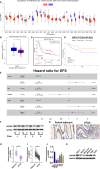
Figure 1
Figure 1
The expression of HADHB was low in STAD tissues and cell lines (A). Differential mRNA expression of HADHB between normal tissues and tumour tissues in TCGA database. (B). The mRNA expression of HADHB between normal tissues and tumour tissues in GEPIA. (C). The relationship between the HADHB gene and overall survival (cutoff = 12.19) of STAD patients using Kaplan-Meier Plotter webtool. (D). The relationship between the HADHB gene and disease-free survival (cutoff = 12.27) of TCGA-STAD patients using R software. (E). Cox risk multivariable analysis of STAD. (F). HADHB expression was determined using 4 pairs of significantly different clinical samples via western blot. (G). HADHB expression was assessed via immunohistochemistry. (H, I). HADHB expression in 40 pairs of clinical STAD tissues was determined using qRT-PCR. (J, K). HADHB expression in STAD cell lines was determined using qRT-PCR and western blot. * P < 0.05, ** P < 0.01.
Figure 2
Figure 2
HADHB upregulation repressed the proliferation and cell cycle progression of HGC27 and AGS cells (A, B). HADHB expression in the transfected HGC27 and AGS cells was determined using qRT-PCR and western blot analysis. (C–F). The viability, proliferation, colony formation, and cell cycle of the transfected AGS and HGC27 cells were determined using the CCK-8 assay, EdU assay, colony formation assay, and flow cytometry analysis, respectively. * P < 0.05, ** P < 0.01.
Figure 3
Figure 3
HADHB upregulation impeded the migration, invasion, and wound healing of HGC27 and AGS cells (A). The invasion and migration of the transfected HGC27 and AGS cells were determined using the transwell assay. (B). The expression of metastasis-related factors (Vimentin, N-cadherin, and E-cadherin) was evaluated using western blot analysis. (C). Cell migration capacity of the transfected AGS and HGC27 cells was assessed using the wound healing assay. * P < 0.05.
Figure 4
Figure 4
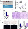
HADHB upregulation impeded tumour growth in xenograft BALB/c nude mice (A). Images of the tumours (4 in each group). (B). Tumour volume was calculated using the formula: tumour volume (V, mm3) = 0.5 × length × width × width. (C). Tumour weight was determined. (D). Histopathological analysis of the collected tumour tissues was performed using H&E staining. (E, F). Tumour cell proliferation and apoptosis were assessed via immunohistochemistry with the Ki67 antibody and TUNEL assay, respectively. * P < 0.05, ** P < 0.01.
Figure 5
Figure 5
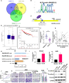
Upregulation of KLF4 abolished the HADHB knockdown-induced tumour promoting effects in AGS cells (A). Venn diagram was generated to screen out the overlapping gene among the three datasets, namely HADHB transcription factor (TF), gastric cancer downregulated genes, and genes positively correlated with HADHB in TCGA. (B, C). Motif analysis of KLF4 and HADHB promotor binding site analysis was conducted using Jaspar database. (D). The mRNA expression of KLF4 between normal tissues and tumour tissues in GEPIA. (E). Overall survival probability of STAD patients was analysed using Kaplan-Meier Plotter (cutoff = 9.01). (F). KLF4 expression in 40 pairs of clinical STAD tissues was determined using qRT-PCR. (G). The correlation between HADHB and KLF4 expression in STAD was derived using Pearson correlation analysis. (H). The regulating relationship between HADHB and KLF4 was determined using the dual luciferase reporter assay. (I). HADHB expression in KLF4-overexpressed AGS and HGC27 cells was determined using qRT-PCR. (J, K). (L). The expression of metastasis-related factors (Vimentin, N-cadherin, and E-cadherin) was evaluated using western blot analysis. The viability, migration, and invasion of the transfected AGS cells were determined using CCK-8 assay and transwell assay, respectively. * P < 0.05, ** P < 0.01.
Figure 6
Figure 6
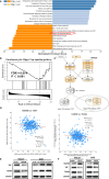
HADHB upregulation restrained the regulation of the Hippo-YAP signalling pathway (A). Pathway enrichment analysis was carried out using GSEA and WebGestalt based on TCGA-STAD database. (B). Enrichment plot of the Hippo-Yap signalling pathway. (C). Pathway view of the Hippo-YAP signalling pathway, with the leading-edge protein highlighted. (D). Analysis of the correlation between HADHB and YAP, as well as HADHB and TEAD4 was performed using the cBioPortal platform. ((E–F)). The expression of HADHB and the Hippo-YAP signalling pathway-related factors (YAP and TEAD4) in the transfected AGS and HGC27 cells was evaluated via western blot.
Figure 7
Figure 7
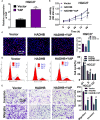
YAP upregulation effectively reversed the HADHB upregulation-induced tumour suppressing effects in HGC27 cells (A). YAP expression in pcDNA3.1-YAP transfected HGC27 cells was measured using qRT-PCR. (B–E). The viability, proliferation, cell cycle progression, migration, and invasion of the transfected HGC27 cells were determined using the CCK-8 assay, EdU assay, flow cytometry analysis, and transwell assay, respectively. * P < 0.05, ** P < 0.01, # P < 0.05, ## P < 0.01.

Similar articles

Cited by

References

    1. Boubrik F, Belmouden A, Kadmiri NE. Potential non-invasive biomarkers of helicobacter pylori-associated gastric cancer. J Gastrointest Cancer (2021) 53(4):1113–20. doi: 10.1007/s12029-021-00734-7 - DOI - PubMed
    1. Yao L, Shi W, Gu J. Micro-RNA 205-5p is involved in the progression of gastric cancer and targets phosphatase and tensin homolog (PTEN) in SGC-7901 human gastric cancer cells. Med Sci Monit (2019) 25:6367–77. doi: 10.12659/MSM.915970 - DOI - PMC - PubMed
    1. Sitarz R, Skierucha M, Mielko J, Offerhaus GJA, Maciejewski R, Polkowski WP. Gastric cancer: epidemiology, prevention, classification, and treatment. Cancer Manag Res (2018) 10:239–48. doi: 10.2147/CMAR.S149619 - DOI - PMC - PubMed
    1. Wu PL, He YF, Yao HH, Hu B. Martrilin-3 (MATN3) overexpression in gastric adenocarcinoma and its prognostic significance. Med Sci Monit (2018) 24:348–55. doi: 10.12659/MSM.908447 - DOI - PMC - PubMed
    1. Johnston FM, Beckman M. Updates on management of gastric cancer. Curr Oncol Rep (2019) 21:67. doi: 10.1007/s11912-019-0820-4 - DOI - PubMed

LinkOut - more resources