. 2022 Dec 8;7(1):69.
doi: 10.1038/s41536-022-00265-0.

Rapid induction and long-term self-renewal of neural crest-derived ectodermal chondrogenic cells from hPSCs

Affiliations

Rapid induction and long-term self-renewal of neural crest-derived ectodermal chondrogenic cells from hPSCs

Pei Shen et al. NPJ Regen Med. .

Abstract

Articular cartilage is highly specific and has limited capacity for regeneration if damaged. Human pluripotent stem cells (hPSCs) have the potential to generate any cell type in the body. Here, we report the dual-phase induction of ectodermal chondrogenic cells (ECCs) from hPSCs through the neural crest (NC). ECCs were able to self-renew long-term (over numerous passages) in a cocktail of growth factors and small molecules. The cells stably expressed cranial neural crest-derived mandibular condylar cartilage markers, such as MSX1, FOXC1 and FOXC2. Compared with chondroprogenitors from iPSCs via the paraxial mesoderm, ECCs had single-cell transcriptome profiles similar to condylar chondrocytes. After the removal of the cocktail sustaining self-renewal, the cells stopped proliferating and differentiated into a homogenous chondrocyte population. Remarkably, after transplantation, this cell lineage was able to form cartilage-like structures resembling mandibular condylar cartilage in vivo. This finding provides a framework to generate self-renewing cranial chondrogenic progenitors, which could be useful for developing cell-based therapy for cranial cartilage injury.


Conflict of interest statement

The authors declare no competing interests.

Figures
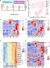
Fig. 1
Fig. 1. Generation of NC-derived progenitor cells from hPSCs.
a Schematic representation of the NC-derived progenitor cell induction process. b PCA indicates that five unique hPSC (H1, Hues9, H9, Hues7 and ihPSC) lines followed similar differentiation trajectories. c Genes encoding ectoderm markers were upregulated during induction. d Genes encoding NC markers were upregulated during induction. e GO analysis highlighted bone and cartilage development, especially cranial skeleton morphogenesis and cranial skeletal system development, in NC-derived progenitor cells. f Genes encoding chondrogenesis markers were upregulated during induction.
Fig. 2
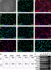
Fig. 2. CSSEDF-expanded cells stably self-renewed and shared markers of CPCs/CSCs.
a A single CSSEDF-expanded cell (passage 5) colony on a Matrigel-coated surface. Scale bars, 50 µm. bd Immunocytochemistry showing that CSSEDF-expanded cells expressed genes identified as neural ectodermal markers, including SIX1, NESTIN and ETS1. Scale bars, 50 µm. e–i Immunocytochemistry showing that CSSEDF-expanded cells (passage 5) expressed genes identified as CPC/CSC markers, including SOX9, RUNX2, SOX5, TWIST1, and CD29. Scale bars, 50 µm. j–l Immunocytochemistry showing that CSSEDF-expanded cells (passage 5) express genes recently identified as TMJ condylar cartilage markers, including FOXC1, FOXC2 and MSX1. Scale bars, 50 µm. m Flow cytometry analysis showing that CSSEDF-expanded cells stably express cell proliferation and CPC/CSC markers after long-term in vitro expansion. n Gene expression of chondrocytic lineage markers by CSSEDF-expanded cells at different passages and patient TMJ condylar cartilage were analysed by PCR. CPCs/CSCs: cartilaginous progenitor cells/cartilaginous stem cells.
Fig. 3
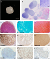
Fig. 3. ECCs can spontaneously differentiate into chondrocytes in vitro.
a Gross appearance of ECCs seeded in a collagen sponge for 8 weeks. Scale bars, 1 mm. b Panoramic scanning showing the formation of cartilage masses in the collagen scaffold (inset: HE staining enlargement). Scale bars, 200 µm. (Inset) Scale bars, 50 µm. ce The chondrocytes were positive for toluidine blue, alcian blue and safranin O staining. Scale bars, 100 µm. fk Immunohistochemical staining showing that the chondrocytes express common chondrocytic markers, including collagen II, collagen X, aggrecan, RUNX2, collagen I, and the articular cartilage marker lubricin. Scale bars, 100 µm.
Fig. 4
Fig. 4. ECCs can generate cartilage in vivo without teratoma formation.
a Schematic representation showing ECCs seeded in a collagen sponge in vitro for 4 weeks and surgically transplanted subcutaneously into the dorsum of NOD-SCID mice. The implants were harvested after 4 weeks and histologically confirmed to be cartilage. (Inset) Enlargement of HE staining. Scale bars, 200 µm. (Inset) Scale bars, 50 µm. b–d Harvested cartilage was positive for safranin O, alcian blue and toluidine blue staining. Scale bars, 100 µm. ej Immunohistochemical staining showing that differentiated chondrocytes express common chondrocytic markers, including collagen II, collagen X, aggrecan, RUNX2, collagen I, and the articular cartilage marker lubricin. Scale bars, 100 µm.
Fig. 5
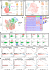
Fig. 5. ECCs have similar single-cell transcriptome profiles to TMJ condylar chondrocytes.
a and b Both TMJ-CC and ECCs were enriched in mandibular condyle and neural crest markers, such as COL1A1, BARX1, FOXC1, MSX1, SIX1, and TWIST1. c UMAP confirmed that ECCs are closer to TMJ-CC than Cp from hiPSCs via the paraxial mesoderm. d Both the TMJ-CC and ECCs exhibited high expression of neural crest markers, while genes involved in mesoderm were clustered in the Cp. e Violin plots showing that TMJ-CC and ECCs presented nearly the same chondrogenic markers. f GSEA suggesting that ECCs had different gene expression from Cp in skeletal system development, ossification, cartilage development, and bone development. g MAPK, calcium, ErbB, and PPAR signalling pathway-associated genes were different between ECCs and Cp.
Fig. 6
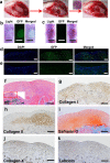
Fig. 6. ECCs can repair knee defects and form layered cartilage structures.
a Schematic representation showing that a full-thickness rat cartilage defect was created. ECCs were seeded in collagen sponges, cultured in vitro for 4 weeks, and then surgically transplanted into cartilage defects. b, d Knees treated with GFP-expressing ECCs loaded on a collagen sponge were harvested 4 weeks after transplantation. GFP expression was detected in the ECC group. b Scale bar, 1 mm. d Scale bar, 50 μm. c, e No GFP expression was observed in the control group transplanted with only the collagen sponge. c Scale bar, 1 mm. e Scale bar, 50 μm. f HE staining showing that the repaired tissue was cartilage-like. (Inset) HE staining at low magnification. Scale bar, 100 μm. (Inset) Scale bar, 200 μm. g Immunohistochemical staining revealing that collagen I was present throughout the cartilaginous cell layers. h Immunohistochemical staining showing that collagen II was predominant in the deep layer. i Safranin O is more significantly positive than the deeper layer. j, k Immunohistochemical staining showing that collagen X and lubricin are expressed in the regenerated cartilage tissue. g–k Scale bar, 100 μm.
Fig. 7
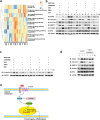
Fig. 7. The activated Wnt signalling pathway affects chondrogenic differentiation of ECCs.
a KEGG analysis of bulk RNA-seq at different induction points. b The transcription factor SOX9, condylar chondrocyte anabolism markers COL2A1 and COL1A1, and the catabolism marker MMP13 were obviously upregulated 3 days after the removal of CHIR99021. c An obvious downregulation of β-catenin was detected by western blotting 7 days after the removal of CHIR99021. d The chondrogenic transcription factors SOX9 and RUNX2 were decreased, as well as COL2A1 and OPN, while MMP13 was enhanced in the presence of CHIR99021 in N2B27. e A scheme for the Wnt signalling pathway involved in chondrogenic differentiation of ECCs.

Similar articles

Cited by

References

    1. Correa D, Lietman SA. Articular cartilage repair: current needs, methods and research directions. Semin. Cell Dev. Biol. 2017;62:67–77. - PubMed
    1. Yamaoka H, et al. Involvement of fibroblast growth factor 18 in dedifferentiation of cultured human chondrocytes. Cell Prolif. 2010;43:67–76. - PMC - PubMed
    1. Barry F, Murphy M. Mesenchymal stem cells in joint disease and repair. Nat. Rev. Rheumatol. 2013;9:584–594. - PubMed
    1. Oldershaw RA, et al. Directed differentiation of human embryonic stem cells toward chondrocytes. Nat. Biotechnol. 2010;28:1187–1194. - PubMed
    1. Craft AM, et al. Generation of articular chondrocytes from human pluripotent stem cells. Nat. Biotechnol. 2015;33:638–645. - PubMed