

The heterogeneity of astrocytes in glaucoma
- PMID: 36466782
- PMCID: PMC9714578
- DOI: 10.3389/fnana.2022.995369
The heterogeneity of astrocytes in glaucoma
Abstract
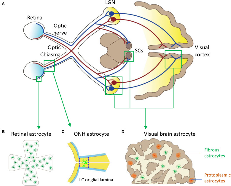
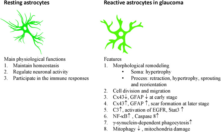
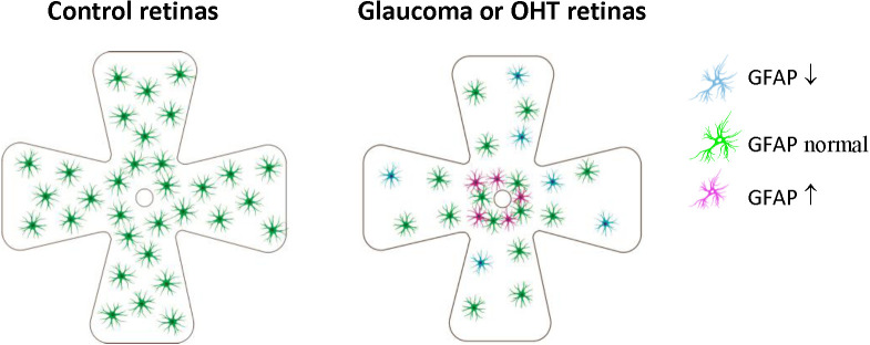
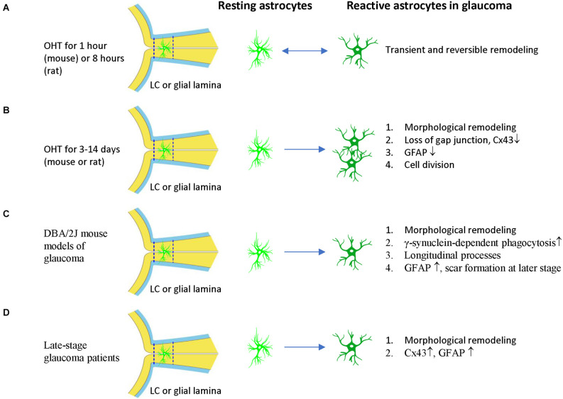
Glaucoma is a leading cause of blindness with progressive degeneration of retinal ganglion cells. Aging and increased intraocular pressure (IOP) are major risk factors. Lowering IOP does not always stop the disease progression. Alternative ways of protecting the optic nerve are intensively studied in glaucoma. Astrocytes are macroglia residing in the retina, optic nerve head (ONH), and visual brain, which keep neuronal homeostasis, regulate neuronal activities and are part of the immune responses to the retina and brain insults. In this brief review, we discuss the activation and heterogeneity of astrocytes in the retina, optic nerve head, and visual brain of glaucoma patients and animal models. We also discuss some recent transgenic and gene knockout studies using glaucoma mouse models to clarify the role of astrocytes in the pathogenesis of glaucoma. Astrocytes are heterogeneous and play crucial roles in the pathogenesis of glaucoma, especially in the process of neuroinflammation and mitochondrial dysfunction. In astrocytes, overexpression of Stat3 or knockdown of IκKβ/p65, caspase-8, and mitochondrial uncoupling proteins (Ucp2) can reduce ganglion cell loss in glaucoma mouse models. Based on these studies, therapeutic strategies targeting the heterogeneity of reactive astrocytes by enhancing their beneficial reactivity or suppressing their detrimental reactivity are alternative options for glaucoma treatment in the future.
Keywords: astrocytes; glaucoma; heterogeneity; inflammation; intraocular pressure; mitochondrial dysfunction; optic nerve head; retinal ganglion cells.
Copyright © 2022 Tang, Chen and Chen.
Conflict of interest statement
The authors declare that the research was conducted in the absence of any commercial or financial relationships that could be construed as a potential conflict of interest.
Figures
Similar articles
-
Astrocytes of the eye and optic nerve: heterogeneous populations with unique functions mediate axonal resilience and vulnerability to glaucoma.Front Ophthalmol (Lausanne). 2023;3:1217137. doi: 10.3389/fopht.2023.1217137. Epub 2023 Jul 21. Front Ophthalmol (Lausanne). 2023. PMID: 37829657 Free PMC article.
-
[Aiming for zero blindness].Nippon Ganka Gakkai Zasshi. 2015 Mar;119(3):168-93; discussion 194. Nippon Ganka Gakkai Zasshi. 2015. PMID: 25854109 Review. Japanese.
-
Potential roles of astrocytes and Müller cells in the pathogenesis of glaucoma.J Pharmacol Sci. 2021 Mar;145(3):262-267. doi: 10.1016/j.jphs.2020.12.009. Epub 2020 Dec 31. J Pharmacol Sci. 2021. PMID: 33602506 Review.
-
Expression and activation of mitogen-activated protein kinases in the optic nerve head in a rat model of ocular hypertension.Mol Cell Neurosci. 2018 Apr;88:270-291. doi: 10.1016/j.mcn.2018.01.002. Epub 2018 Mar 20. Mol Cell Neurosci. 2018. PMID: 29408550
-
Astrocytes of the optic nerve exhibit a region-specific and temporally distinct response to elevated intraocular pressure.Mol Neurodegener. 2023 Sep 27;18(1):68. doi: 10.1186/s13024-023-00658-9. Mol Neurodegener. 2023. PMID: 37759301 Free PMC article.
Cited by
-
Glial metabolic alterations during glaucoma pathogenesis.Front Ophthalmol (Lausanne). 2023 Nov 28;3:1290465. doi: 10.3389/fopht.2023.1290465. eCollection 2023. Front Ophthalmol (Lausanne). 2023. PMID: 38983068 Free PMC article. Review.
-
Inhibition of Galectins and the P2X7 Purinergic Receptor as a Therapeutic Approach in the Neurovascular Inflammation of Diabetic Retinopathy.Int J Mol Sci. 2023 Jun 3;24(11):9721. doi: 10.3390/ijms24119721. Int J Mol Sci. 2023. PMID: 37298672 Free PMC article. Review.
-
Glial Cell Activation and Immune Responses in Glaucoma: A Systematic Review of Human Postmortem Studies of the Retina and Optic Nerve.Aging Dis. 2024 Oct 1;15(5):2069-2083. doi: 10.14336/AD.2024.0103. Aging Dis. 2024. PMID: 38502591 Free PMC article.
-
Evaluation of the LDN-0060609 PERK Inhibitor as a Selective Treatment for Primary Open-Angle Glaucoma: An In Vitro Study on Human Retinal Astrocytes.Int J Mol Sci. 2024 Jan 5;25(2):728. doi: 10.3390/ijms25020728. Int J Mol Sci. 2024. PMID: 38255802 Free PMC article.
-
The Application of Rho Kinase Inhibitors in the Management of Glaucoma.Int J Mol Sci. 2024 May 21;25(11):5576. doi: 10.3390/ijms25115576. Int J Mol Sci. 2024. PMID: 38891764 Free PMC article. Review.
References
Publication types
LinkOut - more resources
Full Text Sources
Miscellaneous