. 2022 Nov 19;25(12):105627.
doi: 10.1016/j.isci.2022.105627. eCollection 2022 Dec 22.

Twisting development, the birth of a potential new gene

Affiliations

Twisting development, the birth of a potential new gene

Nayelli Marsch-Martínez et al. iScience. .

Abstract

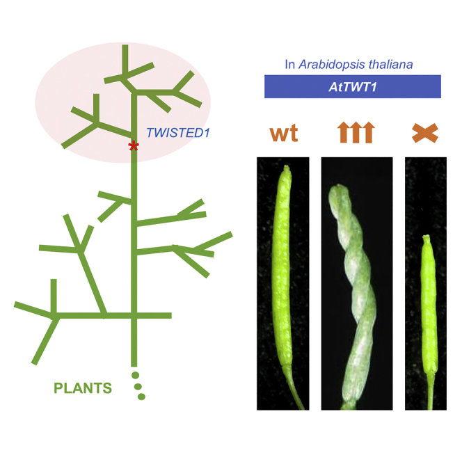
Evolution has long been considered to be a conservative process in which new genes arise from pre-existing genes through gene duplication, domain shuffling, horizontal transfer, overprinting, retrotransposition, etc. However, this view is changing as new genes originating from non-genic sequences are discovered in different organisms. Still, rather limited functional information is available. Here, we have identified TWISTED1 (TWT1), a possible de novo-originated protein-coding gene that modifies microtubule arrangement and causes helicoidal growth in Arabidopsis thaliana when its expression is increased. Interestingly, even though TWT1 is a likely recent gene, the lack of TWT1 function affects A. thaliana development. TWT1 seems to have originated from a non-genic sequence. If so, it would be one of the few examples to date of how during evolution de novo genes are integrated into developmental cellular and organismal processes.

Keywords: Evolutionary biology; Molecular biology; Plant development.


Conflict of interest statement

The authors declare no competing interests.

Figures

None
Graphical abstract
Figure 1
Figure 1
twt1-D confers a twisted growth pattern in different organs (A–J) See also Figures S1 and S2. Wild-type (WT) and twt1-D siliques (A, B), stamens and pistils (C, D), and rosettes (E, F). In (F) yellow arrows indicate right-handed twisting. Electron microscopy image of WT and twt1-D siliques (G, H). Seedlings presenting differences in root architecture (I, J). (K and L) Confocal microscopy image of the upper hypocotyl region of WT (K), and twt1-D (L). (M) Confocal microscopy image of a twt1-D root. (N) Subcellular localization of TWT:GFP protein in the plasma membrane region of root cells (in a plant bearing the gtwt1-D:GFP construct). (O) Schematic representation of the twt1-D genomic region, where the activating I element (AIE) was inserted. Blue lines represent the different regions used for constructs that recapitulated the phenotypes. The four arrows in the AIE represent the enhancer tetramer in the transposon. Dark green boxes represent exons, light green boxes represent UTRs, and triangles indicate the orientation of the gene. The black line represents non-coding genomic regions and introns. (P–S) Phenotypes of stable transgenic lines bearing the constructs depicted in the scheme in (O). Each construct recovers the twt1-D phenotype (right-handed twists indicated by yellow arrows). (T–V) Three-weeks-old WT (T), twt1-D (U), and twt1-D transformed with a construct causing the constitutive overexpression of an antisense version of cTWT1 (35S::antiTWT1) (V). The twt1-D phenotype can be rescued by the 35S::antiTWT1 construct. (W and X) twt1-D (W) and twt1-D 35S::antiTWT1 (X) inflorescences. Scale bars: 2 mm (A, B, P-S, W-X), 1 mm (C, D), 1 cm (E-F, I-J, T-V), 100 μm (G), 200 μm (H), 50 μm (K-M), 5 μm (N).
Figure 2
Figure 2
Microtubule disruption, mutation, and orientation analyses in twt1-D See also Figure S3. (A–F) Effects of propyzamide on helical growth phenotypes in wild-type (WT) (A, C, E) and twt1-D mutants (B, D, F). Seedlings grown on control medium (A, WT; B, twt1-D) or medium containing 0.5 μM (C, WT; D, twt1-D) and 5 μM (E, WT; F, twt1-D) propyzamide for 7 days. (G–J) WT (G, I) and twt1-D mutant (H, J) plants grown on control agar medium (G, H) or medium containing 5 μM propyzamide (I, J) for three weeks. (K) WT (left) and twt1-D plants grown in medium containing 5 μM propyzamide for four weeks. (L) Scheme indicating right- (yellow arrows) and left- (cyan arrows) handed twist. (M–R) twt1-D interaction with tubulin mutants; (M) WT, (N) right-handed twisted TUA5D251N mutant, (O) left-handed twisted TUA4S178Δ mutant, (P) twt1-D, (Q) twt1-D TUA5D251N double mutant exhibiting increased right-handed twisting, and (R) twt1-D TUA4S178Δ double mutant displaying reduced twisting. Cortical microtubules in seedling hypocotyls of GFP:TUA6 (S) and GFP:TUA6 twt1-D (T), where microtubules are oriented to the left with respect to the top of the long axis of the cell. Asterisk indicates the basal part of the cell. Cartoons on the right illustrate the microtubule orientation in the cell (S, T). Scale bars: 2 mm (A–F), 5 mm (G-R), 10 μm (S, T).
Figure 3
Figure 3
Synteny and phylogenetic analyses of TWT1 See also Figure S4 and Table S1. (A) Visualization of genomic regions surrounding the TWT1 gene based on GenBank files and the software clinker and clustermap.js. Syntenic genes are indicated in different colors. Gene percentage identity is presented in gray color scale. (B) Phylogenetic tree inferred by maximum likelihood (ML) of putative TWT1 genes in Brassicaceae species. (C) Left: Phylogenetic tree inferred by ML of Brassicaceae species coding for TWT1. Tarenaya hassleriana from the Brassicaceae sister family Cleomaceae was used as the outgroup. The origin of the TWT1 gene in the phylogeny is indicated with a red asterisk coinciding with the origin of Brassicaceae around 38 MYA. Right: Synteny of TWT1 (in red) and neighboring genes (in other colors). The diamond (red) indicates the non-genic region in T. hassleriana corresponding to the region where TWT is located in the genome of the other species. The tree was reconstructed from a concatenated alignment of the genes coding for GPI inositol-deacylase C (green) and beta-amylase 6 (cyan). In Sinapis alba, these two genes are located in a different contig. Bootstrap values are shown in tree nodes. The bars indicate the number of substitutions per site.
Figure 4
Figure 4
Phenotype and structure of edited TWT1 alleles See also Figure S5. (A) Genomic structure of two twt1 CRISPR-Cas9 edited alleles. (B) TWT1 protein structure and comparison to the proteins produced by the edited alleles. In both cases, a deletion results in a frameshift and premature stop codons that produce smaller proteins than TWT1. The 30 and 4 letters in red represent the amino acids that are different from the original TWT1 amino acids in each allele, due to frameshifts. Asterisks represent stop codons. (C) Ten-day-old twt1-cr2 seedling with dissimilar cotyledons. (D and E) Seventeen-day-old wild-type (WT) (D) and twt1-cr1 (E) plant. (F and G) Twenty-six-day-old WT (F) and twt1-cr1 (G) plants. (H–L) Flower phenotypes of WT and twt1-cr mutants. (M and N) Comparison between WT and mutant siliques. (O and P) Comparison between WT and mutant open siliques with exposed seeds. Scale bars: 1 mm (C, H, I, M-P), 5 mm (D, E), 1 cm (F, G), 0.5 mm (J-L).

Similar articles

Cited by

References

    1. Schlötterer C. Genes from scratch - the evolutionary fate of de novo genes. Trends Genet. 2015;31:215–219. doi: 10.1016/j.tig.2015.02.007. - DOI - PMC - PubMed
    1. Chen S., Krinsky B.H., Long M. New genes as drivers of phenotypic evolution. Nat. Rev. Genet. 2013;14:645–660. doi: 10.1038/NRG3521. - DOI - PMC - PubMed
    1. Long M., Vankuren N.W., Chen S., Vibranovski M.D. New gene evolution: little did we know. Annu. Rev. Genet. 2013;47:307–333. doi: 10.1146/ANNUREV-GENET-111212-133301. - DOI - PMC - PubMed
    1. Ohno S. Springer-Verlag; 1970. Evolution by Gene Duplication.
    1. McLysaght A., Hurst L.D. Open questions in the study of de novo genes: what, how and why. Nat. Rev. Genet. 2016;17:567–578. doi: 10.1038/nrg.2016.78. - DOI - PubMed