. 2023 Nov;340(7):455-468.
doi: 10.1002/jez.b.23183. Epub 2022 Dec 4.

Ganoin and acrodin formation on scales and teeth in spotted gar: A vital role of enamelin in the unique process of enamel mineralization

Affiliations

Ganoin and acrodin formation on scales and teeth in spotted gar: A vital role of enamelin in the unique process of enamel mineralization

Kazuhiko Kawasaki et al. J Exp Zool B Mol Dev Evol. 2023 Nov.

Abstract

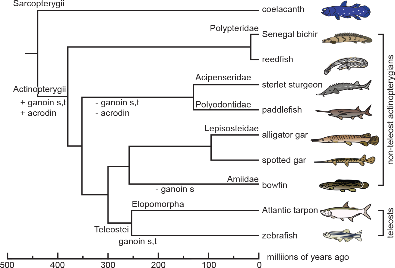
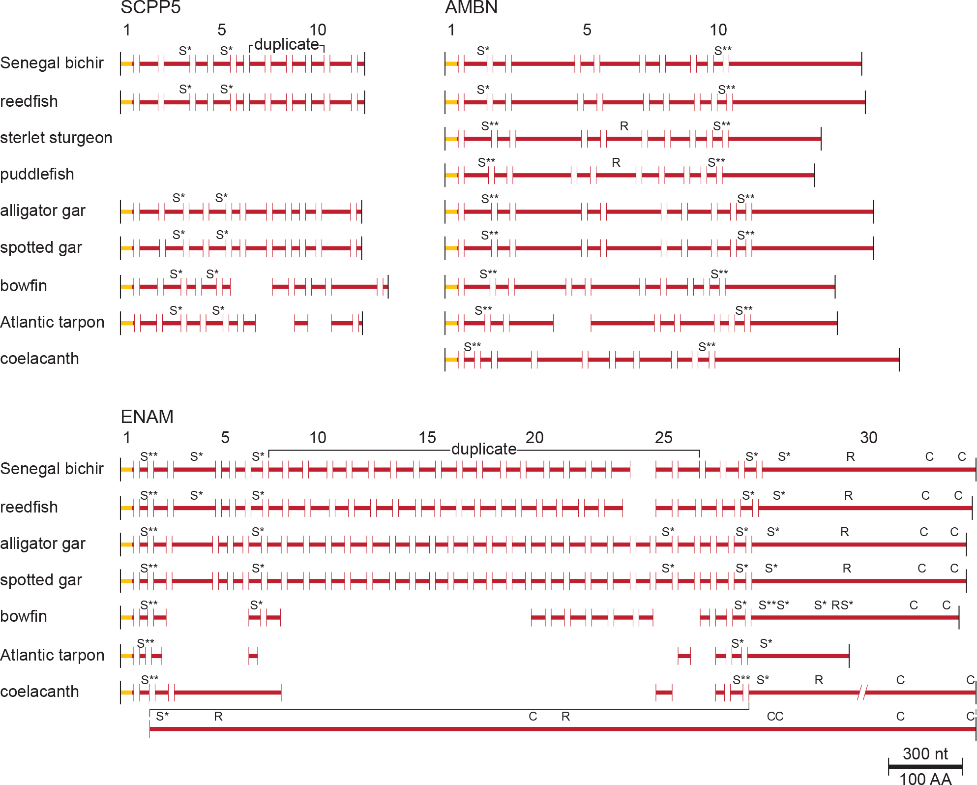
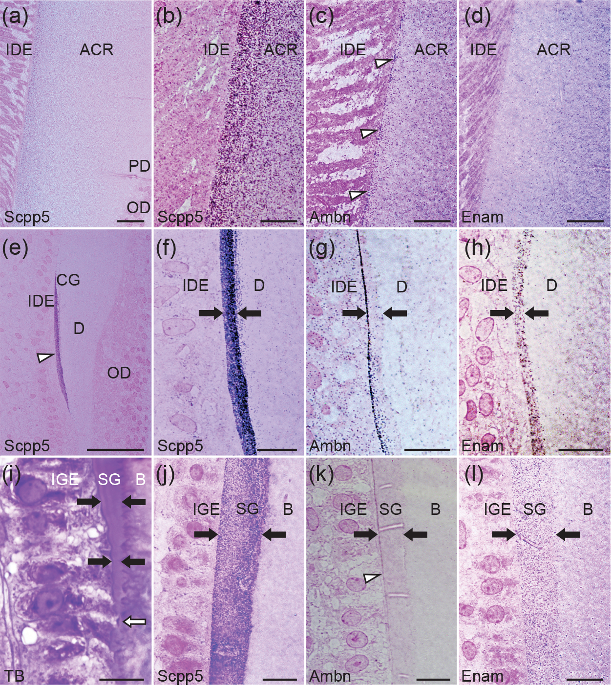
Gars and bichirs develop scales and teeth with ancient actinopterygian characteristics. Their scale surface and tooth collar are covered with enamel, also known as ganoin, whereas the tooth cap is equipped with an enamel-like tissue, acrodin. Here, we investigated the formation and mineralization of the ganoin and acrodin matrices in spotted gar, and the evolution of the scpp5, ameloblastin (ambn), and enamelin (enam) genes, which encode matrix proteins of ganoin. Results suggest that, in bichirs and gars, all these genes retain structural characteristics of their orthologs in stem actinopterygians, presumably reflecting the presence of ganoin on scales and teeth. During scale formation, Scpp5 and Enam were initially found in the incipient ganoin matrix and the underlying collagen matrix, whereas Ambn was detected mostly in a surface region of the well-developed ganoin matrix. Although collagen is the principal acrodin matrix protein, Scpp5 was detected within the matrix. Similarities in timings of mineralization and the secretion of Scpp5 suggest that acrodin evolved by the loss of the matrix secretory stage of ganoin formation: dentin formation is immediately followed by the maturation stage. The late onset of Ambn secretion during ganoin formation implies that Ambn is not essential for mineral ribbon formation, the hallmark of the enamel matrix. Furthermore, Scpp5 resembles amelogenin that is not important for the initial formation of mineral ribbons in mammals. It is thus likely that the evolution of ENAM was vital to the origin of the unique mineralization process of the enamel matrix.

Keywords: SCPP genes; SCPP5; ameloblastin; enamelin; ganoid scales; mineral ribbons.


Conflict of interest statement

Declaration of Competing Interest: The authors declare that they have no known competing financial interests or personal relationships that could have appeared to influence the work reported in this paper.

Figures

Figure 1.
Figure 1.
Phylogenetic relationship of Senegal bichir (Polypterus senegalus), reedfish (Erpetoichthys calabaricus), sterlet sturgeon (Acipenser ruthenus), paddlefish (Polyodon spathula), alligator gar (Atractosteus spatula), spotted gar (Lepisosteus oculatus), bowfin (Amia calva), Atlantic tarpon (Megalops atlanticus), and zebrafish (Danio rerio) among actinopterygians (Actinopterygii). Coelacanth (Latimeria chalumnae, Sarcopterygii) is also shown. Non-teleost actinopterygians comprise five families, Polypteridae, Acipenseridae, Polyodontidae, Lepisosteidae, and Amiidae. Elopomorpha is the basal group of teleosts (Teleostei) (Near et al., 2012; Hughes et al., 2018). The divergence times shown at the bottom are based on a previous study (Hughes et al., 2018). The gain (+) and loss (−) of ganoin in scales (s) and teeth (t) and acrodin (Mikami et al., 2022), indicated along branches, are not in scale.
Figure 2.
Figure 2.
Protein-coding exons of SCPP5, AMBN, and ENAM. The black vertical bars at the beginning and the end of the entire protein-coding exons represent the initiation and termination codons, respectively. The horizontal bars represent the coding regions of the signal peptide (yellow) and the mature protein (red). Each exon is delimited by red vertical bars (splice sites). The number of protein-coding exons for Senegal bichir scpp5, ambn, and enam are indicated on the top. The Ser residue in Ser-Xaa-Glu and Ser-Xaa-pSer (phospho-Ser) is potentially phosphorylated (Tagliabracci et al., 2012; Worby et al., 2021) and is shown by “S*” (“S**” for clustered pSer residues). An integrin-binding sequence (Arg-Gly-Asp) is indicated by “R.” Cys (“C”) residues are not common in most SCPPs but common to ENAM proteins (Kawasaki et al., 2011; Kawasaki and Amemiya, 2014). See the text for “duplicate” exons. The large last protein-coding exon of coelacanth ENAM is shown in the next line. A scale (300 nucleotides (nt)/100 amino acids (AA)) is shown at the bottom. See Figure S1 for encoded amino acid sequences.
Figure 3.
Figure 3.
Light micrographs showing acrodin (a-d), collar ganoin (e-h), and scale ganoin (i-l) formation in spotted gar. For immunohistochemical analysis (a-h and j-l; a and e, overview), reactivity (protein A-gold particles) is detected as purple or magenta dots. (a-d) The middle stage of acrodin mineralization was analyzed using the anti-SCPP5N (a and b), anti-AMBN (c), and anti-ENAM (d) antibodies. (c) Immunoreactivity along the acrodin surface is shown by open arrow heads. (e-h) The secretory stage of collar ganoin was analyzed using the anti-SCPP5N (e and f), anti-AMBN (g), and anti-ENAM (h) antibodies. (e) Immunoreactivity throughout the collar ganoin layer is indicated by an open arrowhead. (f-l) A pair of closed arrows show the thickness of ganoin. (i-l) The secretory stage of scale ganoin formation was analyzed by staining with toluidine blue (i; TB) and by immunohistochemistry using the anti-SCPP5N (j), anti-AMBN (k), and anti-ENAM (l) antibodies. (i) Newly differentiated IGE cells begin to secrete the ganoin matrix (open arrow). The ganoin layer is thick underneath well-differentiated IGE cells (top). (k) Note increasing reactivity above and below the open arrowhead. Negative controls are provided in Figure S2 (A and B). Scale bars: 20 μm (a), 10 μm (b-d and f-l), 50 μm (e). Abbreviations: acrodin (ACR), bone (B), collar ganoin (CG), dentin (D), inner dental epithelial cells (IDE), inner scale ganoin epithelial cells (IGE), odontoblasts (OD), predentin (PD), scale ganoin (SG).
Figure 4.
Figure 4.
Transmission electron micrographs showing acrodin (a-h) and collar ganoin (i-l) formation in spotted gar. For immunohistochemical analysis (a-e and g-l), PAG particles are detected as dots. The middle (a-c) and late (d) mineralization stages of acrodin formation were analyzed using the anti-SCPP5N antibody. (a) The arrow indicates electron-dense fibrous structures (EDFSs). (b) Packed collagen fibrils are formed. (c) Immunoreactivity in granules of IDE cells is indicated by the open arrow. (d) No significant reactivity was detected. The middle (e) and late (g) mineralization stages of acrodin formation were analyzed using the anti-AMBN. PAG particles along the surface region of acrodin (e and g) and in a granule of IDE cells (e) are indicated by open arrowheads. (f) In the late mineralization stage, crystallites (arrow) are found along the surface region of acrodin (f and g, sequential sections). (h) No significant reaction was detected using the anti-ENAM antibody in the middle mineralization stage of acrodin formation. The secretory stage of collar ganoin formation was analyzed using the anti-SCPP5N (i and j; i, collar ganoin and IDE cells; j, collar ganoin and dentin), anti-AMBN (k), and anti-ENAM (l) antibodies. (i) The open arrowhead indicates PAG particles in a granule of IDE cells. (l) Note that collagen fibrils on the right bottom run parallel to the IDE cell surface. Negative controls are shown in Figure S2 (E–H). Sections were stained with TI-Pb (a and c), TI-Pb-PTA (b, d, e and g-l), or Pb (f). Scale bars: 500 nm (a-e, h, and j), 200 nm (f, g, i, k, and l). See the legend of Figure 3 for abbreviations.
Figure 5.
Figure 5.
Transmission electron micrographs showing mineralization of bone and ganoin in scales (a-e) and immunohistochemical analysis of scale formation (f-p) in spotted gar. In the presecretory (a, b), early secretory (c, d), and middle secretory (e) stages of scale ganoin formation, mineral foci are found in the surface zone (SZ; near IGE cells) and the subsurface zone (SSZ) of the bone matrix (B). An overview (a) and an enlarged view of SZ (b) are shown. The ganoin matrix is initially secreted from IGE cells as radiated mineral ribbons accommodated in hemispheres (arrow in c and d). (e) In the middle secretory stage, numerous mineral ribbons, running perpendicular to IGE cells in the ganoin matrix (SG), form bundles that extend from mammillary knobs (arrow). (f-j) The early (f-i) and middle (j) secretory stages of scale ganoin formation were analyzed using the anti-SCPP5N antibody. The positions shown by “#” and “*” in the overview (f) are enlarged (“#” in g and “*” in h). (g) Note that collagen fibers are arranged perpendicular to the distal membrane of IGE cells (arrow). (j) In the middle secretory stage, SZ and SSZ in the bone matrix (B) are separated by a narrow mineral loci-free zone. The early (k), middle (l), and late (m) secretory stages of scale ganoin formation were analyzed using the anti-AMBN antibody. PAG particles were significantly detected only in the late secretory stage in SG near the distal membrane of IGE cells (open arrow). The early (n) and middle (o and p) secretory stages of scale ganoin formation were analyzed using the anti-ENAM antibody. Two different regions (outer region in o; inner region in p) are shown for the middle secretory stage. Negative controls are shown in Figure S2 (K and L). Sections were stained with TI-Pb-PTA. Scale bars: 2.0 μm (a, and e-i), 1.0 μm (b, j, and l), 500 nm (c, d, k, and m-p).

Similar articles

Cited by

References

    1. Al-Hashimi N, Lafont AG, Delgado S, Kawasaki K, & Sire J-Y (2010). The enamelin genes in lizard, crocodile, and frog and the pseudogene in the chicken provide new insights on enamelin evolution in tetrapods. Molecular Biology and Evolution 27:2078–2094. 10.1093/molbev/msq098 - DOI - PubMed
    1. Al-Hashimi N, Sire J-Y, & Delgado S (2009). Evolutionary analysis of mammalian enamelin, the largest enamel protein, supports a crucial role for the 32-kDa peptide and reveals selective adaptation in rodents and primates. Journal of Molecular Evolution 69:635–656. 10.1007/s00239-009-9302-x - DOI - PubMed
    1. Altschul SF, Gish W, Miller W, Myers EW, & Lipman DJ (1990). Basic local alignment search tool. Journal of Molecular Biology 215:403–410. 10.1016/S0022-2836(05)80360-2 - DOI - PubMed
    1. Assaraf-Weill N, Gasse B, Silvent J, Bardet C, Sire J-Y, & Davit-Béal T (2014). Ameloblasts express type I collagen during amelogenesis. Journal Dental Research 93:502–507. 10.1177/0022034514526236 - DOI - PubMed
    1. Bartlett JD, Smith CE, Hu Y, Ikeda A, Strauss M, Liang T et al. (2021). MMP20-generated amelogenin cleavage products prevent formation of fan-shaped enamel malformations. Scientific Reports 11:10570. 10.1038/s41598-021-90005-z - DOI - PMC - PubMed

Publication types

LinkOut - more resources