. 2022 Nov 28;18(11):e1010459.
doi: 10.1371/journal.pgen.1010459. eCollection 2022 Nov.

TGFβ pathway is required for viable gestation of Fanconi anemia embryos

Affiliations

TGFβ pathway is required for viable gestation of Fanconi anemia embryos

Alfredo Rodríguez et al. PLoS Genet. .

Abstract

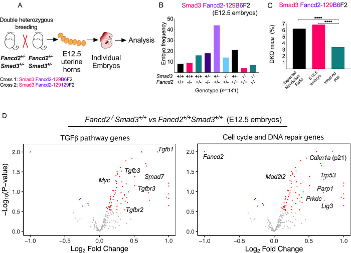
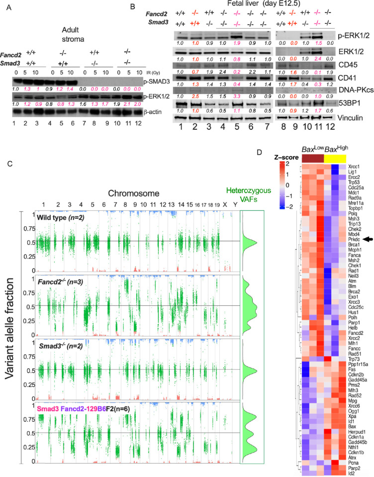
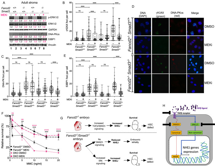
Overexpression of the TGFβ pathway impairs the proliferation of the hematopoietic stem and progenitor cells (HSPCs) pool in Fanconi anemia (FA). TGFβ promotes the expression of NHEJ genes, known to function in a low-fidelity DNA repair pathway, and pharmacological inhibition of TGFβ signaling rescues FA HSPCs. Here, we demonstrate that genetic disruption of Smad3, a transducer of the canonical TGFβ pathway, modifies the phenotype of FA mouse models deficient for Fancd2. We observed that the TGFβ and NHEJ pathway genes are overexpressed during the embryogenesis of Fancd2-/- mice and that the Fancd2-/-Smad3-/- double knockout (DKO) mice undergo high levels of embryonic lethality due to loss of the TGFβ-NHEJ axis. Fancd2-deficient embryos acquire extensive genomic instability during gestation which is not reversed by Smad3 inactivation. Strikingly, the few DKO survivors have activated the non-canonical TGFβ-ERK pathway, ensuring expression of NHEJ genes during embryogenesis and improved survival. Activation of the TGFβ-NHEJ axis was critical for the survival of the few Fancd2-/-Smad3-/- DKO newborn mice but had detrimental consequences for these surviving mice, such as enhanced genomic instability and ineffective hematopoiesis.


Conflict of interest statement

I have read the journal’s policy and the authors of this manuscript have the following competing interests: A.D. D’Andrea is a consultant/advisory board member for Lilly Oncology, Merck-EMD Serono, Intellia Therapeutics, Sierra Oncology, Cyteir Therapeutics, Third Rock Ventures, AstraZeneca, Ideaya Inc., Cedilla Therapeutics Inc., a stockholder in Ideaya Inc., Cedilla Therapeutics Inc., and Cyteir, and reports receiving commercial research grants from Lilly Oncology and Merck-EMD Serono.

Figures

Fig 1
Fig 1. TGFβ pathway inhibitors rescue hematopoiesis in FA models.
(A) TGFβ pathway inhibitors Galunisertib and LSN3301240 increase the clonogenic capacity of Lin- cells from bone marrow of Fancd2-/- mice measured in a CFU assay. Lineage negative primary bone marrow cells from wild-type (WT) or Fancd2-/- mice were cultured in methylcellulose medium containing increasing doses of Galunisertib or LSN3301240 for 7 days and hematopoietic colonies (CFUs) were counted for evaluating clonogenic growth of progenitors. (B) Western blots of the lysates from Lin cells from bone marrow of wild-type and Fancd2-/- mice cultured for 2 hours in the presence of TGFβ1 (5 ng/mL) or TGFβ3 (5 ng/mL) with or without LSN3301240. Levels of phospho-Smad2 (measured by two different antibodies, one against phospho-Smad2 and the other against both phospho-Smad2 and Smad3) are shown. Quantification relative to basal conditions and the loading control is shown below every lane. Pink indicates samples treated with LSN3301240. (C) Stromal cell lines generated from WT and Fancd2-/- mice were cultured in the presence of LSN3301240 and Mitomycin C (MMC) and survival was determined. LSN3301240 partially rescued the MMC sensitivity characteristic of FA cell lines. (D) Schematics showing treatment of WT and Fancd2-/- mice in vivo with pI:pC + LSN3301240. Wild-type (WT) or Fancd2-/- mice (KO) were injected with pI:pC along with LSN3301240 and 48 hrs after the exposure, DNA damage was analyzed in bone marrow LT-HSCs using a comet assay. (E) Comet assay on sorted LT-HSCs showing that pI:pC increases the DNA damage, measured by tail length, of LT-HSCs, whereas co-treatment with LSN3301240 reduces DNA damage in LT-HSCs from Fancd2-/- mice. Data in (A) and (C) are represented as mean ± SEM. Data in (E) are represented as boxplots. p values of 0.01 to 0.05 were considered significant (*), p values of 0.001 to 0.01 were considered very significant (**) and p values of < 0.001 were considered extremely significant (***, ****). See also S1 Fig.
Fig 2
Fig 2. Smad3 loss is synthetic lethal with Fancd2 loss and surviving DKO mice do not have improved adult hematopoiesis.
(A) Two different Smad3+/- and Fancd2+/- mouse strains on two different genetic backgrounds (129/Sv and C57BL/6) were bred to obtain 4 strains of Smad3-/- Fancd2-/- double Knockout (DKO) mice: Smad3 Fancd2-129B6F2; Smad3 Fancd2-129129F2; Smad3 Fancd2-B6B6F2 and Smad3 Fancd2-B6129F2 (See also S2A Fig). (B) Breeding frequency of DKO mice was significantly reduced in all 4 background combinations compared to the expected 6.5% frequency of DKO births from breeding double heterozygotes, suggesting embryonic lethality. For Smad3 Fancd2-129B6F2; Smad3 Fancd2-129129F2; Smad3 Fancd2-B6B6F2 and Smad3 Fancd2-B6129F2, we tested whether the frequency of births is significantly different from an expected frequency of 1 in 16, using the two-sided proportional test. The p-values are adjusted for multiple tests with the Bonferroni method. The adjusted p-values for the four breeds are <0.0001, 0.0028, <0.0001 and 0.0044 respectively, so the birth frequencies are all significantly smaller than 1/16 (i.e. 0.0625). The exact 95% confidence interval for the four breeds are respectively (0.0251, 0.0462), (0.0187, 0.0468), (0.0006, 0.0192) and (0.0011, 0.0325). (C) Reduced frequency of bone marrow hematopoietic stem cell (HSC) numbers in Smad3 Fancd2-129B6F2 mice compared to parental strains. (D) In vitro LTBMC assay showing that bone marrow from adult Smad3 Fancd2-129B6F2 (Fancd2-/-Smad3-/-) mice have a reduced production of hematopoietic progenitors, similar to a Fancd2-/- (Fancd2-/- Smad3+/+) mouse bone marrow genotype. Results are presented as cumulative day 14 CFU-GEMM forming cells. (E) Competitive repopulation capacity of the bone marrow in transplant assays showing that hematopoietic cells derived from adult Smad3 Fancd2-129B6F2 (Fancd2-/-Smad3-/-) mice have a defective hematopoiesis, similar to Fancd2-/- (Fancd2-/- Smad3+/+) mice. (F) Survival of stromal cell lines derived from the bone marrow of adult mice showing that cells from Smad3 Fancd2-129B6F2 (Fancd2-/-Smad3-/-) mice are hypersensitive to MMC. (G) Survival of stromal cell lines derived from bone marrow of adult mice showing that cells from Smad3 Fancd2-129B6F2 (Fancd2-/-Smad3-/-) mice are hypersensitive to irradiation. (H) Western blots of the lysates from stromal cell lines derived from bone marrow of adult mice showing that cells from Smad3 Fancd2-129B6F2 (Fancd2-/-Smad3-/-) mice overexpress the NHEJ proteins DNA-PKcs and 53BP1, and RAD51 a mediator for homologous recombination. A representative blot from three independent experiments is shown. Quantification relative to wild type in basal conditions and the loading control is shown below every lane. Pink indicates irradiated samples (10 Gy). Cell lysis for western blot was performed 4 h after IR. Data in (B) and (C) are represented as bar plots. Data in (D), (E), (F) and (G) are represented as mean ± SEM. p values of 0.01 to 0.05 were considered significant (*), p values of 0.001 to 0.01 were considered very significant (**) and p values of < 0.001 were considered extremely significant (***, ****). See also S2 Fig.
Fig 3
Fig 3. Fancd2-/- embryos overexpress the TGFβ pathway and error-prone DNA repair genes.
(A) Fancd2/Smad3 heterozygous littermates were crossed to breed. Pregnant females were sacrificed at day E12.5 of gestation, uterine horns were dissected, and embryos genotyped. RNA from embryos was analyzed for expression of genes in the TGFβ pathway and DNA repair pathways. (B) Smad3 Fancd2-129B6F2 breeding has a Mendelian distribution at day E12.5, showing that Smad3 Fancd2-129B6F2 embryos can survive until day E12.5 of embryonic development (n = 141 embryos). (C) The frequency of Smad3 Fancd2-129B6F2 embryos at day E12.5 yields the expected Mendelian ratio, however the frequency of weaned Smad3 Fancd2-129B6F2 pups is halved, suggesting later lethality during embryonic development. (D) Gene expression analysis of E12.5 embryos. Left. Compared to WT embryos, Fancd2-/- embryos overexpress TGFβ pathway-related genes and overexpress the ligands for activation of the TGFβ pathway, Tgfb1 and Tgfb3, as well as their receptors Tgfbr2 and Tgfbr3. The oncogene Myc was also found to be overexpressed in Fancd2-/- embryos. Right. Compared to WT embryos, Fancd2-/- embryos overexpress genes related to alternative DNA repair pathways, including Prkdc, Mad2l2 and Parp1, as well as negative regulators of cell cycle progression, including Trp53 (p53) and Cdkn1a (p21). Red dots indicate differentially upregulated genes with respect to wild type embryos. Blue dots indicate differentially downregulated genes with respect to wild type embryos. Data in (B) and (C) are represented as bar plots. Data in (D) are represented as volcano plots. p values of 0.01 to 0.05 were considered significant (*), p values of 0.001 to 0.01 were considered very significant (**) and p values of < 0.001 were considered extremely significant (***, ****). See also S3 Fig.
Fig 4
Fig 4. Smad3 Fancd2-129B6F2 embryos with ERK pathway activity express NHEJ components.
(A) Western blot of the lysates from adult bone marrow stroma cells of mice showing that irradiation activates the canonical phosphorylation of Smad3 in bone marrow stroma of WT and Fancd2-/- mice and activates the non-canonical ERK1/2 phosphorylation in Smad3-/- and DKO mice stroma. Quantifications relative to wild type in basal conditions and the loading control are shown below every lane. Pink indicates the irradiated cultures per cell line. (B) Western blot of the lysates from E12.5 fetal liver showing that a subset of Smad3 Fancd2-129B6F2 embryos have higher levels of the phospho-ERK protein, suggesting activation of the non-canonical TGFβ pathway. High phospho-ERK embryos have a reduced expression of CD45 and CD41 (CD45 and CD41 were used as surrogates for indication of active hematopoiesis in the fetal liver). High phospho-ERK embryos have higher expression of NHEJ related proteins, DNA-PK and 53BP1. Two western blots are depicted showing a total of two wild type fetal livers, two Fancd2-/- fetal livers, two Smad3-/- fetal livers, and six Smad3 Fancd2-129B6F2 fetal livers. Quantifications relative to wild type in basal conditions and the loading control per gel are shown below every lane. Red indicates Fancd2-/- fetal livers, pink indicates Smad3 Fancd2-129B6F2 fetal livers with high phospho-ERK levels. (C) Left. Representative scatter plots of variant allele fractions (VAFs) of the 129/Sv germline variants across the genomes of the analyzed embryos (n = 2 wild type, n = 3 Fancd2-/-, n = 2 Smad3-/-, n = 6 Smad3 Fancd2-129B6F2). Long chromosomal segments with known strain origins are clearly observed. Homozygous reference (C57BL/6) segments are those with VAFs around zero (red); homozygous 129/Sv segments are those with VAFs around one (blue); and heterozygous segments of both strains are those with VAFs between zero and one (green). Wild type and Smad3-/- embryos show a balanced heterozygosity with VAFs of heterozygous sites clustering around 0.5. Fancd2-/- and Smad3 Fancd2-129B6F2 embryos show pronounced genomic instability with VAFs of heterozygous sites deviating markedly from 0.5. Right. Green inset summarizes the density of VAFs in the heterozygous 129/Sv germline. (D) Heatmap for gene expression showing that two types of E12.5 Smad3 Fancd2-129B6F2 embryos exist. Half of the studied E12.5 Smad3 Fancd2-129B6F2 embryos have a BaxHigh gene expression profile (suggesting lethality) and the other half have a BaxLow gene expression profile (suggesting survivorship). When profiled for DNA repair related genes, these two groups of E12.5 Smad3 Fancd2-129B6F2 embryos seem to have mutually exclusive gene expression profiles, with down-regulation of alternative error-prone and low fidelity DNA repair genes in the BaxHigh embryos, an effect similar to what has been previously ascribed to pharmaceutical inhibition of the TGFβ pathway. Black arrowhead indicates Prkdc, the gene codifying for DNA-PKcs. Data in (C) are represented as variant allele frequency (VAF). Data in (D) are represented as heatmap. See also S4 Fig.
Fig 5
Fig 5. Inhibition of ERK in surviving Smad3 Fancd2-129B6F2 mice stroma reduces NHEJ proteins and improves DNA repair.
(A) Western blots of the lysates from bone marrow stromal cells showing that inhibition of ERK phosphorylation with the MEK inhibitor PD0325901 reduces the levels of DNA-PKcs in DKO cells but not in Fancd2-/- cells. A representative blot from three independent experiments is shown. Quantification relative to wild type in basal conditions and the loading control is shown below every lane. Pink indicates samples treated with MEKi PD0325901. (B) Quantitation of γH2AX foci by immunofluorescence showing that the MEK inhibitor PD0325901 reduces the number of γH2AX foci in Fancd2-/- cells and DKO cells. (C) Quantitation of DNA-PKcs foci by immunofluorescence showing that the MEK inhibitor PD0325901 significantly reduces the number of DNA-PKcs foci in DKO cells. (D) Representative images of γH2AX foci and DNA-PKcs foci in Fancd2-/- and DKO stromal cells after exposure with MEK inhibitor PD0325901. (E) Quantitation of 53BP1 foci by immunofluorescence showing that the MEK inhibitor PD0325901 significantly reduces the number of 53BP1 foci in DKO cells. (F) Survival curves of bone marrow stromal cells showing that the MEK inhibitor PD0325901 rescues Fancd2-/- and Smad3 Fancd2-129B6F2 stromal cells from MMC toxicity. Response of the WT and Smad3-/- cell lines is shown in S5D Fig. (G) Working model. Upper panel. FA mouse embryos depend on the canonical TGFβ-SMAD3 pathway for activating the expression of DNA-PKcs and perform NHEJ thus allowing Fancd2-/- embryo survival in absence of a functional FA pathway. Lower panel. Absence of the FA pathway and the canonical SMAD3 pathway is embryonic lethal, however a small fraction of Smad3 Fancd2-129B6F2 embryos activate the non-canonical ERK pathway thus circumventing embryonic death. In both cases surviving mice present with defective hematopoietic stem cells in the bone marrow. (H) Schematics showing that the TGFβ ligands and TGFβ receptors can engage the canonical (SMAD3) or the non-canonical (ERK1/2) pathways and activate expression of NHEJ genes. Upon inhibition of the canonical TGFβ pathway, activation of the non-canonical ERK pathway occurs. This possess the potential emergence of resistance to TGFβ pathway inhibitors or refractory to treatment patients, therefore combining TGFβ pathway inhibitors, to avoid the emergence of resistance, has to be considered for designment of future clinical trials. Data in (B), (C) and (E) are represented as boxplots. Data in (F) are represented as mean ± SEM. p values of 0.01 to 0.05 were considered significant (*), p values of 0.001 to 0.01 were considered very significant (**) and p values of < 0.001 were considered extremely significant (***, ****). See also S5 Fig.

Similar articles

Cited by

References

    1. Fiesco-Roa MO, Giri N, McReynolds LJ, Best AF, Alter BP. Genotype-phenotype associations in Fanconi anemia: A literature review. Blood Rev. 2019;37:100589. Epub 2019/07/29. doi: 10.1016/j.blre.2019.100589 ; PubMed Central PMCID: PMC6730648. - DOI - PMC - PubMed
    1. Garcia-de-Teresa B, Rodriguez A, Frias S. Chromosome Instability in Fanconi Anemia: From Breaks to Phenotypic Consequences. Genes (Basel). 2020;11(12). Epub 2020/12/30. doi: 10.3390/genes11121528 ; PubMed Central PMCID: PMC7767525. - DOI - PMC - PubMed
    1. Rodriguez A, D’Andrea A. Fanconi anemia pathway. Curr Biol. 2017;27(18):R986–R8. Epub 2017/09/28. doi: 10.1016/j.cub.2017.07.043 . - DOI - PubMed
    1. Pontel LB, Rosado IV, Burgos-Barragan G, Garaycoechea JI, Yu R, Arends MJ, et al.. Endogenous Formaldehyde Is a Hematopoietic Stem Cell Genotoxin and Metabolic Carcinogen. Mol Cell. 2015;60(1):177–88. Epub 2015/09/29. doi: 10.1016/j.molcel.2015.08.020 ; PubMed Central PMCID: PMC4595711. - DOI - PMC - PubMed
    1. Ceccaldi R, Parmar K, Mouly E, Delord M, Kim JM, Regairaz M, et al.. Bone marrow failure in Fanconi anemia is triggered by an exacerbated p53/p21 DNA damage response that impairs hematopoietic stem and progenitor cells. Cell Stem Cell. 2012;11(1):36–49. doi: 10.1016/j.stem.2012.05.013 . - DOI - PMC - PubMed

Publication types

Substances