Review
. 2022 Nov 19;12(22):4076.
doi: 10.3390/nano12224076.

Application of Peptides in Construction of Nonviral Vectors for Gene Delivery

Affiliations
Review

Application of Peptides in Construction of Nonviral Vectors for Gene Delivery

Yujie Yang et al. Nanomaterials (Basel). .

Abstract

Gene therapy, which aims to cure diseases by knocking out, editing, correcting or compensating abnormal genes, provides new strategies for the treatment of tumors, genetic diseases and other diseases that are closely related to human gene abnormalities. In order to deliver genes efficiently to abnormal sites in vivo to achieve therapeutic effects, a variety of gene vectors have been designed. Among them, peptide-based vectors show superior advantages because of their ease of design, perfect biocompatibility and safety. Rationally designed peptides can carry nucleic acids into cells to perform therapeutic effects by overcoming a series of biological barriers including cellular uptake, endosomal escape, nuclear entrance and so on. Moreover, peptides can also be incorporated into other delivery systems as functional segments. In this review, we referred to the biological barriers for gene delivery in vivo and discussed several kinds of peptide-based nonviral gene vectors developed for overcoming these barriers. These vectors can deliver different types of genetic materials into targeted cells/tissues individually or in combination by having specific structure-function relationships. Based on the general review of peptide-based gene delivery systems, the current challenges and future perspectives in development of peptidic nonviral vectors for clinical applications were also put forward, with the aim of providing guidance towards the rational design and development of such systems.

Keywords: gene delivery; gene therapy; nonviral vector; peptide; self-assembly.


Conflict of interest statement

The authors declare no conflict of interest.

Figures
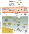
Figure 1
Figure 1
Biological barriers to overcome when using nonviral vectors to deliver nucleic acids in vivo. Nucleic acids bind to peptides through electrostatic interactions, transferring them across the cell membrane via endocytosis, providing endosome escape, and ultimately releasing the associated nucleic acids in the cytoplasm or nucleus. Reprinted with permission from Ref. [20]. Copyright 2018, Elsevier.
Figure 2
Figure 2
Schematic illustration of different cellular pathways involved in gene silencing. Reprinted with permission from Ref. [21]. Copyright 2022, Elsevier.
Figure 3
Figure 3
(a) CHAT peptide condenses pDNA to produce cationic nanoparticles less than 200 nm in diameter. The complex can cross the cell membrane through endocytosis and successfully escape from the endosomes, obtaining high transfection efficiency. Reprinted with permission from Ref. [22]. Copyright 2020, Elsevier. (b) The process of preparation of nanoparticles formed from FA-PC/R8-PC/pDNA complex. Reprinted with permission from Ref. [26]. Copyright 2011, Elsevier. (c) CPPs condense siRNA and deliver it to macrophages. Reprinted with permission from Ref. [30]. Copyright 2018, American Chemical Society.
Figure 4
Figure 4
(a) Bi-functional NGR-10R peptide condenses siRNA to form spherical nanostructures which can enter cells by receptor αvβ3 and CD13 mediated endocytosis. After escaping from the endosomes/lysosomes, siRNA is released into the cytoplasm and loaded by the RISC. Reprinted with permission from Ref. [28]. Copyright 2015, Biomaterials Science. (b) RPgWSC-pDNA complexes can suppress solid tumor growth by silencing BCL2 mRNA. Reprinted with permission from Ref. [35]. Copyright 2017, Elsevier. (c) CRIP28-AuNPs form nanocomplexes with nucleic acids by electrostatic interaction for cellular delivery. Reprinted with permission from Ref. [41]. Copyright 2022, Elsevier.
Figure 5
Figure 5
(a) RALA peptides condense mRNA into nanoparticles, releasing mRNA in dendritic cell cytosol to promote antigen specific T cell proliferation. Reprinted with permission from Ref. [43]. Copyright 2017, Wiley Online Library. (b) RALA peptides form a complex with siRNA to deliver siRNA into the cell and promote the regeneration of blood vessels. Reprinted with permission from Ref. [44]. Copyright 2019, Elsevier. (c) G3 peptide was assembled with siRNA and delivered to cancer cells, where siRNA was released to regulate gene expression in cancer cells. Reprinted with permission from Ref. [50]. Copyright 2021, American Chemical Society.
Figure 6
Figure 6
(a) The REDV-G4 -TAT-G4-NLS peptide assembles with pZNF580 plasmid to form nanocomplexes, which are transported to endothelial cells by the targeting effect of REDV. After the transmembrane and endosomal escape, the complexes enter the nucleus by the action of NLS to promote the expression of pZNF580 plasmid and enhance the revascularization ability of cells. Reprinted with permission from Ref. [59]. Copyright 2017, American Chemical Society. (b) The Cas9/sgRNA plasmid gene delivery system was prepared by the self-assembly method, which can specifically deliver the plasmid to the nuclei of tumor cells by the targeting of NLS, and knock down the protein tyrosine kinase 2 (PTK2) gene to the down-regulated local adhesion kinase (FAK). Reprinted with permission from Ref. [60]. Copyright 2019, American Chemical Society.
Figure 7
Figure 7
(a) Co-assembly of the K3C6SPD short peptide with plasmid DNA develops cocoon-like viral mimics. Reprinted with permission from Ref. [70]. Copyright 2017, German Chemical Society. (b) The mushroom shaped nanostructures SP-CC-PEG created by the synergistic self-assembly of three functional fragments, which has high affinity with DNA by electrostatic interaction, is used to prepare synthetic filamentous viruses. Reprinted with permission from Ref. [71]. Copyright 2013, American Chemical Society. (c) The dumbbell-like peptide, I3V3A3G3K3, binds onto the DNA chain through electrostatic interactions, and then self-associates into β-sheets under hydrophobic interactions and hydrogen bonding, the resulting final formed structure being able to imitate the essence of viral capsid to condense and wrap DNA. Reprinted with permission from Ref. [72]. Copyright 2018, American Chemical Society. (d) NapFFGPLGLAG(CKm)nC peptides, containing the multifunctional segment, self-assemble into stable nanospheres which can encapsulate DNA by interacting with DNA in the interior, and finally realize intracellular delivery and release of genome. Reprinted with permission from Ref. [64]. Copyright 2022, Elsevier.

Similar articles

Cited by

References

    1. Luo M., Lee L.K.C., Peng B., Choi C.H.J., Tong W.Y., Voelcker N.H. Delivering the Promise of Gene Therapy with Nanomedicines in Treating Central Nervous System Diseases. Adv. Sci. 2022;9:2201740. doi: 10.1002/advs.202201740. - DOI - PMC - PubMed
    1. Zhu Y., Shen R., Vuong I., Reynolds R.A., Shears M.J., Yao Z.-C., Hu Y., Cho W.J., Kong J., Reddy S.K., et al. Multistep screening of DNA/lipid nanoparticles and co-delivery with siRNA to enhance and prolong gene expression. Nat. Commun. 2022;13:4282. doi: 10.1038/s41467-022-31993-y. - DOI - PMC - PubMed
    1. Kuriyama N., Yoshioka Y., Kikuchi S., Okamura A., Azuma N., Ochiya T. Challenges for the Development of Extracellular Vesicle-Based Nucleic Acid Medicines. Cancers. 2021;13:6137. doi: 10.3390/cancers13236137. - DOI - PMC - PubMed
    1. Isgrig K., McDougald D.S., Zhu J., Wang H.J., Bennett J., Chien W.W. AAV2.7m8 is a powerful viral vector for inner ear gene therapy. Nat. Commun. 2019;10:427. doi: 10.1038/s41467-018-08243-1. - DOI - PMC - PubMed
    1. Shirley J.L., de Jong Y.P., Terhorst C., Herzog R.W. Immune Responses to Viral Gene Therapy Vectors. Mol. Ther. 2020;28:709–722. doi: 10.1016/j.ymthe.2020.01.001. - DOI - PMC - PubMed

LinkOut - more resources