. 2022 Nov 20;23(22):14431.
doi: 10.3390/ijms232214431.

TGF-β/Smad Signalling Activation by HTRA1 Regulates the Function of Human Lens Epithelial Cells and Its Mechanism in Posterior Subcapsular Congenital Cataract

Affiliations

TGF-β/Smad Signalling Activation by HTRA1 Regulates the Function of Human Lens Epithelial Cells and Its Mechanism in Posterior Subcapsular Congenital Cataract

Xiaolei Lin et al. Int J Mol Sci. .

Abstract

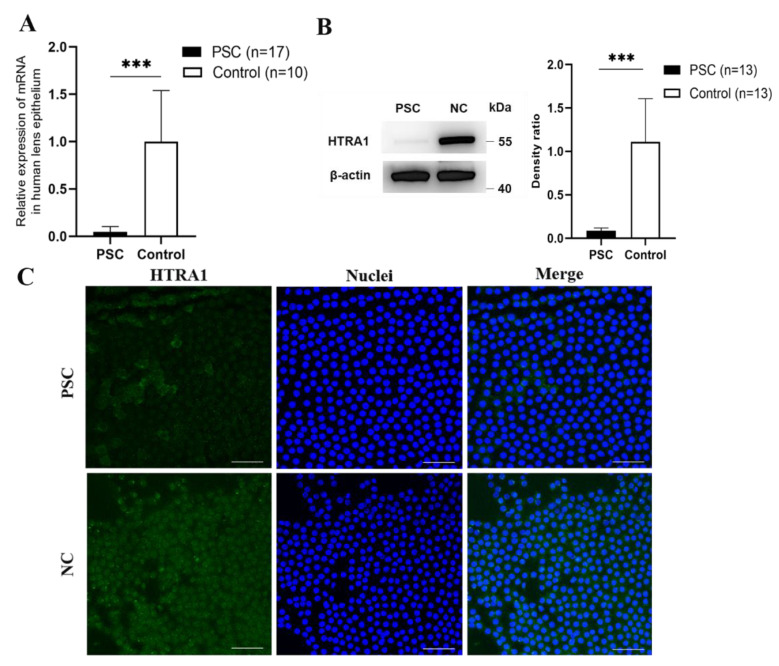
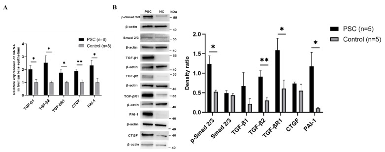
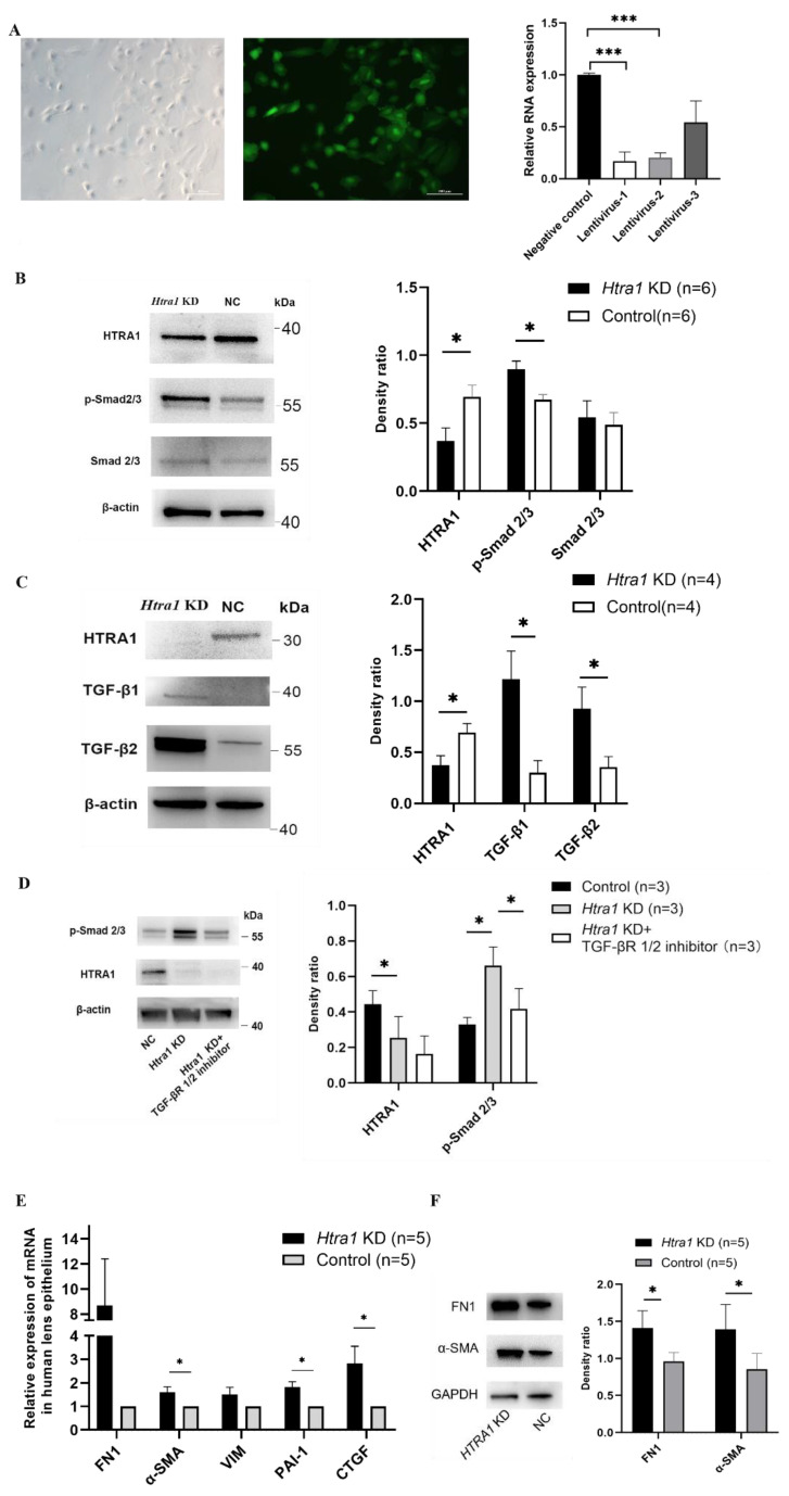
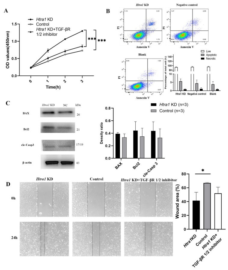
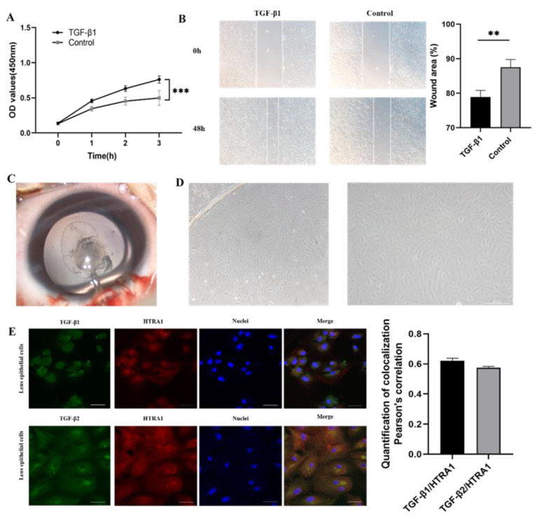
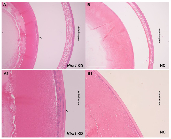
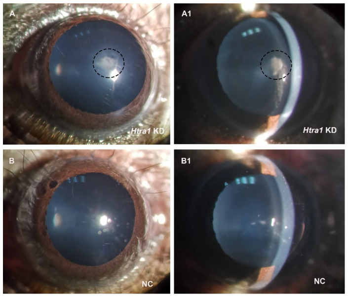
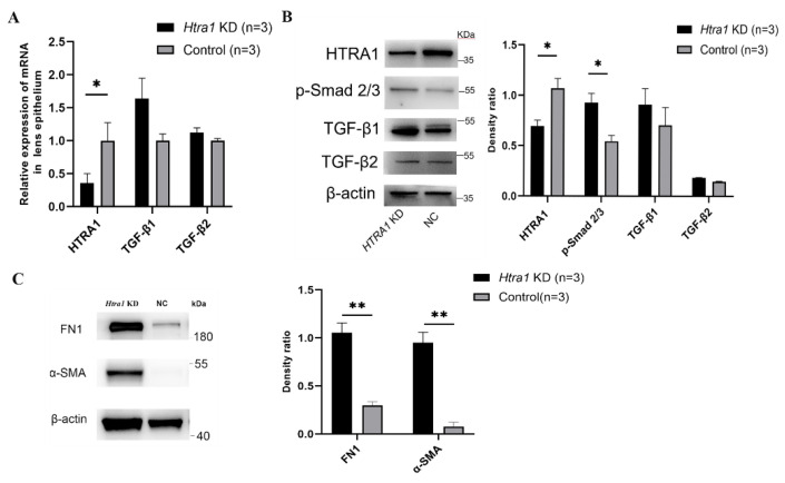
Congenital cataract is the leading cause of blindness among children worldwide. Patients with posterior subcapsular congenital cataract (PSC) in the central visual axis can result in worsening vision and stimulus deprivation amblyopia. However, the pathogenesis of PSC remains unclear. This study aims to explore the functional regulation and mechanism of HTRA1 in human lens epithelial cells (HLECs). HTRA1 was significantly downregulated in the lens capsules of children with PSC compared to normal controls. HTRA1 is a suppression factor of transforming growth factor-β (TGF-β) signalling pathway, which plays a key role in cataract formation. The results showed that the TGF-β/Smad signalling pathway was activated in the lens tissue of PSC. The effect of HTRA1 on cell proliferation, migration and apoptosis was measured in HLECs. In primary HLECs, the downregulation of HTRA1 can promote the proliferation and migration of HLECs by activating the TGF-β/Smad signalling pathway and can significantly upregulate the TGF-β/Smad downstream target genes FN1 and α-SMA. HTRA1 was also knocked out in the eyes of C57BL/6J mice via adeno-associated virus-mediated RNA interference. The results showed that HTRA1 knockout can significantly upregulate p-Smad2/3 and activate the TGF-β/Smad signalling pathway, resulting in abnormal proliferation and irregular arrangement of lens epithelial cells and leading to the occurrence of subcapsular cataract. To conclude, HTRA1 was significantly downregulated in children with PSC, and the downregulation of HTRA1 enhanced the proliferation and migration of HLECs by activating the TGF-β/Smad signalling pathway, which led to the occurrence of PSC.

Keywords: HTRA1; TGF-β; lens epithelial cells; posterior subcapsular congenital cataract.


Conflict of interest statement

The authors declare no conflict of interest.

Figures

Figure 1
Figure 1
HTRA1 expression downregulated in PSC patients. (A) The mRNA level of HTRA1 in anterior lens capsules based on RT-qPCR analyses (p < 0.01). (B) The protein levels of HTRA1 in anterior lens capsules based on Western blotting (p < 0.001). (C) Immunofluorescence images of HTRA1 staining in anterior lens capsules (n = 3). *** p < 0.001, Scale bar: 20 μm.
Figure 2
Figure 2
HTRA1 expression levels in normal human lenses at different layers and for different age groups. (A) The mRNA levels of HTRA1 expressed in central lens epithelial cells, equatorial epithelial cells and fibre cells in normal human lenses. (B) The mRNA levels of HTRA1 expressed in different age groups. (C) Immunofluorescence images of HTRA1 staining in HLECs (n = 3). Scale bar: 10 μm.
Figure 3
Figure 3
TGF-β/Smads signalling activated in lens of PSC patients. (A) The mRNA levels of TGF-β1, TGF-β2, TGF-βR1, CTGF and PAI-1 in human lens epithelial cells based on RT-qPCR analyses (p = 0.015, 0.014, 0.023, 0.002 and 0.027, respectively). (B) The protein expression of TGF-β1, TGF-β2, TGF-βR1, CTGF, PAI-1, Smad2/3 and p-Smad2/3 (p = 0.261, 0.008, 0.040, 0.321, 0.040, 0.487 and 0.017, respectively) based on Western blotting. * p < 0.05, ** p < 0.01.
Figure 4
Figure 4
Downregulation of HTRA1 directly activated the TGF-β/Smad signalling in primary HLECs. (A) Primary HLECs after LV-HTRA1-RNAi lentivirus transfection, the middle image showed the GFP expression, and the right image showed the mRNA levels of HTRA1 after transfection. (B,C) Changes of HTRA1, Smad2/3, p-Smad2/3, TGF-β1 and TGF-β2 gene expression detected by Western blotting (Smad2/3 p > 0.05, p-Smad2/3 p = 0.014, TGF-β1 p = 0.021, TGF-β2 p = 0.039) in primary HLECs collected from PSC eyes after treatment of HTRA1 knockdown (Htra1 KD). (D) Changes of p-Smad2/3 expression detected by Western blotting with HTRA1 knockdown ± TGF-βR1/2 inhibitor treatment (10 ng/mL for 24 h) in primary HLECs (p-Smad2/3 p = 0.042, 0.027, respectively). (E) The mRNA levels of α-SMA, PAI-1 and CTGF elevated in HLECs after treatment of HTRA1 knockdown (p = 0.022, 0.005, and 0.038, respectively). (F) The protein levels of FN-1, and α-SMA in HLECs after treatment of HTRA1 knockdown (p = 0.017 and 0.031, respectively). * p < 0.05, *** p < 0.001.
Figure 5
Figure 5
The proliferation, migration and apoptosis changes of HLECs (SRA 01/04) after treatment of HTRA1 knockdown. (A) CCK-8 assay showed promoted growth ability of HLECs after downregulated expression of HTRA1 (p < 0.001) and was attenuated after treatment of HTRA1 knockdown and the TGF-βR1/2 inhibitor (p < 0.001). (B) Annexin V/7-AAD staining and flow cytometry did not show significant apoptotic cell changes after treatment of HTRA1 knockdown. (C) The protein levels of BAX, Bcl2 and cleaved Caspase-3 in HLECs after treatment of HTRA1 knockdown (p > 0.05). (D) The scratch healing assay showed knockdown of HTRA1 promoted the migration of HLECs (p = 0.022), and was attenuated after treatment of HTRA1 knockdown and the TGF-βR1/2 inhibitor. * p < 0.05, *** p < 0.001.
Figure 6
Figure 6
Elevated proliferation and migration of lens epithelial cells by TGF-β1 and the colocalization between HTRA1 and TGF-β1, as well as TGF-β2. (A) CCK-8 assay of human lens epithelial cells after TGF-β1 treatment (p < 0.001). (B) Scratch healing assay of human lens epithelial cells after TGF-β1 treatment (24 h, p = 0.007). (C) Anterior lens capsules were obtained via capsulorhexis during cataract surgery. (D) Cultured primary lens epithelial cells showed migration from the rim of lens capsules. (E) Immunofluorescence images of HTRA1, TGF-β1, and TGF-β2 staining. The amount of colocalization was analysed. ** p < 0.01, *** p < 0.001, Scale bar: 20 μm.
Figure 7
Figure 7
Images of haematoxylin and eosin staining after knocking down HTRA1 by rAAV injection in mouse eyes. Aberrant proliferation and migration of lens epithelial cells were observed in (A,A1) (Arrow). (A,B): Scale bar: 200 μm, (A1,B1): Scale bar: 50 μm. A and A1, as well as B and B1, were images from the same position.
Figure 8
Figure 8
Images of mouse eyes after treatment of HTRA1 knockdown. (A,A1) showed that downregulation of HTRA1 induced the formation of subcapsular cataract. (A,A1), as well as (B,B1), were images from the same position.
Figure 9
Figure 9
Downregulation of HTRA1 activated the TGF-β/Smad signalling in primary mouse lens epithelial cells. (A) Changes of HTRA1, TGF-β1 and TGF-β2 gene expression detected by RT-qPCR (HTRA1 p = 0.045) in primary mouse lens epithelial cells after treatment of HTRA1 knockdown. (B) Changes of HTRA1, p-Smad2/3, TGF-β1 and TGF-β2 gene expression detected by Western blotting (HTRA1 p = 0.029, p-Smad 2/3 p = 0.024) in primary mouse lens epithelial cells after treatment of HTRA1 knockdown. (C) The protein levels of FN-1 and α-SMA in primary mouse lens epithelial cells after treatment of HTRA1 knockdown (p = 0.002 and 0.002, respectively). * p < 0.05, ** p < 0.01.

Similar articles

Cited by

References

    1. Mohammadpour M., Shaabani A., Sahraian A., Momenaei B., Tayebi F., Bayat R., Mirshahi R. Updates on managements of pediatric cataract. J. Curr. Ophthalmol. 2019;31:118–126. doi: 10.1016/j.joco.2018.11.005. - DOI - PMC - PubMed
    1. Churchill A., Graw J. Clinical and experimental advances in congenital and paediatric cataracts. Philos. Trans. R. Soc. B Biol. Sci. 2011;366:1234–1249. doi: 10.1098/rstb.2010.0227. - DOI - PMC - PubMed
    1. Li J., Chen X., Yan Y., Yao K. Molecular genetics of congenital cataracts. Exp. Eye Res. 2020;191:107872. doi: 10.1016/j.exer.2019.107872. - DOI - PubMed
    1. Santana A., Waiswo M. The genetic and molecular basis of congenital cataract. Arq. Bras. Oftalmol. 2011;74:136–142. doi: 10.1590/S0004-27492011000200016. - DOI - PubMed
    1. Rechsteiner D., Issler L., Koller S., Lang E., Bähr L., Feil S., Rüegger C.M., Kottke R., Toelle S.P., Zweifel N., et al. Genetic Analysis in a Swiss Cohort of Bilateral Congenital Cataract. JAMA Ophthalmol. 2021;139:691. doi: 10.1001/jamaophthalmol.2021.0385. - DOI - PMC - PubMed

Substances