Review
. 2022 Nov 16;23(22):14188.
doi: 10.3390/ijms232214188.

Neurodegenerative Diseases: From Dysproteostasis, Altered Calcium Signalosome to Selective Neuronal Vulnerability to AAV-Mediated Gene Therapy

Affiliations
Review

Neurodegenerative Diseases: From Dysproteostasis, Altered Calcium Signalosome to Selective Neuronal Vulnerability to AAV-Mediated Gene Therapy

Tam T Quach et al. Int J Mol Sci. .

Abstract

Despite intense research into the multifaceted etiology of neurodegenerative diseases (ND), they remain incurable. Here we provide a brief overview of several major ND and explore novel therapeutic approaches. Although the cause (s) of ND are not fully understood, the accumulation of misfolded/aggregated proteins in the brain is a common pathological feature. This aggregation may initiate disruption of Ca++ signaling, which is an early pathological event leading to altered dendritic structure, neuronal dysfunction, and cell death. Presently, ND gene therapies remain unidimensional, elusive, and limited to modifying one pathological feature while ignoring others. Considering the complexity of signaling cascades in ND, we discuss emerging therapeutic concepts and suggest that deciphering the molecular mechanisms involved in dendritic pathology may broaden the phenotypic spectrum of ND treatment. An innovative multiplexed gene transfer strategy that employs silencing and/or over-expressing multiple effectors could preserve vulnerable neurons before they are lost. Such therapeutic approaches may extend brain health span and ameliorate burdensome chronic disease states.

Keywords: CRMP3/DPYSL4; calcium signaling; dendritic dystrophy; dysproteostasis; gene therapy; neurodegeneration; neuronal vulnerability.


Conflict of interest statement

The authors declare that they have no conflict of interest.

Figures

Figure 1
Figure 1
Proposed pathways depicting the potential role of misfolded proteins and the contribution of ER/Mitochondria/Nucleus in the pathogenesis of major ND. Accumulated unfolded/misfolded proteins in the ER may either be degraded by ERAD or activate the UPR, which induces a set of transcriptional and translational events to the nucleus through activation of 2 transcription factors (ATF6 and XbP1s), an unspliced X-box binding protein1 (XbP1u) and mitochondria via IRE1α/PERK/ATF6α, 3 ER transmembrane protein sensors to restore ER homeostasis via adaptive mechanisms. Conversely, if ER stress persists chronically at high levels, a terminal UPR signal induces cell death via the activation of CHOP/GADD34/DR5/BCL2 when damage is irreversible. Chronic ER stress and defects in UPR signaling are contributors to ND. In addition to UPR, the excessive transfer of Ca++ to the mitochondria also leads to enhanced ROS production (oxidative stress) and mitoproteases (BAX/BAK-dependent apoptosome) activation. Depending on the proteostasis context in ND subtypes and their pathological conditions, ERS may trigger distinct signaling pathways. Proteasomes are multicatalytic protease complexes that selectively degrade target proteins into peptide fragments to maintain protein homeostasis.
Figure 2
Figure 2
Neuronal Ca++ homeostasis via Pore/Receptor-operated Ca++ channels, Ca++ binding proteins, gene transcription, and ER/Mitochondria interactions. The complex signaling pathways regulating neuronal Ca++ concentration and organization require various membrane Ca++-conducting channels, intracytoplasmic organelles such as ER and mitochondria, and a great number of Ca++ buffering/dependent proteins including calretinin, parvalbumin, calbindin, and kinases. (Ca++)ic is determined by the balance between Ca++ influx (left side) and efflux (right side) and is buffered in the cytosol by mitochondria through VDAC, MCU, and ER with the help of SERCA while RyR and IP3R mediate ER Ca++ efflux. (Ca++)ic also regulates the expression of target genes. Ca++ released from the ER interacts with mitochondria through MAM and contributes to the activation of the Tricyclic Acid Cycle (TAC) to stimulate ATP synthesis, whereas the excessive transfer of Ca++ to mitochondria leads to ROS and BAX/BAK-dependent apoptosome. Inter-organelles do not act as autonomous units but as interconnected hubs that engage in extensive communication through membrane contacts. The proteins within MAM—a central hub involved in different fundamental cell processes—play important roles in maintaining MAM stability, Ca++ transport, and apoptosis: 1a, b: REEP1: Receptor expression-enhancing protein 1 (REEP1); 2a: PTPIP51; 2b: VABP; 3a: GrP75; 3b: IP3R; 4a: Protein tyrosine phosphatase interacting protein1 (PTPIP51); 4b: Motile sperm domain-containing protein 2 (MOSPD2); 5a: MFN1; 5b: MFN2; 6a,b: MFN2. Yet, the depletion of the Ʃ−1 receptor leads to abnormal Ca++ signaling between ER and mitochondria, and the disruption of ATP production.
Figure 3
Figure 3
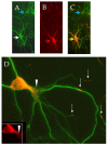
CRMP3-mediated dendritic activity. Representative CRMP3-transfected neurons were immunostained for dendritic marker MAP2 ((A) white arrow and Flag-CRMP3; (B) red arrow). Overlay images of transfected neurons are in orange ((C) yellow arrow; untransfected neuron: blue arrow). Flag-CRMP3 transfected neurons are characterized by an increase in lamellopodial/dendritic formation ((D), white arrows heads). Interestingly, the protein did not exhibit passive lateral diffusion but presented as consistent puncta over long distances in the soma and the dendrites with some extending up to the dendritic tips suggesting an active transport of the protein into dendrites ((D) white arrows).
Figure 4
Figure 4
Schematic diagram of the structure of AAV vector mediating gene transfer and therapeutic protein delivery in neurons. Devoid of rep/cap genes, the 145 multipalindromic nucleotides of ITR are the only viral origin sequences needed to guide genome replication and packaging during vector production because ITR can form a T-shaped hairpin structure through Watson–Crick base pairing to initiate DNA replication and produce rec-AAVs. Viral ORFs are replaced by a transgene with its regulatory elements. Once the transgene expression cassette is optimized, the next step involves the production of vector stocks. Rec-AAV vectors can be produced at high yields by transient triple transfection in mammalian cells (i.e., HEK293 cells) with three plasmids: the first plasmid containing the transgene of interest, the second plasmid containing Rep and Cap, and a third plasmid encoding for adenoviral helper genes. The purification of rec-AAV vectors is performed by either column chromatography or gradient centrifugation. Overall, rec-AAV vectors are capable of delivering transgenes into the CNS. They are bound to cell surface receptors, then integrated into the cytosol. Following the endosomal escape, they are uncoated and transported into the nucleus. These single-stranded forms are then converted to double-stranded DNA via host cell DNA polymerases for transcription. This conversion can also be achieved by strand annealing of the plus and minus strands that may coexist in the nucleus. The double-stranded DNA vector can form circularized episomes and persist in the nucleus or undergo integration into the host chromosomes.

Similar articles

Cited by

References

    1. Voet S., Srinivasan S., Lamkanfi M., van Loo G. Inflammasomes in neuroinflammatory and neurodegenerative diseases. EMBO Mol. Med. 2019;11:e10248. doi: 10.15252/emmm.201810248. - DOI - PMC - PubMed
    1. Hetz C., Zhang K., Kaufman R.J. Mechanisms, regulation and functions of the unfolded protein response. Nat. Rev. Mol. Cell Biol. 2020;21:421–438. doi: 10.1038/s41580-020-0250-z. - DOI - PMC - PubMed
    1. Schrank S., Barrington N., Stutzmann G.E. Calcium-Handling Defects and Neurodegenerative Disease. Cold Spring Harb. Perspect. Biol. 2020;12:a035212. doi: 10.1101/cshperspect.a035212. - DOI - PMC - PubMed
    1. Rossmann M.P., Dubois S.M., Agarwal S., Zon L.I. Mitochondrial function in development and disease. Dis. Models Mech. 2021;14:dmm048912. doi: 10.1242/dmm.048912. - DOI - PMC - PubMed
    1. Oakes S.A., Papa F.R. The role of endoplasmic reticulum stress in human pathology. Annu. Rev. Pathol. 2015;10:173–194. doi: 10.1146/annurev-pathol-012513-104649. - DOI - PMC - PubMed

Grants and funding

This research received no external funding.