

Microglia: Friends or Foes in Glaucoma? A Developmental Perspective
- PMID: 36426733
- PMCID: PMC9801300
- DOI: 10.1093/stcltm/szac077
Microglia: Friends or Foes in Glaucoma? A Developmental Perspective
Abstract
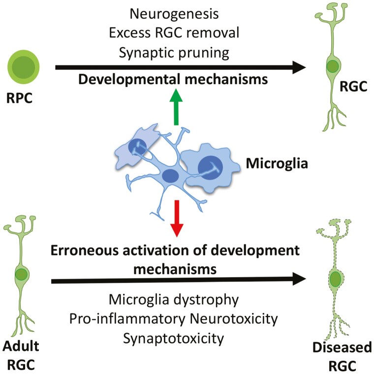
Glaucoma is the most prevalent form of optic neuropathy where a progressive degeneration of retinal ganglion cells (RGCs) leads to irreversible loss of vision. The mechanism underlying glaucomatous degeneration remains poorly understood. However, evidence suggests that microglia, which regulate RGC numbers and synaptic integrity during development and provide homeostatic support in adults, may contribute to the disease process. Hence, microglia represent a valid cellular target for therapeutic approaches in glaucoma. Here, we provide an overview of the role of microglia in RGC development and degeneration in the backdrop of neurogenesis and neurodegeneration in the central nervous system and discuss how pathological recapitulation of microglia-mediated developmental mechanisms may help initiate or exacerbate glaucomatous degeneration.
Keywords: degeneration; glaucoma; microglia; neurogenesis; retinal ganglion cell.
© The Author(s) 2022. Published by Oxford University Press.
Figures
Similar articles
-
Assessment of retinal ganglion cell damage in glaucomatous optic neuropathy: Axon transport, injury and soma loss.Exp Eye Res. 2015 Dec;141:111-24. doi: 10.1016/j.exer.2015.06.006. Epub 2015 Jun 9. Exp Eye Res. 2015. PMID: 26070986 Review.
-
DRP1 inhibition rescues retinal ganglion cells and their axons by preserving mitochondrial integrity in a mouse model of glaucoma.Cell Death Dis. 2015 Aug 6;6(8):e1839. doi: 10.1038/cddis.2015.180. Cell Death Dis. 2015. PMID: 26247724 Free PMC article.
-
Detection of early neuron degeneration and accompanying microglial responses in the retina of a rat model of glaucoma.Invest Ophthalmol Vis Sci. 2002 Sep;43(9):2962-8. Invest Ophthalmol Vis Sci. 2002. PMID: 12202516
-
Time-dependent retinal ganglion cell loss, microglial activation and blood-retina-barrier tightness in an acute model of ocular hypertension.Exp Eye Res. 2015 Jul;136:59-71. doi: 10.1016/j.exer.2015.05.010. Epub 2015 May 20. Exp Eye Res. 2015. PMID: 26001526
-
Commonalities of optic nerve injury and glaucoma-induced neurodegeneration: Insights from transcriptome-wide studies.Exp Eye Res. 2021 Jun;207:108571. doi: 10.1016/j.exer.2021.108571. Epub 2021 Apr 15. Exp Eye Res. 2021. PMID: 33844961 Free PMC article. Review.
Cited by
-
Neuroprotection of Retinal Ganglion Cells Suppresses Microglia Activation in a Mouse Model of Glaucoma.Invest Ophthalmol Vis Sci. 2023 Jun 1;64(7):24. doi: 10.1167/iovs.64.7.24. Invest Ophthalmol Vis Sci. 2023. PMID: 37318444 Free PMC article.
-
Loss of Stim2 in zebrafish induces glaucoma-like phenotype.Sci Rep. 2024 Oct 18;14(1):24442. doi: 10.1038/s41598-024-74909-0. Sci Rep. 2024. PMID: 39424970 Free PMC article.
-
Human microglia-derived proinflammatory cytokines facilitate human retinal ganglion cell development and regeneration.Stem Cell Reports. 2024 Aug 13;19(8):1092-1106. doi: 10.1016/j.stemcr.2024.06.009. Epub 2024 Jul 25. Stem Cell Reports. 2024. PMID: 39059376 Free PMC article.
-
Regulation of disease-associated microglia in the optic nerve by lipoxin B4 and ocular hypertension.Mol Neurodegener. 2024 Nov 20;19(1):86. doi: 10.1186/s13024-024-00775-z. Mol Neurodegener. 2024. PMID: 39568070 Free PMC article.
-
Regulation of Diseases-Associated Microglia in the Optic Nerve by Lipoxin B4 and Ocular Hypertension.bioRxiv [Preprint]. 2024 Oct 28:2024.03.18.585452. doi: 10.1101/2024.03.18.585452. bioRxiv. 2024. Update in: Mol Neurodegener. 2024 Nov 20;19(1):86. doi: 10.1186/s13024-024-00775-z. PMID: 38562864 Free PMC article. Updated. Preprint.
References
-
- Tham YC, Li X, Wong TY, Quigley HA, Aung T, Cheng CY. et al. . Global prevalence of glaucoma and projections of glaucoma burden through 2040: a systematic review and meta-analysis. Ophthalmology. 2014;121:2081-2090. - PubMed
Publication types
MeSH terms
Grants and funding
LinkOut - more resources
Full Text Sources
Medical
