. 2022 Dec 20;13(6):e0296122.
doi: 10.1128/mbio.02961-22. Epub 2022 Nov 21.

The Obligate Intracellular Bacterial Pathogen Anaplasma phagocytophilum Exploits Host Cell Multivesicular Body Biogenesis for Proliferation and Dissemination

Affiliations

The Obligate Intracellular Bacterial Pathogen Anaplasma phagocytophilum Exploits Host Cell Multivesicular Body Biogenesis for Proliferation and Dissemination

Curtis B Read et al. mBio. .

Abstract

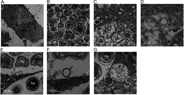
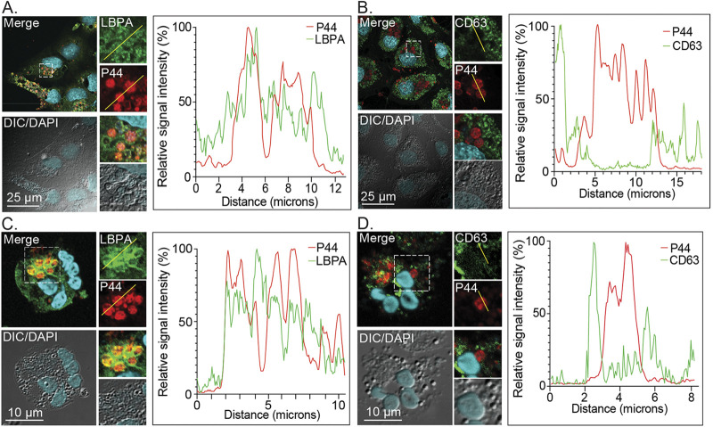
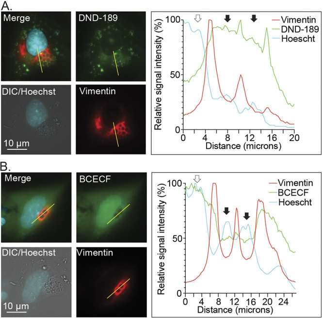
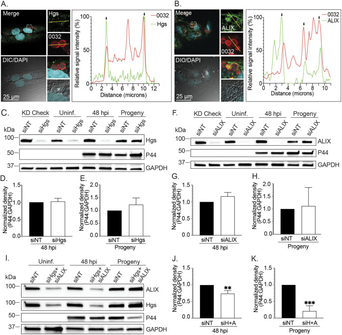
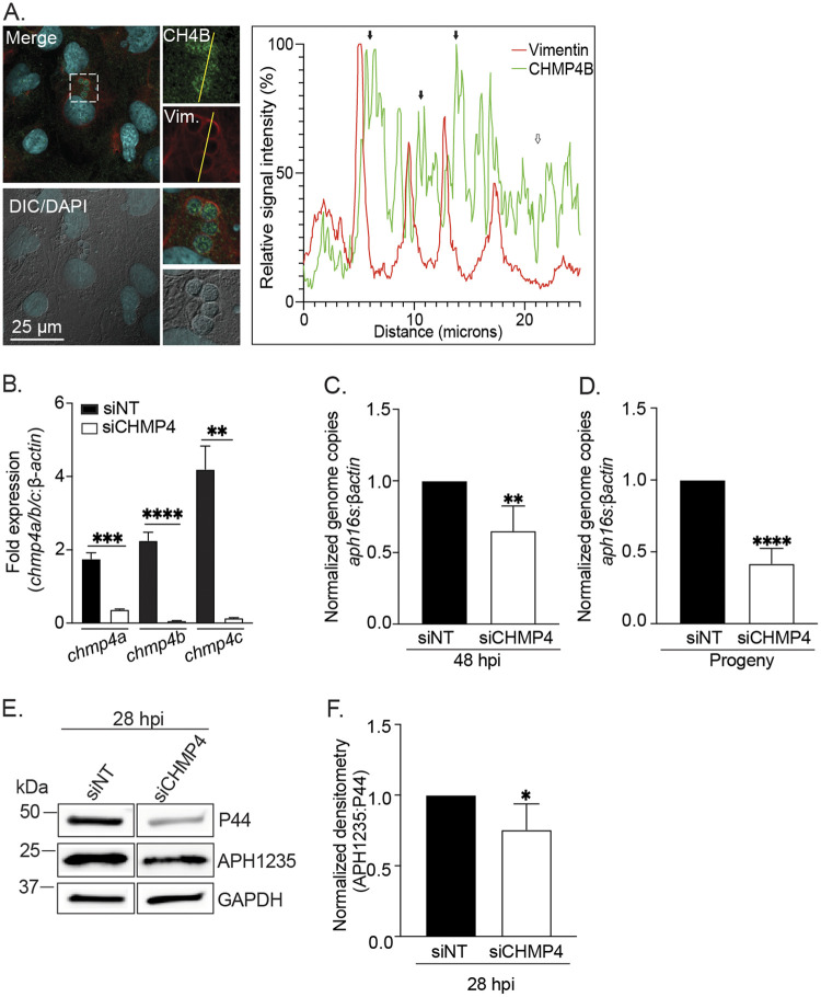
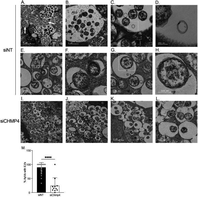
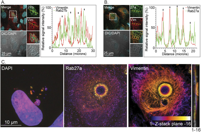
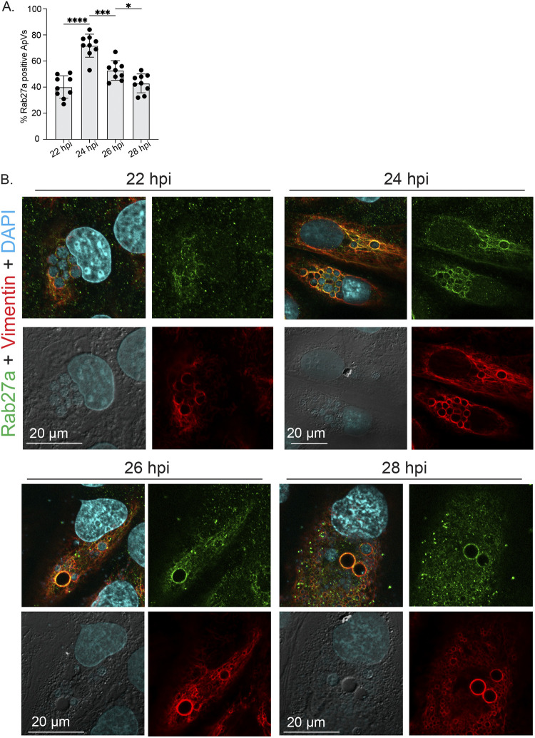
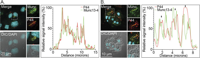
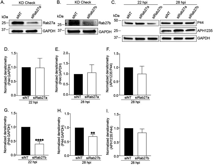
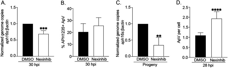
Anaplasma phagocytophilum is the etiologic agent of the emerging infection, granulocytic anaplasmosis. This obligate intracellular bacterium lives in a host cell-derived vacuole that receives membrane traffic from multiple organelles to fuel its proliferation and from which it must ultimately exit to disseminate infection. Understanding of these essential pathogenic mechanisms has remained poor. Multivesicular bodies (MVBs) are late endosomal compartments that receive biomolecules from other organelles and encapsulate them into intralumenal vesicles (ILVs) using endosomal sorting complexes required for transport (ESCRT) machinery and ESCRT-independent machinery. Association of the ESCRT-independent protein, ALIX, directs MVBs to the plasma membrane where they release ILVs as exosomes. We report that the A. phagocytophilum vacuole (ApV) is acidified and enriched in lysobisphosphatidic acid, a lipid that is abundant in MVBs. ESCRT-0 and ESCRT-III components along with ALIX localize to the ApV membrane. siRNA-mediated inactivation of ESCRT-0 and ALIX together impairs A. phagocytophilum proliferation and infectious progeny production. RNA silencing of ESCRT-III, which regulates ILV scission, pronouncedly reduces ILV formation in ApVs and halts infection by arresting bacterial growth. Rab27a and its effector Munc13-4, which drive MVB trafficking to the plasma membrane and subsequent exosome release, localize to the ApV. Treatment with Nexinhib20, a small molecule inhibitor that specifically targets Rab27a to block MVB exocytosis, abrogates A. phagocytophilum infectious progeny release. Thus, A. phagocytophilum exploits MVB biogenesis and exosome release to benefit each major stage of its intracellular infection cycle: intravacuolar growth, conversion to the infectious form, and exit from the host cell. IMPORTANCE Anaplasma phagocytophilum causes granulocytic anaplasmosis, a globally emerging zoonosis that can be severe, even fatal, and for which antibiotic treatment options are limited. A. phagocytophilum lives in an endosomal-like compartment that interfaces with multiple organelles and from which it must ultimately exit to spread within the host. How the bacterium accomplishes these tasks is poorly understood. Multivesicular bodies (MVBs) are intermediates in the endolysosomal pathway that package biomolecular cargo from other organelles as intralumenal vesicles for release at the plasma membrane as exosomes. We discovered that A. phagocytophilum exploits MVB biogenesis and trafficking to benefit all aspects of its intracellular infection cycle: proliferation, conversion to its infectious form, and release of infectious progeny. The ability of a small molecule inhibitor of MVB exocytosis to impede A. phagocytophilum dissemination indicates the potential of this pathway as a novel host-directed therapeutic target for granulocytic anaplasmosis.

Keywords: Anaplasma phagocytophilum; ESCRT; Munc13-4; Rab27a; exocytosis; exosome; intralumenal vesicle; multivesicular body; multivesicular endosome; nutritional virulence; obligate intracellular bacterium; rickettsia; vacuolar pathogen.


Conflict of interest statement

The authors declare no conflict of interest.

Figures

FIG 1
FIG 1
The ApV contains intralumenal vesicles as visualized by TEM. Panels (A), (B), (E), and (F) are transmission electron micrographs of A. phagocytophilum infected RF/6A cells. Panels (C), (D), and (G) are transmission electron micrographs of infected RF/6A cells after incubation with BSA-gold particles for 4 h at 37°C. White hatched boxes in the images in (A), (C), and (E) are enlarged in (B), (D), and (F), respectively. Black arrows in (A) delineate individual ApVs. Black arrows in (B) denote individual bacteria. White arrows in (B) indicate ILVs. Black arrows in (D) point to BSA-gold particles associated with ILVs in close apposition to A. phagocytophilum organisms within an ApV. (E) and (F) A newly formed ILV undergoing scission from the ApV membrane (black arrow in [F]) and a nascent invagination of the ApV membrane (white arrow in [F]) are presented. (G) An MVB containing endocytosed BSA-gold (black arrow) is observed adjacent to an ApV. Scale bars are presented on individual images. Electron micrographs shown are representative of three independent experiments.
FIG 2
FIG 2
The ApV is enriched in LBPA and lacks CD63. RF/6A cells (A) and (B) and human neutrophils (C) and (D) were infected with A. phagocytophilum for 24 h or 20 h, respectively, after which they were fixed and immunolabeled with antibodies against LBPA (A) and (C), CD63 (B) and (D), and A. phagocytophilum P44 (A) to (D). The cells were stained with DAPI and visualized using LSCM and differential interference contrast microscopy (DIC). The graphs by each panel of micrographs represent the relative signal intensity profiles of green and red pixels along the yellow line (moving left to right) normalized to the highest fluorescence intensity per channel. Scale bars, 25 μm. Results shown are representative of two to four experiments with similar results.
FIG 3
FIG 3
The ApV lumen is acidified. A. phagocytophilum infected RF/6A cells expressing mCherry-vimentin were incubated with LysoSensor Green DND-189 (A) or BCECF (B), stained with Hoechst, and imaged using live cell fluorescence and DIC microscopy. The graphs next to each set of micrographs present the relative signal intensity profiles of red, green, and cyan pixels along the yellow line (moving left to right) normalized to the highest fluorescence intensity per channel. White arrows denote host cell nuclei. Black arrows indicate individual ApVs. Scale bars, 10 μm. Results are representative of at least three independent experiments with similar results.
FIG 4
FIG 4
Hgs and ALIX are recruited to the ApV and benefit A. phagocytophilum replication and infectious progeny production. (A) and (B) Hgs and ALIX colocalize with the ApV membrane-localized effector, APH0032. RF/6A cells were infected with A. phagocytophilum for 24 h, after which they were fixed and immunolabeled with antibodies against Hgs (A), ALIX (B), and A. phagocytophilum APH0032 (0032) (A) and (B). The cells were stained with DAPI and examined using LSCM and DIC microscopy. The graphs by each set of micrographs represent the relative signal intensity profiles of green and red pixels along the yellow line (moving left to right) normalized to the highest fluorescence intensity per channel. Black arrows demarcate the ApV membrane. Scale bars, 25 μm. (C) to (K) Knockdown of both Hgs and ALIX hinders A. phagocytophilum proliferation and infectious progeny generation. RF/6A cells were treated with siRNA targeting Hgs (siHgs) (C) to (E), ALIX (siALIX) (F) to (H), both Hgs and ALIX (siH+A) (I) to (K), or non-targeting siRNA (siNT) (C) to (K). At 48 h, one set of each condition was collected to confirm knockdown of the protein of interest (KD check). The other two sets were either mock infected (Uninf.) or incubated with A. phagocytophilum DC organisms for 48 h (48 hpi). All samples were examined by Western blotting using antibodies specific for Hgs, ALIX, P44, and GAPDH. The A. phagocytophilum load was quantified as the mean (±SD) normalized ratio of P44:GAPDH densitometric signals from triplicate samples per condition. To assess for infectious progeny production and release, media from siNT- or target-specific siRNA-treated cells that had been infected with A. phagocytophilum was collected at 48 h postinfection and added to naive RF/6A cells that had not been treated with siRNA. This time point corresponds to when infectious DC bacteria would be present in the media if the bacterial developmental cycle had proceeded normally. Twenty-four h later, recipient cells were assessed for Hgs, ALIX, P44, and GAPDH levels and the A. phagocytophilum load (Progeny). Statistically significant (**, P < 0.01; ***, P < 0.001) are indicated. Data shown are representative of three independent experiments.
FIG 5
FIG 5
CHMP4 is recruited to the ApV and is critical for infectious progeny release, but not RC-to-DC conversion. (A) CHMP4B localizes to the ApV. RF/6A cells were infected with A. phagocytophilum for 24 h and screened with CHMP4B and vimentin (Vim.) antibodies, stained with DAPI to denote host cell nuclei and A. phagocytophilum nucleoids, and examined using LSCM and DIC microscopy. Scale bar, 25 μm. The graph presents the relative signal intensity profiles of green and red pixels along the yellow line (moving right to left) normalized to the highest fluorescence intensity per channel. (B) and (C) CHMP4 benefits A. phagocytophilum intracellular proliferation. RF/6A cells were treated with an siRNA cocktail targeting CHMP4A, CHMP4B, and CHMP4C (siCHMP4) or siNT. (B) Target knockdown was confirmed by RT-qPCR as the normalized ratios of chmp4a, chmp4b, and chmp4c transcript levels to β-actin transcript levels per triplicate samples. (C) siNT-treated and CHMP4 knockdown cells were incubated with A. phagocytophilum DC organisms. At 48 h, the cells were subjected to qPCR to determine the bacterial load as the normalized ratio of the A. phagocytophilum 16S rRNA gene (aph16S) to β-actin. (D) CHMP4 is important for A. phagocytophilum infectious progeny release. To assess for infectious progeny production and release, media from siNT-treated and CHMP4 knockdown cells that had been infected with A. phagocytophilum was collected at 48 hpi and added to naive RF/6A cells. This time point corresponds to when infectious DC bacteria would be present in the media if the bacterial developmental cycle had proceeded normally. Twenty-four h later, the recipient cells were subjected to qPCR to determine the normalized aph16S:β-actin ratio. (E) and (F) CHMP4 knockdown does not impair A. phagocytophilum RC-to-DC conversion. siNT-treated and CHMP4 knockdown cells that had been infected with A. phagocytophilum were examined at 28 hpi by Western blotting (E) to determine RC-to-DC conversion as the mean (±SD) normalized ratio of APH1235:P44 densitometric signals (F). Statistically significant (*, P < 0.05; **, P < 0.01; ***, P < 0.001; ****, P < 0.0001) are indicated. Data presented are representative of 3 to 5 independent experiments.
FIG 6
FIG 6
ESCRT-III is essential for ILV delivery into and development of the ApV. siNT-treated RF/6A cells (A) to (H) and CHMP4A/B/C knockdown (siCHMP4) cells (I) to (K) were infected with A. phagocytophilum and examined by TEM at 24 h. Panels (C) to (H) are transmission electron micrographs of infected control cells after incubation with BSA-gold particles for 4 h at 37°C. White hatched boxes in the images in (C), (E), (G), and (K) are enlarged in (D), (F), (H), and (L), respectively. Black arrows and white arrows in (A) denote ApVs harboring DCs or RCs, respectively. Black arrows in (D), (F), and (H) point to BSA-gold particles. Scale bar sizes are indicated. Data are representative of 2 experiments. (M) The percentage of ApVs with ILVs is significantly reduced in siCHMP4 cells. The mean (±SD) percentage of ApVs having ILVs in siCHMP4 and siNT cells was calculated by examining more 200 ApVs in TEM images acquired from grids in different Z-plane sections in two separate experiments. Each gray or black circle corresponds to the percentage of ILV-positive ApVs in individual siNT or siCHMP4 cells. Statistical significance (****, P < 0.0001) is indicated.
FIG 7
FIG 7
Rab27 isoforms localize to the ApV. At 24 h post-synchronous infection, A. phagocytophilum infected RF/6A cells were immunolabeled with antibodies against Rab27b (A) or Rab27a (B) and (C) and vimentin (A) to (C), stained with DAPI to visualize host cell nuclei and bacterial nucleoids (A) to (C), and examined by LSCM (A) to (C) and DIC microscopy (A) and (B). Scale bar sizes are indicated. The graphs in (A) and (B) represent the relative signal intensity profiles of green and red pixels along the yellow line (moving left to right) normalized to the highest fluorescence intensity per channel. The Z-projection in (C) is an array of the 16 successive Z-plane images from bottom to top that are presented in Fig. S3. A color code, which is indicated at the bottom of the vimentin panel, enabled for assessment of the three-dimensional depth of Rab27a, vimentin, the host cell nucleus, and bacterial nucleoids. For the Z-stack that was used to produce the Z-projection, plane 1 is the bottom-most focal plane while plane 16 is at the top of the stack. An orthogonal view of the Z-stack is provided to the right of the Z-projection. Please note the positions of the Rab27a-positive ApV and its released A. phagocytophilum organisms at the plasma membrane. Data are representative of three experiments with similar results.
FIG 8
FIG 8
Peak Rab27a localization to ApVs precedes reinfection. RF/6A cells that had been synchronously infected with A. phagocytophilum were immunolabeled with antibodies against Rab27a and vimentin, stained with DAPI to visualize host cell nuclei and bacterial nucleoids, and examined by LSCM and DIC microscopy at 22, 24, 26, and 28 hpi. (A) The mean (±SD) percentage of Rab27a-positive ApVs was calculated from the total number of ApVs from 100 infected cells examined per time point. Each black circle corresponds to the percentage of Rab27a-positive ApVs in individual cells. (B) Representative confocal micrographs and DIC images. Statistically significant (*, P < 0.05; ***, P < 0.001; ****, P < 0.0001) are indicated. Data are representative of three experiments with similar results each performed in triplicate.
FIG 9
FIG 9
Munc13-4 localizes to the ApV. RF/6A cells and human neutrophils were infected with A. phagocytophilum for 24 h or 20 h, respectively, after which they were fixed, immunolabeled with antibodies against Munc13-4 (Munc) and A. phagocytophilum P44, stained with DAPI, and examined using LSCM and DIC microscopy. Scale bar sizes are indicated. The graphs represent the relative signal intensity profiles of green and red pixels along the yellow line (moving left to right) normalized to the highest fluorescence intensity per channel. Results are representative of 2 to 3 experiments with similar results.
FIG 10
FIG 10
siRNA-mediated knockdown of Rab27b, but not Rab27a inhibits the A. phagocytophilum infection cycle. (A) and (C) to (F) Rab27a knockdown does not impair the A. phagocytophilum infection cycle within the first 28 h. RF/6A cells were treated with Rab27a-specific siRNA (siRab27a) or non-targeting siRNA (siNT). Target knockdown was confirmed at by Western blotting at 48 h (A). Rab27a knockdown and siNT control-treated cells were infected with A. phagocytophilum DC organisms and assessed at the indicated time points by Western blotting (C) and densitometry to determine the bacterial load (D) and (E) and RC-to-DC conversion (F) as the mean (±SD) normalized ratios of P44:GAPDH and APH1235:P44 densitometric signals, respectively. (B), (C), (G) to (I) Rab27b knockdown reduces the A. phagocytophilum load, but not RC-to-DC conversion. RF/6A cells treated with Rab27b-targeting siRNA (siRab27b) or siNT cells were examined by Western blotting for target knockdown at 48 h (B). Rab27b knockdown and control cells were infected with DC organisms and assessed by Western blotting (C) and densitometry to determine the bacterial load (G) and (H) and RC-to-DC conversion (I). Statistically significant (**, P < 0.01; ****, P < 0.0001) are indicated. Data presented are representative of 5 separate experiments.
FIG 11
FIG 11
Nexinhib20 inhibits the A. phagocytophilum infection cycle by impeding infectious progeny release. (A) to (C) Nexinhib20 treatment reduces the overall A. phagocytophilum load by inhibiting DC release, but not RC-to-DC conversion. HL-60 cells that had been synchronously infected with A. phagocytophilum were treated with Nexinhib20 or vehicle control (DMSO) starting at 24 h. Six h later, which corresponds to 30 hpi, aliquots were examined by qPCR to determine the bacterial load as the normalized aph16S:β-actin ratio (A) or immunofluorescence microscopy screening to determine the mean (±SD) percentage of ApVs harboring bacteria that were positive for the DC-specific marker, APH1235 (B). Also at 30 h, media from DMSO- and Nexinhib20-treated cells, which normally would contain naturally liberated infectious DC organisms, was transferred to naive HL-60 cells. Twenty-four h later, the A. phagocytophilum DNA load in the recipient cells was quantified using qPCR (C). (D) Nexinhib20 inhibits ApV exit. HL-60 cells were synchronously infected with A. phagocytophilum and treated with Nexinhib20 or DMSO beginning at 20 h. After 8 h, which corresponds to 28 hpi, the cells were immunolabeled with P44 antibody and the normalized percentage of infected cells and mean (±SD) number of ApVs per cell were determined. Statistically significant (**, P < 0.01; ***, P < 0.001; ****, P < 0.0001) are indicated. Data are representative of three experiments.

Similar articles

Cited by

References

    1. Ismail N, McBride JW. 2017. Tick-borne emerging infections: ehrlichiosis and anaplasmosis. Clin Lab Med 37:317–340. doi:10.1016/j.cll.2017.01.006. - DOI - PubMed
    1. Matos AL, Curto P, Simões I. 2022. Moonlighting in Rickettsiales: expanding virulence landscape. Trop Med Infect Dis 7. doi:10.3390/tropicalmed7020032. - DOI - PMC - PubMed
    1. Bakken JS, Dumler JS. 2015. Human granulocytic anaplasmosis. Infect Dis Clin North Am 29:341–355. doi:10.1016/j.idc.2015.02.007. - DOI - PMC - PubMed
    1. Dixon DM, Branda JA, Clark SH, Dumler JS, Horowitz HW, Perdue SS, Pritt BS, Sexton DJ, Storch GA, Walker DH. 2021. Ehrlichiosis and anaplasmosis subcommittee report to the Tick-borne Disease Working Group. Ticks Tick Borne Dis 12:101823. doi:10.1016/j.ttbdis.2021.101823. - DOI - PubMed
    1. Dumic I, Jevtic D, Veselinovic M, Nordstrom CW, Jovanovic M, Mogulla V, Veselinovic EM, Hudson A, Simeunovic G, Petcu E, Ramanan P. 2022. Human granulocytic anaplasmosis-a systematic review of published cases. Microorganisms 10:1433. doi:10.3390/microorganisms10071433. - DOI - PMC - PubMed

MeSH terms

Substances

LinkOut - more resources