. 2022 Nov 3:13:1029269.
doi: 10.3389/fimmu.2022.1029269. eCollection 2022.

Oncolytic virus driven T-cell-based combination immunotherapy platform for colorectal cancer

Affiliations

Oncolytic virus driven T-cell-based combination immunotherapy platform for colorectal cancer

Mathieu J F Crupi et al. Front Immunol. .

Abstract

Colorectal cancer is the third most diagnosed cancer and the second leading cause of cancer mortality worldwide, highlighting an urgent need for new therapeutic options and combination strategies for patients. The orchestration of potent T cell responses against human cancers is necessary for effective antitumour immunity. However, regression of a limited number of cancers has been induced by immune checkpoint inhibitors, T cell engagers (TCEs) and/or oncolytic viruses. Although one TCE has been FDA-approved for the treatment of hematological malignancies, many challenges exist for the treatment of solid cancers. Here, we show that TCEs targeting CEACAM5 and CD3 stimulate robust activation of CD4 and CD8-positive T cells in in vitro co-culture models with colorectal cancer cells, but in vivo efficacy is hindered by a lack of TCE retention in the tumour microenvironment and short TCE half-life, as demonstrated by HiBiT bioluminescent TCE-tagging technology. To overcome these limitations, we engineered Bispecific Engager Viruses, or BEVirs, a novel tumour-targeted vaccinia virus platform for intra-tumour delivery of these immunomodulatory molecules. We characterized virus-mediated TCE-secretion, TCE specificity and functionality from infected colorectal cancer cells and patient tumour samples, as well as TCE cytotoxicity in spheroid models, in the presence and absence of T cells. Importantly, we show regression of colorectal tumours in both syngeneic and xenograft mouse models. Our data suggest that a different profile of cytokines may contribute to the pro-inflammatory and immune effects driven by T cells in the tumour microenvironment to provide long-lasting immunity and abscopal effects. We establish combination regimens with immune checkpoint inhibitors for aggressive colorectal peritoneal metastases. We also observe a significant reduction in lung metastases of colorectal tumours through intravenous delivery of our oncolytic virus driven T-cell based combination immunotherapy to target colorectal tumours and FAP-positive stromal cells or CTLA4-positive Treg cells in the tumour microenvironment. In summary, we devised a novel combination strategy for the treatment of colorectal cancers using oncolytic vaccinia virus to enhance immune-payload delivery and boost T cell responses within tumours.

Keywords: CEA; CTLA4; FAP; T cell engager; oncolytic virus.


Conflict of interest statement

We declare that JB has an interest in Turnstone Biologics, which develops the oncolytic vaccinia virus as an OV platform. LP, MH, JD, AP, CB, DS and MB have worked for Turnstone Biologics. JB, CB, DS and MB are shareholders in Turnstone Biologics. The remaining authors declare that the research was conducted in the absence of any commercial or financial relationships that could be construed as a potential conflict of interest.

Figures

Figure 1
Figure 1
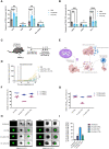
Generation and validation of TCEs targeting CRC cells to encode into oncolytic vaccinia virus as an optimal delivery system. (A) Two novel TCEs were designed by linking a scFv that binds human CEA on the surface of cancer cells to a scFv that recognizes CD3ε on either murine T cells (αCEA:mCD3) or human T cells (αCEA:hCD3) which can be CD4 or CD8 positive. (B) HEK293T cells were transiently transfected with αCEA TCE constructs or pcDNA3.1 empty vector control (EV) and incubated for 48 h. TCE-containing supernatants were collected, spun down to remove cell debris, and concentrated using centrifugal filter units with a 10 kDa molecular weight cutoff. Samples were quantified by BCA assay to load 10 µg of supernatant per lane, separated by SDS-PAGE, and immunoblots were probed with a His antibody to detect His-tagged TCEs. No His tag was detected for the EV control as expected. (C) HT-29 cells were co-cultured with human PBMCs (E:T = 5:1) and αCEA TCE (αCEA:hCD3) or αCEA CTRL (αCEA:mCD3) at indicated concentrations. Resazurin assay was performed at 72 h to determine cancer cell viability after TCE treatment. Results show relative % ± SEM. (D) COLO 205 cells were co-cultured with human PBMCs (E:T = 5:1) and αCEA TCE (αCEA:hCD3) or αCEA CTRL (αCEA:mCD3) at indicated concentrations. Resazurin assay was performed at 72 h to determine cancer cell viability after TCE treatment. Results show relative % ± SEM. (E) In co-cultures with HT-29 cells that express CEA and Jurkat CD69-tdTomato reporter cells (J69; T cells were modified by CRISPR to express tdTomato under the control of the CD69 promoter), the addition of TCEs (1 µg) leads to the visualization of tdTomato-positive Jurkat J69 cells (E:T = 1:1). Scale bar = 400 µm. (F) Three patient-derived CEA-positive colorectal cancer cell lines and other CEA-positive cells (BxPC-3, HT-29, COLO 205) activate J69 cells in co-cultures with αCEA TCE (αCEA:hCD3), but not with αCEA CTRL (αCEA:mCD3) or CEA-negative control cell lines (MIA PaCa-2, HCT 116). Results show MFI ± standard error of the mean (SEM); Two-way ANOVA with Sidak’s correction for multiple comparisons. (G) HT-29, COLO 205, SW620 and MC38WT spheroids were infected at an MOI 1 of oncolytic vaccinia virus (Copenhagen/Cop strain) VV-CTRL, oncolytic Vesicular Stomatitis virus (VSVΔ51), oncolytic Herpes simplex virus (HSV), oncolytic Measles virus (MeV), or oncolytic Adenovirus (AdV). Spheroids were imaged at 48 hpi to detect transgene expression of enhanced green fluorescent protein (eGFP) or red fluorescent protein (RFP). All colorectal cancer spheroids expressed abundant eGFP levels after VV-CTRL infection, compared to other viruses. (H) Spheroid viability was assessed in triplicate by resazurin assay at 120 hpi, relative to uninfected controls. VV-CTRL decreased cell viability of HT-29, COLO205, SW620 and MC38WT spheroids. vaccinia virus was able to infect all the colorectal cancer cell lines as spheroids. VV-CTRL decreased cell viability of HT-29, COLO205, SW620 and MC38WT spheroids. Other viruses also decreased spheroid viability to a lesser extent. Of note, MeV did not significantly change the viability of MC38WT spheroids, as expected, since this oncolytic virus do not infect murine cancer cell lines but served as a control. Results show relative % ± SEM; Two-way ANOVA.
Figure 2
Figure 2
TCEs can activate T cells recruited to the tumour microenvironment by oncolytic vaccinia virus. (A) Two doses of vaccinia B14R-deleted (Cop B14R-) or oncolytic VV-CTRL, but not PBS control, both led to recruitment of CD8-positive T cells into MC38WT tumours at 5 days post-intratumoural injections. In contrast, NK cells (CD3- CD49b+) were not recruited into tumours. Results show the frequency of CD45+ immune cells in % ± SEM, as determined by flow cytometry; Two-way ANOVA. (B) Virally recruited T cells did not express CD69 or CD25 activation markers, and T cells also did not express PD-1 as a marker of T cell exhaustion at this time post-infection. Results show the frequency of CD45+ CD3+ immune cells in % ± SEM, as determined by flow cytometry; Two-way ANOVA. (C) C57BL/6J mice bearing MC38CEA subcutaneous tumours (implanted at 5E5 cells on day 0) were treated with 3 doses of 10 µg of αCEA TCE with or without 3 doses of VV-CTRL at 1E7 pfu at days 6, 8, and 10. (D) Average MC38CEA tumour volumes overtime, showing improved efficacy in decreasing tumour burden with the combination treatment of VV-CTRL and αCEA TCE, compared to PBS control and αCEA TCE alone; Two-way ANOVA. (E) Schematic illustrating a Bispecific-Engager Virus (BEVir), a genetically modified oncolytic vaccinia strain (Cop 3p-5p-/VV-CTRL) encoding one of our validated TCEs targeting CEA and either human or murine T cells. Upon infection of CRC cell lines, the oncolytic vaccinia recruits T cells into the TME that can be activate by TCEs that recognize CEA on uninfected cells. T-cell mediated cell death can lead to a bystander killing effect. (F) Co-cultures with HT-29 cancer cells were infected 24 h after seeding at MOI 0.1. Inoculation medium was removed after 2 h and new medium was added containing human effector cells (E:T = 5:1, or 0:1 as a negative control). Cell viability was assessed by resazurin assay and decreased the most in the presence of human effector cells (PBMCs) and VV-αCEA TCE (αCEA:hCD3), compared to conditions with parental VV-CTRL or VV-αCEA CTRL (αCEA:mCD3). Results show relative % ± SEM. (G) Co-cultures with MC38CEA cancer cells were infected 24 h after seeding at MOI 0.01. Inoculation medium was removed after 2 h and new medium was added containing murine effector cells (E:T = 5:1, or 0:1 as a negative control). Cell viability was assessed by resazurin assay and decreased the most in the presence of murine effector cells (splenocytes) and VV-αCEA TCE (αCEA:mCD3), compared to conditions with parental VV-CTRL or VV-αCEA CTRL (αCEA:hCD3). Results show relative % ± SEM. (H) HT-29-NLuc spheroids were grown in a methylcellulose matrix for 2 d before infecting with VV-CTRL, VV-αCEA TCE (αCEA:hCD3), or VV-αCEA CTRL (αCEA:mCD3). At 48 hpi, we added PBMCs (E:T = 10:1) or no PBMCs (E:T = 0:1) as a control, as indicated. At 96 hpi, spheroids were imaged, and we observed EGFP transgene expression. Scale bar = 500 µm. (I) NLuc release in media, as a surrogate measure for HT-29 cell death, was measured. A significant increase in luminescence was detected for VV-αCEA TCE with PBMCs added, compared to other virus controls and no PBMC conditions. Results show relative % ± SEM.
Figure 3
Figure 3
Antitumour efficacy of VV encoding TCE in a human xenograft model. (A) Athymic nude mice bearing subcutaneous HT-29 tumours were injected intratumourally with 3 doses of viruses at 1E7 pfu on days 19, 20 and 21. Approximately 5 h after virus injections, mice were also co-injected intratumourally with freshly isolated PBMCs at 1E7 per condition, or PBS control, on day 19 only, or co-injected intratumourally with freshly isolated PBMCs at 1E7 per condition, or PBS, on both days 19 and 21. (B) Antitumour efficacies of VV-CTRL, VV-αCEA TCE (αCEA:hCD3), and VV-αCEA CTRL (αCEA:mCD3) viruses were evaluated in combination with one or two doses of PBMCs. Treatment with viruses alone showed significantly prolonged survival (40-50% cures), compared to PBS treatment and regardless of PBMC presence. Highest survival rates were observed in mice treated with VV-αCEA TCE and PBMCs, with 100% survival of mice treated with 3 doses of VV-αCEA TCE and two doses of PBMCs. (C) Mice bearing HT-29 tumours co-injected with VV-αCEA TCE and two doses of PBMCs had the most significant decrease in average tumour volumes. (D) T cells isolated from HT-29 tumours 24 h after the last dose of injection with VV-αCEA TCE and PBMCs showed an increase in expression of the T cell activation marker CD69, while injection of VV-αCEA CTRL had little effect relative to PBS treatment. (E) Vaccinia virus was detected in HT-29 tumours 24 hpi by immunohistochemistry (IHC) in conditions treated with VV-αCEA TCE (αCEA:hCD3) or VV-αCEA CTRL (αCEA:mCD3), but not PBS control. Scale bar = 200 µm. (F) Active caspase-3 was predominantly detected in HT-29 tumours 24 hpi by IHC in conditions co-treated with VV-αCEA TCE (αCEA:hCD3) and PBMCs, compared to other virus and no PMBC conditions. Scale bar = 200 µm.
Figure 4
Figure 4
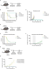
Antitumour efficacy of VV encoding TCE in immunocompetent mouse models leads to cures and immunologic memory. (A) C57BL/6J mice bearing subcutaneous MC38CEA tumours were injected intratumourally with 3 doses of viruses at 1E7 pfu, or PBS as control, at days 6, 8 and 10. (B) Antitumour efficacies of VV-CTRL, VV-αCEA TCE (CEA:mCD3), and VV-αCEA CTRL (αCEA:hCD3) viruses were assessed in immunocompetent mice. Mice bearing MC38CEA tumours treated with viruses showed significantly prolonged survival compared to PBS controls. Of note, mice treated with VV-αCEA TCE were completely cleared of tumours and 100% cured. (C) Mice bearing MC38CEA tumours treated with VV-αCEA TCE (CEA:mCD3) had the most significant decrease in average tumour volumes. (D) C57BL/6J mice bearing subcutaneous MC38WT tumours were injected intratumourally with 3 doses of viruses at 1E7 pfu, or PBS as control, at days 6, 8 and 10. (E) Antitumour efficacies of VV-CTRL, VV-αCEA TCE (CEA:mCD3), and VV-αCEA CTRL (αCEA:hCD3) viruses were assessed in immunocompetent mice. Mice bearing MC38WT tumours treated with virus showed improved survival, but no difference between VV-CTRL, VV-αCEA TCE (CEA:mCD3) groups. (F) Mice bearing MC38WT tumours treated with VV-αCEA TCE (CEA:mCD3) showed no difference in average tumour volumes between VV-CTRL, VV-αCEA TCE (CEA:mCD3) groups. (G) Mice cured of MC38CEA after treatment with VV-αCEA TCE were re-challenged with subcutaneous MC38CEA and MC38WT bilateral tumours (5E5 cells) injected at day 100. (H) All rechallenged mice rejected MC38CEA and MC38WT tumour engraftment compared to naïve mice controls.
Figure 5
Figure 5
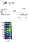
VV encoding TCE leads to abscopal effects and different cytokine profiles in tumours. (A) C57BL/6J mice bearing subcutaneous MC38CEA bilateral tumours were injected unilaterally and intratumourally with 3 doses of viruses at 1E7 pfu, or PBS as control, at days 6, 8 and 10. (B) Mice treated with VV-αCEA TCE (CEA:mCD3) showed increased survival, and 60% of these mice were cured after unilateral treatment, compared to groups treated with PBS, VV-CTRL or VV-αCEA CTRL (CEA:hCD3). (C) Mice treated with VV-αCEA TCE (CEA:mCD3) showed regression of both simultaneously engrafted tumours, compared to groups treated with PBS, VV-CTRL or VV-αCEA CTRL (CEA:hCD3). (D) We performed a murine cytokine assay using tumour lysates from subcutaneous MC38CEA tumours in mice treated with VV-αCEA TCE, VV-CTRL or PBS. Of note, differential expression of cytokines was most evident in tumour samples. We detected more abundant levels of sICAM-1, CXCL10, CXCL1, CCL5, TIMP-1 and TNFα in tumours treated with VV-αCEA TCE, compared to tumours treated with VV-CTRL or PBS. We also identified unique signatures after VV-αCEA TCE treatment, including increased levels of CCL1, IFNγ, IL-1a, IL-1b, TIMP-1, and TREM-1.
Figure 6
Figure 6
Combination viro-immunotherapy for aggressive CRC peritoneal carcinomatosis and lung metastasis models. (A) C57BL/6N mice that express human CTLA4 instead of murine CTLA4 on T cells and bear intraperitoneal MC38CEA tumours, were injected intraperitoneally with 4 doses of viruses at 1E8 pfu, or PBS as control, at days 3, 4, 5 and 6. (B) Antitumour efficacies of the following combinations were assessed compared to PBS alone: VV-αCEA TCE (αCEA:mCD3) on days 3 and 5, in combination with VV-αCEA CTRL (αCEA:hCD3) on days 4 and 6; VV encoding αCTLA4 (which recognizes human CTLA4; VV-αCTLA4) on days 3 and 5, in combination with VV-αCEA CTRL (αCEA:hCD3) on days 4 and 6; VV-αCEA TCE (αCEA:mCD3) on days 3 and 5, in combination with VV-αCTLA4 on days 4 and 6. Mice bearing MC38CEA tumours treated with viruses showed significantly prolonged survival in this aggressive peritoneal carcinomatosis model, and 66% of mice were cured after the combination treatment of VV-αCEA TCE and VV-αCTLA4. (C) Treated mice were dissected upon endpoint or at day 100 for cured mice to confirm no tumours. (D) Following intravenous injection of MC38CEA cells (1E6) in human CTLA4 C57BL/6N mice, we administered 2 doses of viruses at 1E8 pfu, or PBS as control, and harvested the lungs at day 15 for staining. (E) In the CRC metastasis model, VV-αCEA TCE, VV-αFAP TCE, and VV-αCTLA4 monotherapies significantly reduced the number of metastatic nodules in the lungs compared to VV-CTRL or VV-αCEA CTRL, and especially PBS control. Combinations of VV-αCEA TCE with either VV-αFAP TCE or VV-αCTLA4 demonstrated synergy in further decreasing the number of lung metastases. Some of the lungs of the mice treated with VV-αCEA TCE and VV-αCTLA4 were completely cleared of metastases. Results show number of metastatic nodules ± SEM; One-way ANOVA. (F) Images of metastases (white spots) in lungs harvested from mice at day15, after being inflated with india ink and fixed. (G) Schematic illustrating the combination of viro-immunotherapies targeting CEA on cancer cells and FAP on stromal cell populations (CAFs) by TCE, and Tregs through immune checkpoint inhibitor (ICI) αCTLA4 to boost T cell responses for the treatment of CRC.

Similar articles

Cited by

References

    1. Holowatyj AN, Perea J, Lieu CH. Gut instinct: a call to study the biology of early-onset colorectal cancer disparities. Nat Rev Cancer (2021) 21:339–40. doi: 10.1038/s41568-021-00356-y - DOI - PMC - PubMed
    1. Russo M, Crisafulli G, Sogari A, Reilly NM, Arena S, Lamba S, et al. . Adaptive mutability of colorectal cancers in response to targeted therapies. Sci (80- ) (2019) 366:1473–80. doi: 10.1126/science.aav4474 - DOI - PubMed
    1. André T, Shiu K-K, Kim TW, Jensen BV, Jensen LH, Punt C, et al. . Pembrolizumab in Microsatellite-Instability–high advanced colorectal cancer. N Engl J Med (2020) 383:2207–18. doi: 10.1056/nejmoa2017699 - DOI - PubMed
    1. Cercek A, Lumish M, Sinopoli J, Weiss J, Shia J, Lamendola-Essel M, et al. . PD-1 blockade in mismatch repair–deficient, locally advanced rectal cancer. N Engl J Med (2022) 386:2363–76. doi: 10.1056/nejmoa2201445 - DOI - PMC - PubMed
    1. Twumasi-Boateng K, Pettigrew JL, Kwok YYE, Bell JC, Nelson BH. Oncolytic viruses as engineering platforms for combination immunotherapy. Nat Rev Cancer (2018) 18:419–32. doi: 10.1038/s41568-018-0009-4 - DOI - PubMed

Publication types

Substances