. 2022 Nov 18;13(1):7076.
doi: 10.1038/s41467-022-34875-5.

Structural basis for recognition of transcriptional terminator structures by ProQ/FinO domain RNA chaperones

Affiliations

Structural basis for recognition of transcriptional terminator structures by ProQ/FinO domain RNA chaperones

Hyeong Jin Kim et al. Nat Commun. .

Abstract

The ProQ/FinO family of RNA binding proteins mediate sRNA-directed gene regulation throughout gram-negative bacteria. Here, we investigate the structural basis for RNA recognition by ProQ/FinO proteins, through the crystal structure of the ProQ/FinO domain of the Legionella pneumophila DNA uptake regulator, RocC, bound to the transcriptional terminator of its primary partner, the sRNA RocR. The structure reveals specific recognition of the 3' nucleotide of the terminator by a conserved pocket involving a β-turn-α-helix motif, while the hairpin portion of the terminator is recognized by a conserved α-helical N-cap motif. Structure-guided mutagenesis reveals key RNA contact residues that are critical for RocC/RocR to repress the uptake of environmental DNA in L. pneumophila. Structural analysis and RNA binding studies reveal that other ProQ/FinO domains also recognize related transcriptional terminators with different specificities for the length of the 3' ssRNA tail.


Conflict of interest statement

The authors declare no competing interests.

Figures

Fig. 1
Fig. 1. Defining the determinants of RocC/RocR interaction in vitro and in vivo.
a Predicted folding of wild-type RocRSL3 using RNAfold web server. b EMSA binding assay for RocC1-126, RocC14-126, and RocC24-126 vs 5’ radiolabeled RocRSL3 (n = 3). Binding affinity of RocRSL3 with different tail lengths was tested with RocC14-126. The error bars are standard error of the mean (SEM). c Role of the N-terminus of RocC for RocRSL3 binding in vitro and for uptake of DNA (transformation) in vivo. The transformation was assessed in Legionella strains containing either WT RocC, a mutant in which rocC translation is disrupted (rocCTAA), a RocR deletion (ΔrocR), or different deletions at the N-terminus (RocCΔN14—deletion of a.a. 1–13; RocCΔN19 – deletion of a.a. 1-18; RocCΔN24—deletion of a.a. 1−23). Binding measurements were carried out with a fluorescence polarization (FP) assay using RocC1−126 or the indicated N-terminal deletion mutants, with FAM-labeled RocRSL3 as a target. The orange histograms indicate the KD values measured by FP (n = 3) and the blue histograms indicate the transformability of the indicated mutant or strain. Transformability is the ratio of the number of CFUs counted on selective medium divided by the number of CFUs counted on a non-selective medium. NS indicates mutants where RNA binding could not be detected. # indicates a mutant that was not tested in vitro. The error bars are the standard error of the mean (SEM). Transformation experiments were repeated at least twice on two independent clones of each mutant and with two types of donor DNA, FP experiments were repeated three times. d EMSA binding assay for RocC14−126 with 5’ radiolabeled RocRSL3 with various substitution mutants. The error bars are the standard error of the mean (SEM) (n = 3). Source data are provided as a Source Data file.
Fig. 2
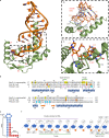
Fig. 2. Structural analysis of the ProQ/FinO domain of RocC bound to a RocRSL3 variant.
a Crystal structure of RocC14−126/RocR9bp-tet complex. Dotted boxes indicate the main interactions between protein–RNA. b, c are zoomed-in views from (a). Black dotted lines indicate hydrogen bonding between protein and RNA. Green dotted lines indicate hydrogen bonding for the base pairing. d Structure-based sequence alignment of the ProQ/FinO domains of RocC, FinO, and ProQ. Yellow highlights in RocC indicate residues in contact with RocR9bp-tet in the crystal structure. Asterisks indicate highly conserved residues across the three proteins. Orange highlights in the FinO sequence indicate residues that show strong cross-linking with SLII of FinP. Magenta letters in the ProQ sequence show residues that are critical for RNA binding in 3-hybrid screening. Cyan highlights in ProQ indicate vital residues for ProQ function. The indicated secondary structure is derived from the RocC/RocR crystal structure. e Schematic diagram of the RocR9bp-tet variant, which is crystallized with RocC. The red circles indicate the nucleotides in direct contact with the protein. f Schematic diagram of RocC-RocR interactions. The purple line indicates the region of double-stranded RNA structure; green lines indicate protein motifs in contact with RNA. Dotted lines show molecular interactions between protein and RNA.
Fig. 3
Fig. 3. Measurement of binding interactions between RocC14−126 and either RocR9bp-tet or a single-stranded RNA.
a ITC analysis of RocC14-126 with RocR9bp-tet. b ITC analysis of RocC14−126 with a 10 nucleotide single-stranded RNA. c ITC analysis of RocC14−126 with RocRSL3 containing a terminal 3’-phosphate (RocRP). ac Each experiment was repeated independently three times with similar results. Source data are provided as a Source Data file.
Fig. 4
Fig. 4. Effects of RocC point mutations on RocR binding in vitro and transformability in vivo.
a FP binding assay for 5’ FAM-labeled RocRSL3 with different RocC14−126 point mutants (n = 3). The error bars are standard error of the mean (SEM). b, c Surface representation of RocC14−126 alone (b) and with RocR (c) colored to indicate the positions of mutated residues. d A graph displaying RNA-binding affinities and transformation efficiencies for the indicated strains and RocC mutants are shown. Orange histograms indicate the KD values measured by FP (n = 3), and the blue histograms indicate the relative transformability of the indicated mutant or strain. Transformability is the ratio of the number of CFUs counted on selective medium divided by the number of CFUs counted on non-selective medium. # indicates a mutant which could not be purified due to low protein solubility. ND indicates mutants where RNA binding could not be detected. Values for the individual transformation measurements are shown (experiments were repeated at least twice on two independent clones), and the standard error of the mean (SEM) from three independent measurements are show for the FP binding data. Source data are provided as a Source Data file.
Fig. 5
Fig. 5. Specificities for 3’ tail length among FinO domain-containing proteins.
ac FP binding assay for various tail lengths of 5’ FAM-labeled RocRSL3 with RocC14-126 (n = 3). a With FinO45-186 (b), or with ProQ1-130. ND indicates mutants where RNA binding was too weak to determine a KD. ND indicates mutants where RNA binding could not be detected. The error bars are the standard error of the mean (SEM). d Models for the interaction of RNAs with different 3’ single-stranded tail lengths to RocC14−126. Left panel: RNA with a 3-nucleotide tail. Center panel: RNA with a 5-nucleotide tail (as in RocR). Right panel: RNA with an 8-nucleotide tail. Source data are provided as a Source Data file.
Fig. 6
Fig. 6. ProQ/FinO domain proteins bind transcriptional terminator structures to regulate RNA-RNA interactions.
a Many ProQ/FinO domain proteins, such as RocC, facilitate in trans RNA association between sRNAs and target mRNAs. The ProQ/FinO domain specifically binds the transcriptional terminator of the RocR sRNA, stabilizing it against degradation. Key to RNA-RNA association is the recognition of the sRNA seed sequence with its complementary region (the RocR box) in the target mRNA. The C-terminal region of RocC is required to facilitate the recognition and translational repression of target mRNAs such as comEA. b Many plasmid-encoded ProQ/FinO domain proteins, such as FinO, regulate in cis sense-antisense RNA interactions. Similar to RocC, the ProQ/FinO domain of FinO also stabilizes the antisense RNA FinP against degradation. Initial RNA-RNA interactions are thought to involve loop–loop kissing interactions, which then proceed to duplex formation between the two RNAs, resulting in translational repression of the traJ mRNA target. In this case, the flexible N-terminal region of FinO is thought to be key in facilitating RNA-RNA interactions.

Similar articles

Cited by

References

    1. Gottesman S, Storz G. Bacterial small RNA regulators: versatile roles and rapidly evolving variations. Cold Spring Harb. Perspect. Biol. 2011;3:1–16. doi: 10.1101/cshperspect.a003798. - DOI - PMC - PubMed
    1. Vogel J, Luisi BF. Hfq and its constellation of RNA. Nat. Rev. Microbiol. 2011;9:578–589. doi: 10.1038/nrmicro2615. - DOI - PMC - PubMed
    1. Updegrove TB, Zhang A, Storz G. Hfq: The flexible RNA matchmaker. Curr. Opin. Microbiol. 2016;30:133–138. doi: 10.1016/j.mib.2016.02.003. - DOI - PMC - PubMed
    1. Santiago-Frangos A, Woodson SA. Hfq chaperone brings speed dating to bacterial sRNA. Wiley Interdiscip. Rev. RNA. 2018;9:1–16. doi: 10.1002/wrna.1475. - DOI - PMC - PubMed
    1. Mark Glover JN, et al. The FinO family of bacterial RNA chaperones. Plasmid. 2015;78:79–87. doi: 10.1016/j.plasmid.2014.07.003. - DOI - PubMed

Publication types

MeSH terms

Substances