Review
. 2022 Nov 15;135(22):jcs259769.
doi: 10.1242/jcs.259769. Epub 2022 Nov 18.

The mechanical cell - the role of force dependencies in synchronising protein interaction networks

Affiliations
Review

The mechanical cell - the role of force dependencies in synchronising protein interaction networks

Benjamin T Goult et al. J Cell Sci. .

Abstract

The role of mechanical signals in the proper functioning of organisms is increasingly recognised, and every cell senses physical forces and responds to them. These forces are generated both from outside the cell or via the sophisticated force-generation machinery of the cell, the cytoskeleton. All regions of the cell are connected via mechanical linkages, enabling the whole cell to function as a mechanical system. In this Review, we define some of the key concepts of how this machinery functions, highlighting the critical requirement for mechanosensory proteins, and conceptualise the coupling of mechanical linkages to mechanochemical switches that enables forces to be converted into biological signals. These mechanical couplings provide a mechanism for how mechanical crosstalk might coordinate the entire cell, its neighbours, extending into whole collections of cells, in tissues and in organs, and ultimately in the coordination and operation of entire organisms. Consequently, many diseases manifest through defects in this machinery, which we map onto schematics of the mechanical linkages within a cell. This mapping approach paves the way for the identification of additional linkages between mechanosignalling pathways and so might identify treatments for diseases, where mechanical connections are affected by mutations or where individual force-regulated components are defective.

Keywords: Cytoskeleton; Disease-associated mutations; Force-dependent switches; Mechanosensing; Mechanotransduction; Motor proteins; Talin.


Conflict of interest statement

Competing interests The authors declare no competing or financial interests.

Figures

Fig. 1.
Fig. 1.
The complex mechanical linkages that scaffold and form the cells machinery. An artistic illustration of a cell connected to a neighbouring cell (bottom) and tightly connected to the extracellular matrix (light grey; right). The matrix contains fibrous molecules, such as collagen and fibronectin, and also flexible molecules, such as hyaluronan. On the intracellular side, cellular components are tightly interlinked, and the mechanical connections formed by the cytoskeleton connect the cell membrane, cellular organelles and the nucleus. (1–3) Three of the mechanosensory complexes that connect the exterior of the cell to the cytoskeleton. (1) the integrin-mediated focal adhesions, (2) the cadherin-mediated adherens junctions and (3) the desmosome. Two of the three major cytoskeletal systems, actin and microtubules are shown and these form mechanical linkages that couple complexes 1–3 to each other and to cellular organelles, such as the mitochondria, and via the LINC complex to the nucleus (4). Mechanosensitive proteins localised at the ends of these linkages provide an array of binary switches, indicated by a ‘1/0’ and a green ‘light switch’, that can be operated by the force-generation machinery of the cell. The mechanical coupling of disparate parts of the cell enables long-range communication both within and between cells and so we present the idea of the cell as a complex array of interconnected, mechanically-operated switches functioning as a machine. Illustration generated by Iiris Mustonen, Tampere University, Finland.
Fig. 2.
Fig. 2.
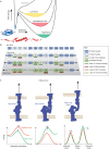
Mechanical load applied on a protein modulates the free energy landscape of folding. (A) Mechanoregulated proteins can be considered as mechanoswitches, adopting multiple metastable states. In the absence of mechanical load, a protein folds into a certain well-defined structure (blue); however, other conformational states can be populated upon mechanical load (shown in red). In the presence of force, these states can be energetically equally favourable, or even lower in terms of the free energy compared to the native state as illustrated by the free-energy landscape shown. At low force (yellow), thermodynamics still favours the relaxed (blue) conformation, but the probability for transitioning to the partially unfolded state (red) is already high. In such a landscape, a coexistence force condition (red) can exist, where the relaxed and partially unfolded states are both expected to be populated with similar probability. Finally, above a certain force threshold (green), the partially unfolded conformation is the energetically favoured state. At high force, the protein will become fully unfolded. (B) Talin has a highly complex free-energy landscape, as its 13 rod domains (R1–R13) all exhibit this switch-like behaviour in response to mechanical force. (C) The architecture of a bond defines its behaviour under mechanical load. Three different interaction mechanisms are illustrated. In the case of a slip bond (left), the lifetime of the bond decreases when mechanical load is applied and can be seen as a decrease of activation energy required for the dissociation. In the case of catch bond (middle), the bond lifetime increases under mechanical load, observed as an increase in the activation energy needed for the dissociation event. In the case of large biological macromolecules such as proteins, the bonds often display a catch–slip bond behaviour (right), in that they exhibit catch bond behaviour under low force, but increased force triggers slip-bond behaviour. In the case of the energy landscape for catch–slip bonds, two force-dependent unbinding pathways have to be considered: at low forces, the catch-bond behaviour is dominant, resulting in an increase in the activation energy (Ea) under applied load (unbinding to the left in the energy diagram), whereas at higher forces, the slip-bond behaviour leads to decrease in the activation energy (unbinding to the right). Panel A reprinted with permission from Stannard et al. (2021). Copyright 2021 American Chemical Society. Panel B reprinted from Mykuliak et al. (2018) where it was published under a CC-BY 4.0 license. Panel C reproduced from Huppa and Schütz (2016) with permission from Elsevier.
Fig. 3.
Fig. 3.
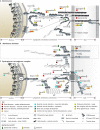
The mechanical linkages between the extracellular matrix and the nucleus. A simplified schematic of the linkages that a cell makes, via its integrin receptors, to the extracellular matrix (right). The ECM components, collagen, fibronectin, laminin and fibrinogen are shown, which are recognised by different integrin isoforms. Middle, on the cytoplasmic face of the integrin–ECM linkages, complex adhesive structures assemble, which are dynamic and mechanoresponsive. Left, the cytoskeletal connections emanating from the adhesion sites directly couple to nuclear envelope proteins, providing a direct mechanical coupling between the outside of the cell and the nucleus. Signals from the surface of the nucleus are propagated into the nucleus to alter the expression patterns and accessibility of genes. The association of the proteins with various diseases is indicated with the colour code as defined in the Key (top section). We stress that the disease associations presented are not exhaustive, particularly as new associations are being discovered all the time. However, even with this abridged dataset, the disease mappings clearly highlight the central role the mechanical machinery has in the correct functioning of the cell and how defects in the balance of these systems at the cellular level manifest as diverse disease states at the organismal level. Key: the protein domains involved for assembling mechanical linkages are defined in the key (middle section). Domains not indicated are as follows: CH, calponin homology; KASH, KASH domain; P, plectin homology. The symbols used are based on the structural features of the domains employing the structural data available in the Protein Data Bank (see also Box 2). The colour of the switch symbol for a protein (lower section) indicates the currently available evidence for mechanical switch properties. For details, please see Tables S1–S3.
Fig. 4.
Fig. 4.
Mechanical linkages the cell makes to the outside world. (A) Desmosomes are specialised adhesive protein complexes responsible for maintaining the mechanical integrity of tissues. Complex protein networks link the ECM and neighbouring cells with the nucleus and cytoskeletal components. (B) The membrane skeleton is a specialised part of the cytoskeleton in close proximity of the cell membrane with a unique protein composition. (C) The dystroglycan–sarcoglycan complex forms a critical link between the cytoskeleton and ECM. The association of the proteins shown with various diseases is indicated with the colour code as defined in the Key (top section). Key: the protein domains involved for assembling mechanical linkages are defined in the key (middle section). Domains not indicated are as follows: ANK, ankyrin repeat; C, cadherin repeat; SU, calponin binding; Death, death domain; D, Desmoglein repeat; DG, dynamin type G domain; KASH, KASH domain; GED, GTPase effector domain; GAR, microtubule-binding domain; PDZ, PDZ domain; PH, Pleckstrin homology; P, plectin homology; SH3, SH3 domain; WW, WW domain; ZU5, ZU5 domain. IF, intermediate filaments; MT, microtubules. For details, please see Tables S1–S3.

Similar articles

Cited by

References

    1. Acsadi, G., Moore, S. A., Chéron, A., Delalande, O., Bennett, L., Kupsky, W., El-Baba, M., Le Rumeur, E. and Hubert, J. F. (2012). Novel mutation in spectrin-like repeat 1 of dystrophin central domain causes protein misfolding and mild Becker muscular dystrophy. J. Biol. Chem. 287, 18153-18162. 10.1074/jbc.M111.284521 - DOI - PMC - PubMed
    1. Amberger, J. S., Bocchini, C. A., Scott, A. F. and , Hamosh, A. (2019). OMIM.org: leveraging knowledge across phenotype-gene relationships. Nucleic Acids Res. 47, D1038-D1043. - PMC - PubMed
    1. Atherton, P., Lausecker, F., Carisey, A., Gilmore, A., Critchley, D., Barsukov, I. and Ballestrem, C. (2020). Relief of talin autoinhibition triggers a force-independent association with vinculin. J. Cell Biol. 219, e201903134. 10.1083/jcb.201903134 - DOI - PMC - PubMed
    1. Azizi, L., Cowell, A. R., Mykuliak, V. V., Goult, B. T., Turkki, P. and Hytönen, V. P. (2021). Cancer associated talin point mutations disorganise cell adhesion and migration. Sci. Rep. 11, 347. 10.1038/s41598-020-77911-4 - DOI - PMC - PubMed
    1. Azizi, L., Varela, L., Turkki, P., Mykuliak, V. V., Korpela, S., Ihalainen, T., Church, J., Hytonen, V. P. and Goult, B. T. (2022). Talin variant P229S compromises integrin activation and associates with multifaceted clinical symptoms. Hum. Mol. Genet. ddac163. - PMC - PubMed

Publication types

MeSH terms