. 2022 Dec;54(12):1933-1945.
doi: 10.1038/s41588-022-01214-9. Epub 2022 Nov 17.

Retrotransposon activation during Drosophila metamorphosis conditions adult antiviral responses

Affiliations

Retrotransposon activation during Drosophila metamorphosis conditions adult antiviral responses

Lu Wang et al. Nat Genet. 2022 Dec.

Abstract

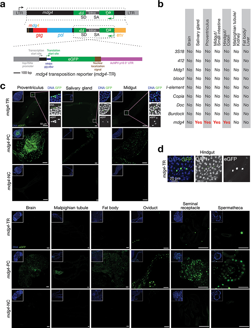
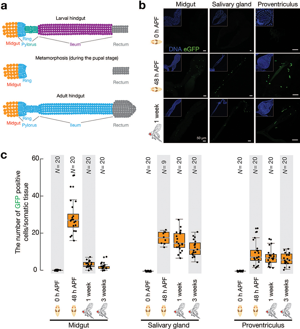
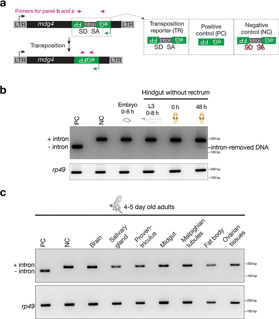
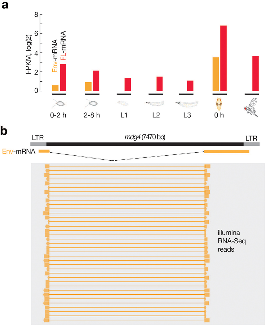
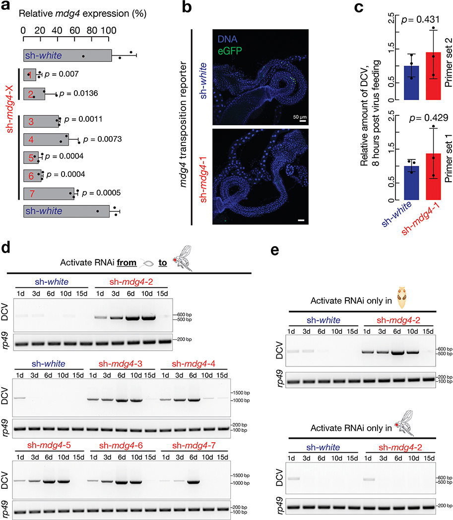
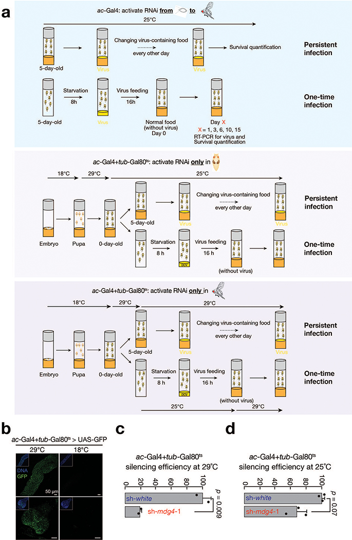
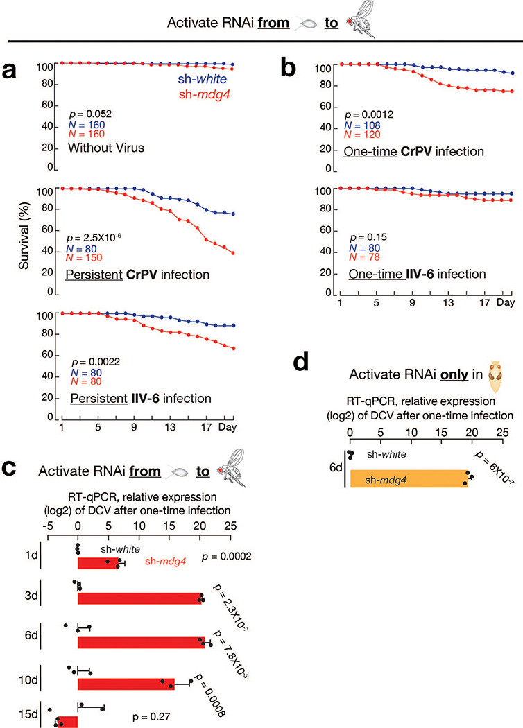
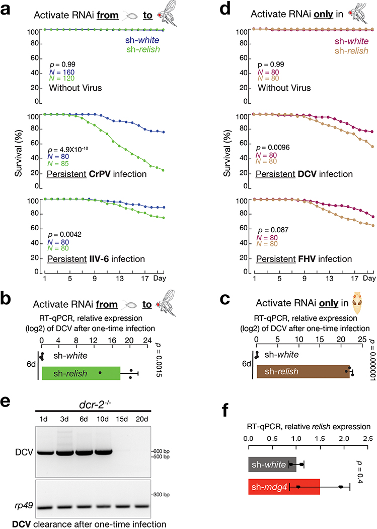
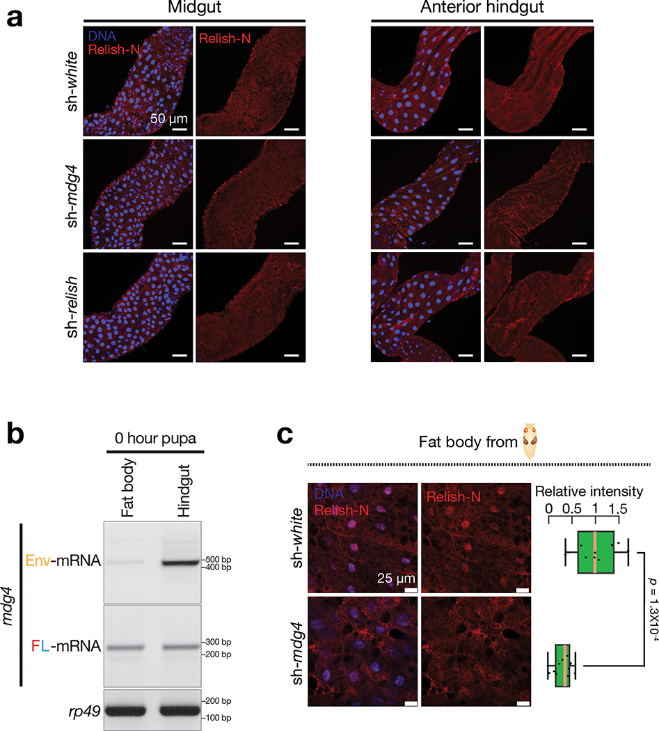
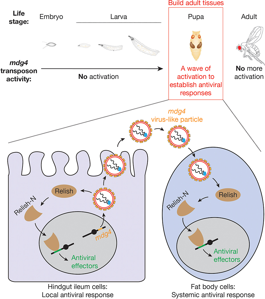
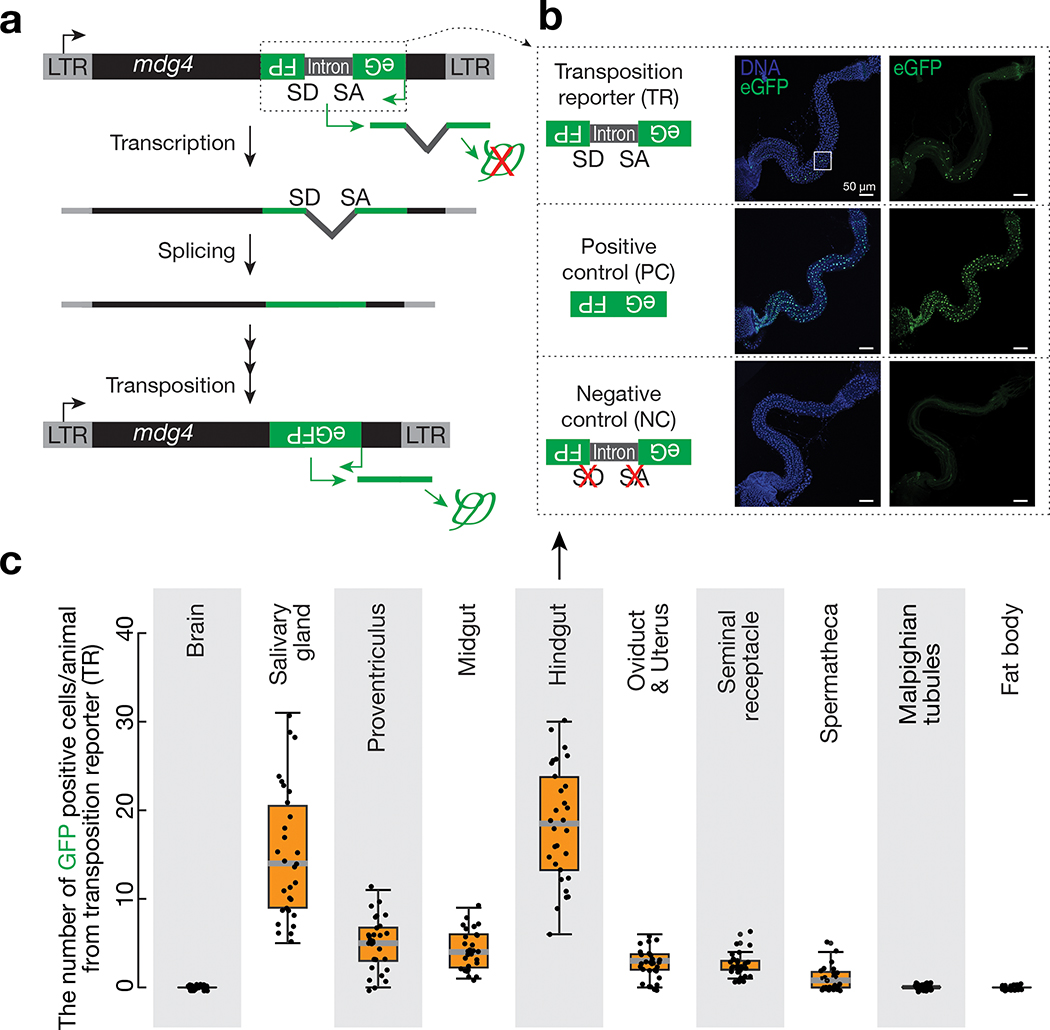
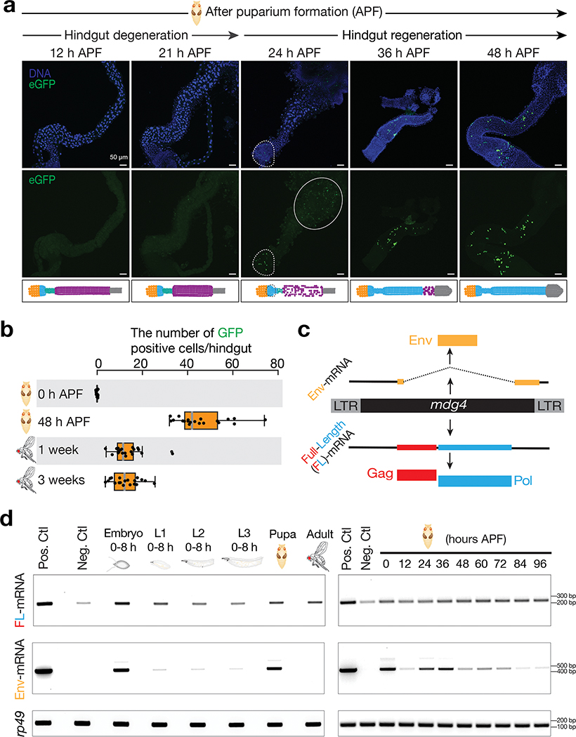
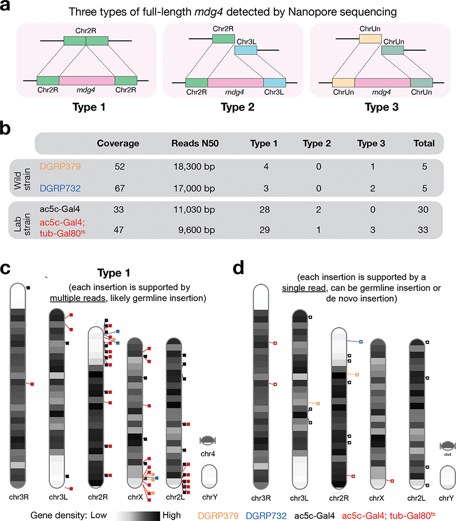
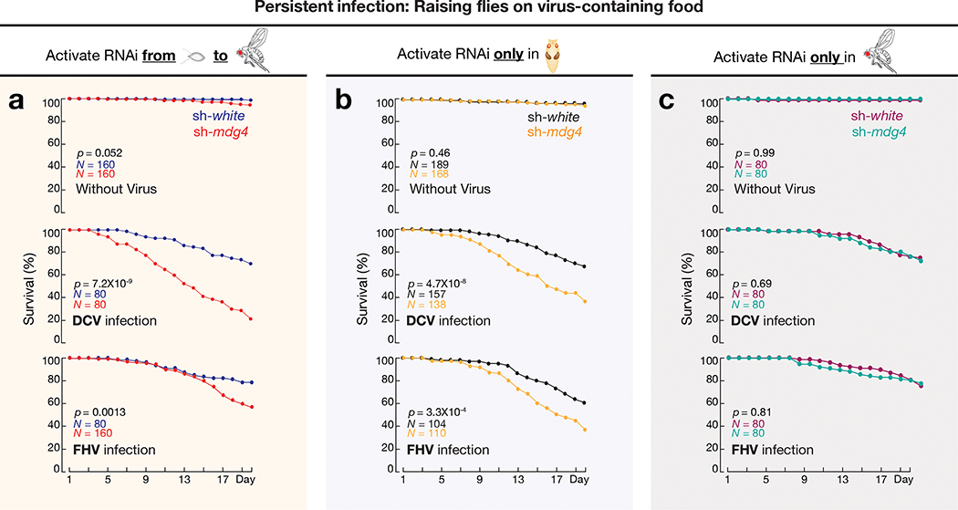
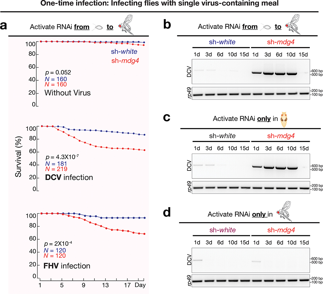
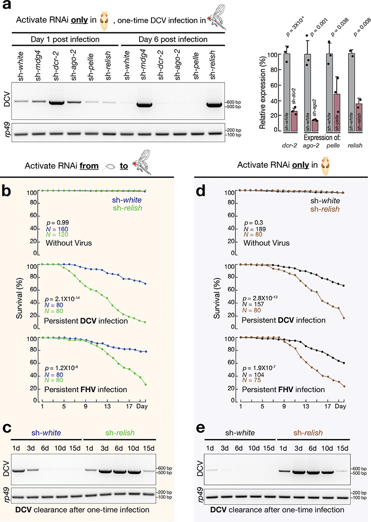
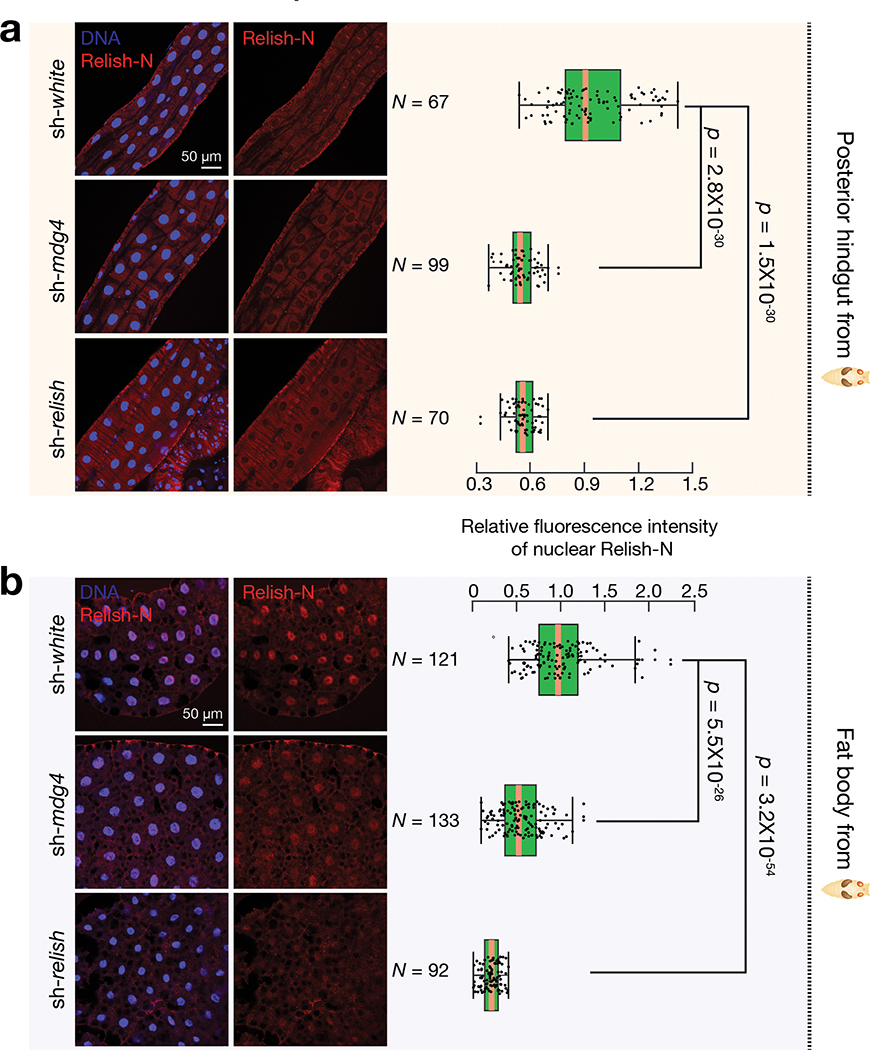
Retrotransposons are one type of mobile genetic element that abundantly reside in the genomes of nearly all animals. Their uncontrolled activation is linked to sterility, cancer and other pathologies, thereby being largely considered detrimental. Here we report that, within a specific time window of development, retrotransposon activation can license the host's immune system for future antiviral responses. We found that the mdg4 (also known as Gypsy) retrotransposon selectively becomes active during metamorphosis at the Drosophila pupal stage. At this stage, mdg4 activation educates the host's innate immune system by inducing the systemic antiviral function of the nuclear factor-κB protein Relish in a dSTING-dependent manner. Consequently, adult flies with mdg4, Relish or dSTING silenced at the pupal stage are unable to clear exogenous viruses and succumb to viral infection. Altogether, our data reveal that hosts can establish a protective antiviral response that endows a long-term benefit in pathogen warfare due to the developmental activation of mobile genetic elements.


Conflict of interest statement

COMPETING INTERESTS

The authors declare no competing interests.

Figures

Extended Data Fig. 1 |
Extended Data Fig. 1 |. Monitoring retrotransposon mobilization in somatic cells via a transposition reporter.
a, Detailed schematic design of eGFP transposition reporter to monitor mdg4 mobilization. b, Summary of mobilization events from different somatic tissues for 9 retrotransposon families, as assayed by corresponding eGFP reporter. No: no eGFP positive cells are detected; Yes: eGFP positive cells can be detected. c, Detecting eGFP signals in somatic tissues from positive control, negative control, and mdg4 transposition reporter in 2–4-day-old adult flies. Note: Positive control construct gives low number of eGFP positive cells in brain and malpighian tubules, indicating that transcription of mdg4 is suppressed in these tissues. Three independent biological replicates were performed. d, Zoom-in display of the box region in Fig. 1b. In DAPI channels, green arrows point to the nuclei that have GFP expression.
Extended Data Fig. 2 |
Extended Data Fig. 2 |. mdg4 selectively mobilizes in the regenerating tissues during metamorphosis.
a, Schematic of Drosophila hindgut. Both larval and adult hindgut include the pylorus, ileum and rectum. During pupal stage metamorphosis, the pylorus and ileum from larval stage degenerate; the anterior part of pylorus (ring) regenerates to produce adult pylorus and ileum. b, Detecting eGFP positive cells from mdg4 transposition reporter in midgut, salivary gland and proventriculus at different stages. c, The box plot shows the number of eGFP positive cells from mdg4 transposition reporter in midgut, salivary gland and proventriculus.
Extended Data Fig. 3 |
Extended Data Fig. 3 |. Probing transposition events by PCR.
a, Transposition events generate intron-removed DNA, which produces a short PCR product. b, Probing mobilization events at different developmental stages. Only the DNA from 48 hours pupal hindguts can harbor enough mobilization events to be detected by this PCR assay. c, Probing mobilization events from different adult tissues. These tissues either have no––or too few––mobilization events to be detected by this method. Three independent biological replicates were performed for b and c.
Extended Data Fig. 4 |
Extended Data Fig. 4 |. RNA-Seq to measure transcripts from mdg4.
a, Bar graph to display the abundance of full-length and Env mRNAs from mdg4. Full-length mdg4 transcripts are constantly expressed at all stages. Env mRNAs can be detected from early stage embryos and pupal stage, but not adult stage. b, IGV browser screenshot to display the representative sequencing reads that support the expression of Env mRNAs.
Extended Data Fig. 5 |
Extended Data Fig. 5 |. Multiple RNAi constructs were designed to silence mdg4.
a, RT-qPCR to quantify the expression of mdg4 upon suppression by using one of the 7 RNAi constructs based on the two-tailed t-test. Each sh-RNA construct was driven by ac-Gal4. Flies were raised at 25°C and newly eclosed flies were used to extract RNA. Data are normalized to rp49 (RpL32) expression; the bars report mean ± standard deviation for three biological replicates (apply to all RT-qPCR data from this manuscript). b, Visualizing mdg4 transposition events (GFP positive cells) in hindgut from either white or mdg4 suppressed 2–4-day-old adults. Flies carrying sh-mdg4-5 construct were used for Fig. 6. sh-mdg4-1 was used in the rest of the experiments. The findings from it were validated by using other constructs (shown in Extended Data Fig. 11). c, RT-qPCR to measure the amount of DCV from fly bodies after feeding animals virus for 8 hours based on the two-tailed t-test. Data are normalized to rp49 (RpL32) expression; the bars report mean ± standard deviation for three biological replicates. d, By activating mdg4 RNAi from embryonic to adult stage, RT-PCR experiments were performed to monitor the amount of DCV in fly bodies after one-time infection. Together with Extended Data Fig. 5a, these data indicate RNAi efficiency negatively correlates with the robustness of antiviral response. e, By using sh-mdg4-2 to silence mdg4 at the stage specific manner, similar findings were made as Fig. 5. Three independent biological replicates were performed for sh-mdg4-2 in d and e, one time experiment was performed for sh-mdg4-3, 4, 5, 6 and 7 in d.
Extended Data Fig. 6 |
Extended Data Fig. 6 |. Schematic design of the virus-feeding assays used in this study.
a, Top panel: Schematic design to achieve RNAi from embryonic to adult stage. Middle panel: Schematic design to achieve RNAi only at pupal stage. At lower temperature, Gal80 inhibits Gal4 activity. At 29°C, Gal80 becomes inactive and cannot suppress Gal4. Bottom panel: Schematic design to achieve RNAi only at adult stage. b, Validating ac-Gal4+tub-Gal80ts system by driving UAS-GFP expression at high temperature (29°C) and low temperature (18°C) in larval midgut and proventriculus. This experiment was only performed once. c, RT-qPCR to measure the mdg4 silencing efficiency for the ac-Gal4+tub-Gal80ts system at 29°C based on the two-tailed t-test. Newly eclosed flies raised at 29°C during pupal stage were used to extract RNA. Data are normalized to rp49 (RpL32) expression; the bars report mean ± standard deviation for two biological replicates. d, RT-qPCR to measure the mdg4 silencing efficiency for the ac-Gal4+tub-Gal80ts system at 25°C based on the two-tailed t-test. Adult flies being shifted to 25°C for 5 days were used to extract RNA. Data are normalized to rp49 (RpL32) expression; the bars report mean ± standard deviation for three biological replicates.
Extended Data Fig. 7 |
Extended Data Fig. 7 |. mdg4 activation renders hosts protection from virus infection.
a, By activating mdg4 RNAi from embryonic to adult stage, the survival rates were measured by raising flies on CrPV- or IIV-6-containing food for 20 days. sh-white flies served as controls. b, By activating mdg4 RNAi from embryonic to adult stage, the survival rates were measured after infecting adult files with different viruses with a single meal. c, RT-qPCR to quantify the fold change of DCV mRNA in sh-mdg4 flies at different time points after one-time infection, relative to sh-white controls based on the two-tailed t-test. d, RT-qPCR to quantify the fold change of DCV mRNA in sh-mdg4 flies on day 6 after one-time infection, relative to sh-white controls based on the two-tailed t-test. The bars in panel c and d report mean ± standard deviation for three biological replicates. Comparison of survival curves was completed using a Cox proportional-hazards model for panels a and b.
Extended Data Fig. 8 |
Extended Data Fig. 8 |. Relish activation renders hosts protection from virus infection.
a, By activating Relish RNAi from embryonic to adult stage, the survival rates were measured by raising flies on CrPV- or IIV-6-containing food for 20 days. sh-white flies served as controls. b, RT-qPCR to quantify the fold changes of DCV mRNA in sh-relish flies on day 6 after one-time infection, relative to sh-white controls based on the two-tailed t-test. c, RT-qPCR to quantify the fold changes of DCV mRNA in sh-relish flies on day 6 after one-time infection, relative to sh-white controls based on the two-tailed t-test. The bars in panel B and C report standard deviation for three biological replicates. d, By activating Relish RNAi only at adult stage, the survival rates were measured by raising flies on DCV- or FHV-containing food for 20 days. sh-white flies served as controls. e, RT-PCR experiments to monitor the amount of DCV in adult dcr-2 mutant (dcr-2L811fsX) flies after one-time infection. Two independent biological replicates were performed. f, RT-qPCR to quantify relish expression upon mdg4 depletion in fly pupae. Data are normalized to rp49 (RpL32) expression; the bars report mean ± standard deviation for 2 biological replicates. Expression was compared using a two-tailed t-test. Comparison of survival curves was completed using a Cox proportional-hazards model for panels a and d.
Extended Data Fig. 9 |
Extended Data Fig. 9 |. mdg4 products promote the translocation of Relish-N into nucleus.
a, By performing immuno-staining with the Relish-N antibody, which can detect both full-length and N-terminal fragment of Relish, very low–if any–signals were detected in midgut and anterior part of hindgut from sh-white, sh-mdg4, or sh-relish early pupae. b, RT-PCR to examine the levels of mdg4 full-length and Env transcripts in fat body cells. Two independent biological replicates were performed for a and b. c, Immuno-staining to detect the nuclear Relish-N signals in the fat body cells from sh-white and sh-mdg4 early pupae. While the animals for Fig. 7 were raised in germ-free condition, the pupae examined for this figure were non-germ-free. In both conditions, silencing mdg4 resulted in a significant decrease of the nuclear Relish-N in fat body cells. Box plots report the minimum, maximum, median, and interquartile ranges of the data. A two-tailed t-test was used to compare the relative intensities of each genotype. The data were collected from 3 individual animals per genotype.
Extended Data Fig. 10 |
Extended Data Fig. 10 |. A model to depict the activity of transposon during animal development and how its activation prepares the host for antiviral responses.
Our data indicate that the mdg4 retrotransposon selectively becomes active during metamorphosis to prepare the antiviral responses.
Fig. 1 |
Fig. 1 |. Monitoring mdg4 mobilization in somatic cells via a transposition reporter.
a, Schematic design of an eGFP reporter to monitor mdg4 mobilization. The eGFP reporter is inserted in the non-coding sequence of mdg4 in the antisense orientation. eGFP is disrupted by an intron, which is in the same direction as mdg4 but opposite direction as eGFP. The intron cannot be spliced during eGFP transcription. mdg4 mobilization, which generates a new copy of the transgene without the disrupting intron, results in eGFP expression. b, mdg4 transposition reporter produces eGFP in hindgut cells in 2–4-day-old adult flies. An intronless construct is used as a positive control to test the potential sensitivity. A construct with mutated splicing acceptor and donor sites serves as a negative control. The boxed region is displayed in Extended Data Figure 1d to show eGFP signals are from nuclei (eGFP has nuclear localization signal sequences). c, The box plot shows the number of eGFP positive cells from mdg4 transposition reporter among different tissues from 2–4-day-old adult flies (N = 30). Box plots report the minimum, maximum, median, and interquartile ranges of the data.
Fig. 2 |
Fig. 2 |. mdg4 selectively mobilizes in the regenerating tissues during metamorphosis.
a, mdg4 mobilization in Drosophila during hindgut development at the pupal stage via eGFP transposition reporter. No eGFP is expressed during hindgut degeneration (12 h and 21 h APF). eGFP positive cells can be detected in the newly formed hindgut (24 h, 36 h, and 48 h APF). Dashed circle highlights eGFP signals. Solid circle depicts autofluorescence from the dying cells. Extended Data Figure 2a depicts the cell-type dynamics of hindgut during metamorphosis. b, The box plot shows the number of eGFP-positive cells per fly from mdg4 transposition reporter at different stages (N = 20). Box plots report the minimum, maximum, median, and interquartile ranges of the data. c, Diagram depicts the transcripts and proteins from mdg4. d, RT-PCR experiments to measure the expression of full-length and Env mRNAs from reporter-carrying flies. Pos. Ctl.: positive control, ovaries with Piwi being depleted in follicle cells. Neg. Ctl.: negative control, ovaries with mdg4 being depleted in both germ cells and follicle cells. APF: after puparium formation. Similar findings were made when using RNA-seq to quantify the full-length and Env mRNAs from endogenous mdg4 (Extended Data Fig. 4). Three independent biological replicates were performed.
Fig. 3 |
Fig. 3 |. Nanopore sequencing detected full-length copies of mdg4 from both wild and laboratory strains.
a, Cartoon display of 3 types of full-length mdg4 detected. Type 1: Full-length mdg4 flanked by coordinated sequences from the reference genome. Type 2: Full-length mdg4 flanked by discordant sequences from the reference genome, likely reflecting the polymorphisms between the reference genome and the genome of individual strain. Type 3: Full-length mdg4 flanked unassembled genome sequences. b, A table to summarize the sequencing depth, N50 of the sequencing reads, and the number of full-length mdg4 detected from each strain. c, Chromosome ideogram to display the chromosomal positions of type 1 full-length mdg4. Each event reported in panels b and c is supported multiple reads, likely reporting germline mdg4 copies. d, Chromosome ideogram to display the chromosomal positions of full-length mdg4 detected by a single read, likely reporting de novo insertions or germline insertions with low regional coverage.
Fig. 4 |
Fig. 4 |. mdg4 activation at pupal stage safeguards adult flies upon persistent viral infections.
a, By activating mdg4 RNAi from embryonic to adult stage, the survival rates were measured by raising flies on virus-containing food for 20 days. sh-white flies served as controls. All viral infection assays from this manuscript were performed with at least two biological replicates. Each replicate contains 20 males and 20 females. b, With mdg4 only silenced at pupal stage, the survival rates were measured by raising flies on virus-containing food for 20 days. c, With mdg4 only suppressed at adult stage, the survival rates were measured by raising flies on virus-containing food for 20 days. Flies used in panel a have one copy of the mdg4 reporter. Flies used in panels b and c only have endogenous copies of mdg4. Comparison of survival curves for all panels was completed using a Cox proportional-hazards model.
Fig. 5 |
Fig. 5 |. mdg4 activation at pupal stage safeguards adult flies upon one-time viral infections.
a, By activating mdg4 RNAi from embryonic to adult stage, the survival rates were measured after infecting adult files with DCV or FHV from single meal. Comparison of survival curves was completed using a Cox proportional-hazards model. b, By activating mdg4 RNAi from embryonic to adult stage, RT-PCR experiments were performed to monitor the amount of DCV in fly bodies after one-time infection. c, With mdg4 only silenced at pupal stage, RT-PCR experiments were performed to monitor the amount of DCV in adult flies after one-time infection. d, With mdg4 only silenced at adult stage, RT-PCR experiments were performed to monitor the amount of DCV in fly bodies after one-time infection. Three independent biological replicates were performed for panels b, c, and d.
Fig. 6 |
Fig. 6 |. Relish activation at pupal stage protects adult flies from viral infections.
a, With each key immune factor depleted only at pupal stage, adult flies were challenged with DCV by one-time feeding. The amount of DCV in fly bodies on day 1 and day 6 after infection was measured by RT-PCR. Right panel: the silencing efficiency of each RNAi construct was measured by RT-qPCR based on the two-tailed t-test. Newly eclosed flies were used to extract RNA. Data are normalized to rp49 (RpL32) expression; the bars report mean ± standard deviation for three biological replicates for sh-dcr2, sh-ago2, sh-pelle and two biological replicates for sh-relish. b, Upon activating Relish RNAi from embryonic to adult stage, the survival rates were measured by raising flies on virus-containing food for 20 days. sh-white flies served as controls. c, Upon activating Relish RNAi from embryonic to adult stage, RT-PCR experiments were performed to monitor the amount of DCV in fly bodies after one-time infection. d, With Relish only silenced at pupal stage, the survival rates were measured by raising flies on virus-containing food for 20 days. e, With Relish only silenced at pupal stage, RT-PCR experiments were performed to monitor the amount of DCV in adult flies after one-time infection. Three independent biological replicates were performed for panels c and e. Comparison of survival curves was completed using a Cox proportional-hazards model for panels b and d.
Fig. 7 |
Fig. 7 |. mdg4 triggers Relish activation in both hindgut and fat body at pupal stage.
a, Immunostaining to detect the nuclear Relish-N signals in the posterior part of hindgut from sh-white, sh-mdg4, or sh-relish early pupae. The box plot shows the relative fluorescence intensity of nuclear Relish-N signals in the posterior part of hindgut. b, Immunostaining to detect the nuclear Relish-N signals in the fat body cells from sh-white, sh-mdg4, or sh-relish early pupae. Flies were raised on germ-free condition; Similar findings were made on non-germ-free condition (Extended Data Fig. 9c). The data were collected from 2 biological replicates with 3 animals per replicate. The box plot shows the relative fluorescence intensity of nuclear Relish-N signals in the fat body cells. Silencing mdg4 resulted in a significant decrease of the nuclear Relish-N in both hindgut and fat body cells. Box plots report the minimum, maximum, median, and interquartile ranges of the data. Two-tailed t-tests were used to evaluate the difference between each pair of groups.
Fig. 8 |
Fig. 8 |. dSTING triggers Relish activation at the pupal stage for adult anti-viral responses.
a, RT-qPCR to measure the RNAi silencing efficiency of dSTING based on the two-tailed t-test. Data are normalized to rp49 (RpL32) expression; the bars report mean ± standard deviation for three biological replicates. b, Immunostaining to detect the nuclear Relish-N signals in the posterior part of hindgut (top panel) and fat body cells (bottom panel) from sh-white and sh-dSTING early pupae. Silencing dSTING resulted in a significant decrease of the nuclear Relish-N in both hindgut and fat body cells. The flies were raised on germ-free condition. The data were collected from 2 biological replicates with 3 animals per replicate. Box plots report the minimum, maximum, median, and interquartile ranges of the data. A two-tailed t-test was used to compare the relative intensities of each group. c, With dSTING only silenced at pupal stage, the survival rates were measured by raising flies on virus-containing food for 20 days. Comparison of survival curves was completed using a Cox proportional-hazards model. d, With dSTING only silenced at pupal stage, RT-PCR experiments were performed to monitor the amount of DCV in adult flies after one-time infection. Three independent biological replicates were performed.

Comment in

Similar articles

Cited by

References

    1. Wells JN & Feschotte C A Field Guide to Eukaryotic Transposable Elements. Annu Rev Genet (2020). - PMC - PubMed
    1. Kazazian HH Jr. & Moran JV Mobile DNA in Health and Disease. N Engl J Med 377, 361–370 (2017). - PMC - PubMed
    1. Feschotte C & Gilbert C Endogenous viruses: insights into viral evolution and impact on host biology. Nat Rev Genet 13, 283–96 (2012). - PubMed
    1. Garfinkel DJ, Boeke JD & Fink GR Ty element transposition: reverse transcriptase and virus-like particles. Cell 42, 507–17 (1985). - PubMed
    1. Boeke JD, Garfinkel DJ, Styles CA & Fink GR Ty elements transpose through an RNA intermediate. Cell 40, 491–500 (1985). - PubMed

Publication types

LinkOut - more resources