. 2022 May;1(2):pgac045.
doi: 10.1093/pnasnexus/pgac045. Epub 2022 Apr 14.

Quantitation of SARS-CoV-2 neutralizing antibodies with a virus-free, authentic test

Affiliations

Quantitation of SARS-CoV-2 neutralizing antibodies with a virus-free, authentic test

Johannes Roessler et al. PNAS Nexus. 2022 May.

Erratum in

Abstract

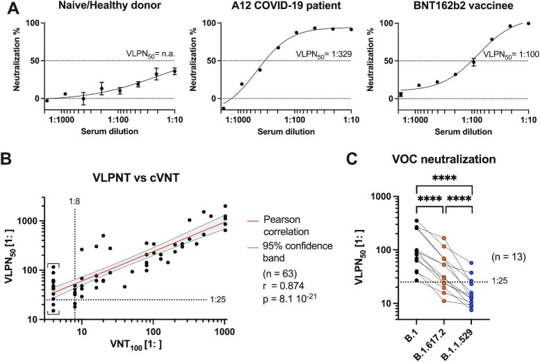
Neutralizing antibodies (NAbs), and their concentration in sera of convalescents and vaccinees are a correlate of protection from COVID-19. The antibody concentrations in clinical samples that neutralize SARS-CoV-2 are difficult and very cumbersome to assess with conventional virus neutralization tests (cVNTs), which require work with the infectious virus and biosafety level 3 containment precautions. Alternative virus neutralization tests currently in use are mostly surrogate tests based on direct or competitive enzyme immunoassays or use viral vectors with the spike protein as the single structural component of SARS-CoV-2. To overcome these obstacles, we developed a virus-free, safe and very fast (4.5 h) in vitro diagnostic test based on engineered yet authentic SARS-CoV-2 virus-like-particles (VLPs). They share all features of the original SARS-CoV-2 but lack the viral RNA genome and thus are non-infectious. NAbs induced by infection or vaccination, but also potentially neutralizing monoclonal antibodies can be reliably quantified and assessed with ease and within hours with our test, because they interfere and block the ACE2-mediated uptake of VLPs by recipient cells. Results from the VLP neutralization test (VLPNT) showed excellent specificity and sensitivity and correlated very well with a cVNT using fully infectious SARS-CoV-2. The results also demonstrated the reduced neutralizing capacity of COVID-19 vaccinee sera against variants of concern of SARS-CoV-2 including omicron B.1.1.529, BA.1.

Keywords: Omicron; SARS-CoV-2; diagnostics; virus neutralization test; virus-like particle.


Conflict of interest statement

Competing interests The authors D.P., R.Z., and W.H. report that the Helmholtz Zentrum Muenchen has filed a patent application relating to SARS-CoV-2 virus neutralization assays. The application lists the authors as inventors.

Figures

Fig. 1.
Fig. 1.
Cryo-EM images of SARS-CoV-2 VLPs (S+ VLPs). The images show different SARS-CoV-2 VLPs (S+ VLPs) of approximately 60 to 150 nm in diameter recorded by cryo-EM. The particles bear the characteristic corona of radial, dense spike-like proteins protruding from the envelopes’ intact lipid bilayer, which are characteristic for trimers of the viral glycoprotein of coronaviruses, spike (S), as observed for SARS-CoV-2 virions. The particle in the right panel shows elongated structures (white arrows), which might correspond to spike protein protrusions lying down on the vesicle surface, likely caused by surface tension effects prior to rapid freezing of the sample. White scale bars indicate 100 nm.
Fig. 2.
Fig. 2.
Spike WB analyses of protein lysates from S+ VLPs and SARS-CoV-2 virus stock. WB analyses of S+ VLPs and extracellular vesicles (EVs) produced in or spontaneously released from HEK293T cells, and SARS-CoV-2 virus stock produced from infected Vero E6 cells are shown. Antibodies are directed against the S1 or S2 domains or recognize the intact, full-length (FL) spike molecule SFL. The analyses confirm the presence of spike protein in various states in S+ VLPs and SARS-CoV-2 virion preparations but not in EVs which served as negative control. (A) and (B) S2 and S1 specific monoclonal respective polyclonal antibodies detect both spike domains in cell-free preparations of S+ VLPs as well as SFL protein (left panels of A and B). The S2 domain specific antibody also detects trimeric SFL (SFL3) and spike complexes of higher order under nonreducing (nonred) conditions. In SARS-CoV-2 virus stock (right panels of A and B) the antibodies detect SFL protein and the S1 domain in panels A and B but not the S2 domain. (C) Mono- and trimeric SFL protein complexes in S+ VLPs (left panel) and SARS-CoV-2 virus stock (right panel) detected with 43A11, a monoclonal antibody that recognizes full-length spike (SFL) exclusively.
Fig. 3.
Fig. 3.
Spike-specific, quantitative sandwich ELISA, and NTA of S+ VLP preparations and heat-inactivated SARS-CoV-2 virus stock. To quantify spike protein in biological samples, a sandwich ELISA was established with two mAbs (43A11 and 55E10) that recognize two orthogonal, nonoverlapping epitopes in SFL protein. Mean values with standard deviations are indicated. (A) Calibration of the sandwich ELISA with a commercially available recombinant (rec.) S protein standard, encompassing the extracellular domain (ECD) of spike. The calibration curve of three independent replicates allows for calculating the amount of S protein in samples within the linear range of optical density (OD) values (0.7 ≤ OD ≤ 1.7, r2 > 0.99). The detection limit of this assay was estimated to correspond to 3 ng mL−1 recombinant S protein. (B) and (C) Concentrated (conc.) and nonconcentrated S+ VLPs from supernatants of transiently transfected HEK293T cells were analyzed for their amount of SFL protein. Based on the linear regression function in panel A, the concentration (c) of S was calculated (ng mL−1) of three technical replicates in the linear OD range and compared with inactivated SARS-CoV-2 virus stock with a known ct value (15.3) of its vRNA copies according to RT-qPCR. Controls are solvent (PBS) and EVs without a viral FP (∆vFP EVs) harvested from cell culture medium of HEK293T cells transiently transfected with expression plasmids coding for M, N, E, and CD63∼HiBiT but omitting S. (D) NTA of three independent preparations of unconcentrated and concentrated S+ VLPs (S, M, N, E, and CD63∼HiBiT) and S+ EVs (S, CD63∼HiBiT omitting M, N, and and E) from cell culture medium of transiently transfected HEK293T cells are shown. For comparison, NTA data from heat-inactivated SARS-CoV-2 virus stock from infected Vero E6 cells are provided.
Fig. 4.
Fig. 4.
Nano flow technology of S+ VLPs and heat-inactivated SARS-CoV-2 virus stock. (A) HEK293T cells were transfected with S, M, N, E, and CD63∼HiBiT or with M, N, E, and CD63∼HiBiT but without S to produce S+ VLPs or control ∆vFP EVs, respectively. After two rounds of low-speed centrifugation, cell culture supernatants containing either S+ VLPs or control ∆vFP EVs were stained with the membrane permeable dye CTV, which exhibits fluorescence only upon its uptake followed by esterase activation within the lumen of intact membranous vesicles. A heat-inactivated SARS-CoV-2 virus stock was also stained with CTV for comparison. Subsequently, samples were counter-stained for the presence of surface spike protein using the monoclonal anti-S antibody 43A11. The samples were diluted and analyzed using a CytoFLEX LX flow cytometer. (B) Panels in the top row show recorded events according to their sideward scatter (SCC-H) using a violet excitation laser (y-axis) and CTV staining (x-axis). CTV+ events were gated as shown to identify subcellular, intact particles (S+ VLPs, ∆vFP EVs, and SARS-CoV-2 virus) to distinguish them from instrument noise seen in the PBS control. CTV+ events were analyzed for their staining with the anti-S antibody 43A11 coupled to AlexaFluor488 (bottom row of panels). 37.5% S+ particles were identified in the preparation of S+ VLPs, 10% S+ particles were identified in SARS-CoV-2 virus stock and fewer than 1% in control ∆vFP EVs. The low fraction of CTV-positive events in SARS-CoV-2 virus stock (0.11%) compared with preparations of S+ VLPs (0.61%) and ∆vFP EVs (0.37%) might be the consequence of a reduced esterase activity in virions (and EVs) after heat inactivation at 56°C for 15 min to inactivate viral infectivity.
Fig. 5.
Fig. 5.
VLPNT. (A) Engineered VLPs were generated in vitro by transient cotransfection of HEK293T cells with an optimized ratio of expression plasmids encoding the four SARS-CoV-2 structural proteins S, M, N, E, and a chimeric membrane anchored activator peptide (CD63∼HiBiT). The resulting particles were termed S+ VLPs and obtained from conditioned cell culture medium 3 days after DNA transfection. (B) Schematic view of a SARS-CoV-2 virion with the four structural proteins S, M, N, and E and the viral genome of positive sense, single-stranded RNA [(+)ssRNA] complexed with N. (C) Basic steps of VLP entry and reconstitution of nano Luciferase (nLuc). Similar to infection with SARS-CoV-2, spike, the trimeric viral FP in the envelope of S+ VLPs (Fig. 1) mediates attachment (step 1) to the host cell receptor ACE2, triggering either proteolytic processing by TMPRSS2 and direct fusion at the plasma membrane (step 2a) or endocytosis (step 2b), cleavage by CTSL and subsequent fusion with the endosomal membrane (step 3). Fusion of the S+ VLP envelope with cellular membranes via both pathways expose the HiBiT activator peptide to make contact with N-myristoylated LgBiT (NM∼LgBiT), which is stably expressed in the cytoplasm of the ACE2+ target cell. Upon in situ reconstitution of the functional nano Luciferase (nLuc*) reporter addition of substrate will induce bioluminescence, which can be quantified in a standard luminometer in 96-well cluster plates. (D) To test body fluids for the content of neutralizing SARS-CoV-2 antibodies (NAbs), S+ VLPs are preincubated with serial dilutions of the samples for 30 min. Suitable medical samples are sera of COVID-19 patients, vaccinated or naïve individuals or other body fluids such as saliva or nasal excretions. SARS-CoV-2 NAbs will interfere with all steps of S+ VLP attachment to ACE2, receptor-mediated intake, endosomal fusion of the VLP envelope with the endosome, and escape to the cytoplasm. Target cells are U251MG cells engineered to express both ACE2 and NM∼LgBiT (LgBiT). Upon encounter with S+ VLP-borne CD63∼HiBiT, NM∼LgBiT is reconstituted into a fully functional nLuc reporter enzyme, which can be quantitated. Neutralizing SARS-CoV-2 antibodies reduce or even block the delivery of the CD63∼HiBiT activator entirely, which can be quantified in a standard clinical laboratory with aid of a luminometer and within 4.5 h. A freely accessible scientific animation narrates the principle of the VLPNT (https://youtu.be/6wckXobT_bM).
Fig. 6.
Fig. 6.
Specificity and tropism of S+ VLPs. (A) Two cell line derivatives of U251MG cells, which express NM∼LgBiT with or without human ACE2 receptor (ACE2+ or ACE2) were incubated with S+ VLPs carrying CD63∼HiBiT, with EVs without a viral FP (ΔvFP EVs) obtained from supernatants of HEK293T cells after transient transfection of expression plasmids encoding M, N, E, and CD63∼HiBiT (but not S) or with EVs from HEK293T cells after transient transfection of expression plasmids encoding CD63∼HiBiT and protein G of the vesicular stomatitis virus (VSV-G+ EVs). The specificity of spike-mediated, ACE2-dependent fusion of all three particle classes was validated measuring luciferase activities upon reconstitution of the split nLuc in the indicated cell types. Data are based on at least four independent experiments. P-values of independent t tests are indicated (ns; not significant). (B) Inhibitor studies with chloroquine, an inhibitor of endosomal acidification and of CTSL and camostat-mesylate, a TMPRSS2 inhibitor, are shown using S+ VLPs and ACE2+ U251MG cells. DMSO and PBS served as negative controls for camostat-mesylate and chloroquine, respectively. Mean values of three biological replicates are displayed with error bars indicating standard deviations.
Fig. 7.
Fig. 7.
Correlation of VLPNT and cVNT data and VOC cross-neutralization using sera from COVID-19 patients and vaccinees. (A) VLPNT neutralization data with dilutions of sera obtained from three individuals (a naïve healthy donor, the COVID-19 patient A12, and a BNT162b2 vaccinee) are shown. The graphs are examples and include mean neutralization results from three independent biological replicates. Serum dilution which resulted in half maximal signal reduction, equivalent to 50% neutralization, was termed VLPN50 titer. (B) Correlation of VLPN50 titers from the VLPNT vs. VNT100 titers obtained in a cVNT using infectious SARS-CoV-2. Pearson correlation data (sample size n, coefficient r, and P-value) of 63 sera from confirmed COVID-19 patients are shown and the linear relationship is indicated. Results below the dotted horizontal line denote sera, which scored negative in the VLPNT. Results left of the dotted vertical line denote sera which scored below the LOD (1:8) in the cVNT; these VNT100 values were defined as 1:4 and indicated by square brackets. (C) VLPNT with two SARS-CoV-2 VOCs compared with the B.1 strain. S+ VLPs were harvested from supernatants of HEK293T cells transiently transfected with expression plasmids encoding either B.1 (S: Wuhan-2019, D614G), B.1.617.2 (Delta-VOC), or B.1.1.529 (BA.1, Omicron-VOC) S protein together with M, N, E, and CD63∼HiBiT. NAbs in sera of 13 COVID-19 vaccinees (after prime-boost vaccination) were cross-neutralizing, but less potent in neutralizing B.1.617.2 compared with B.1. The majority of serum samples however, failed to neutralize the B.1.1.529 variant effectively. Data derived from 13 samples were analyzed using a matched one-way ANOVA, with Tukey's multi comparison test and a single pooled variance. Results are indicated: ****P ≤ 0.0001.

Similar articles

Cited by

References

    1. Wu F, et al. 2020. A new coronavirus associated with human respiratory disease in China. Nature. 579:265–269. - PMC - PubMed
    1. Hu B, Guo H, Zhou P, Shi ZL. 2021. Characteristics of SARS-CoV-2 and COVID-19. Nat Rev Microbiol. 19:141–154. - PMC - PubMed
    1. Mittal A, et al. 2020. COVID-19 pandemic: insights into structure, function, and hACE2 receptor recognition by SARS-CoV-2. PLoS Pathog. 16:e1008762. - PMC - PubMed
    1. Hofmann H, Pohlmann S. 2004. Cellular entry of the SARS coronavirus. Trends Microbiol. 12:466–472. - PMC - PubMed
    1. Stevens CS, Oguntuyo KY, Lee B. 2021. Proteases and variants: context matters for SARS-CoV-2 entry assays. Curr Opin Virol. 50:49–58. - PMC - PubMed