. 2022 Oct 26;8(2):veac101.
doi: 10.1093/ve/veac101. eCollection 2022.

Multiple waves of viral invasions in Symbiodiniaceae algal genomes

Affiliations

Multiple waves of viral invasions in Symbiodiniaceae algal genomes

L Felipe Benites et al. Virus Evol. .

Abstract

Dinoflagellates from the family Symbiodiniaceae are phototrophic marine protists that engage in symbiosis with diverse hosts. Their large and distinct genomes are characterized by pervasive gene duplication and large-scale retroposition events. However, little is known about the role and scale of horizontal gene transfer (HGT) in the evolution of this algal family. In other dinoflagellates, high levels of HGTs have been observed, linked to major genomic transitions, such as the appearance of a viral-acquired nucleoprotein that originated via HGT from a large DNA algal virus. Previous work showed that Symbiodiniaceae from different hosts are actively infected by viral groups, such as giant DNA viruses and ssRNA viruses, that may play an important role in coral health. Latent viral infections may also occur, whereby viruses could persist in the cytoplasm or integrate into the host genome as a provirus. This hypothesis received experimental support; however, the cellular localization of putative latent viruses and their taxonomic affiliation are still unknown. In addition, despite the finding of viral sequences in some genomes of Symbiodiniaceae, viral origin, taxonomic breadth, and metabolic potential have not been explored. To address these questions, we searched for putative viral-derived proteins in thirteen Symbiodiniaceae genomes. We found fifty-nine candidate viral-derived HGTs that gave rise to twelve phylogenies across ten genomes. We also describe the taxonomic affiliation of these virus-related sequences, their structure, and their genomic context. These results lead us to propose a model to explain the origin and fate of Symbiodiniaceae viral acquisitions.

Keywords: Symbiodiniaceae evolution; Symbiodinium virus; algal virus; dinoflagellate virus; virus horizontal gene transfer.


Figures

Figure 1.
Figure 1.
The main characteristics of genes in Symbiodiniaceae genomes that were acquired by viral horizontal gene transfer (vHGT). Cladogram depicting phylogenetic relationships of Symbiodiniaceae genomes (adapted from González-Pech et al. 2021) in which vHGTs were identified (left); the hosts associated with each Symbiodiniaceae species are represented by colored circles (top left key). Assembly and estimated genome sizes are shown in gigabase pairs (Gbp). The total number of vHGTs and their putative taxonomic origins (at the order level; center key), the CDS percent Guanine–Cytosine (GC%) content, the average number of introns, and the total number of associated protein domains are shown for the vHGTs identified in each genome. The CDS GC% content and average number of introns is shown separately for the vHGTs and the background Symbiodiniaceae sequences (i.e. all Symbiodiniaceae CDS or genes [respectively] excluding the vHGTs). Abbreviations: Brevioloum minutum (Bmin); Cladocopium sp. C92 (CC92); Cladocopium goreaui (Cgor); Fugacium kawagutii (Fkaw); Symbiodinium linucheae CCMP2456 (Slin_CCMP2456); S. microadriaticum (Smic); S. microadriaticum 04–503SCI.03 (Smic_04-503SCI.03); S. microadriaticum CassKB8 (Smic_CassKB8); S. natans CCMP2548 (Snat_CCMP2548); S. necroappetens CCMP2469 (Snec_CCMP2469); S. pilosum CCMP2461 (Spil_CCMP2461); S. tridacnidorum (Stri); S. tridacnidorum CCMP2592 (Stri_CCMP2592); DNA/RNA pol (DNA/RNA polymerase superfamily); DUF4116 (Domain of unknown function DUF4116); None (None predicted); RdRp (RNA-dependent RNA polymerase); RNA hel core (RNA virus helicase core domain); R-dRP (RNA-directed RNA polymerase); R-dRp core (RNA-directed RNA polymerase catalytic domain); RNA hel (Viral (Superfamily 1) RNA helicase); Vir hel (Viral helicase1) (Supplementary Tables S2 and S12).
Figure 2.
Figure 2.
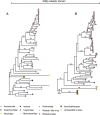
Phylogenetic profile of vHGT’s grouped by the category ‘Unclassified RNA viruses’: RNA-dependent RNA polymerase (RdRps-like group 1 [A, B, C] and RdRp-like group 2 [D]), Major capsid protein (MCPs-like [E, F, G]), viral RNA helicase (H), and polyprotein coding for replicases including RNA-dependent RNA polymerase region (I). Sequence names are not shown in the trees to enhance readability. The taxonomic affiliation of each sequence in the trees is represented by a colored circle, with the colors described in the legend at the bottom of the figure. Symbiodiniaceae vHGTs are highlighted with thicker borders. Only bootstrap support values ≥90 per cent are shown. Complete names and branch support in Supplementary Fig. S5.
Figure 3.
Figure 3.
Phylogenetic profile of vHGT’s grouped by the category ‘Pimascovirales’: Symbiodiniaceae vHGT restriction endonuclease-like, viral restriction endonucleases and GVMAG homologs. Sequence names are not shown in the trees to enhance readability. Taxonomic affiliation of each sequence in the tree is represented by a colored circle, with the colors described in the legend at the bottom of the figure. Symbiodiniaceae vHGTs are highlighted with thicker borders. Only bootstrap support values ≥90 per cent are shown. Complete names and branch support in Supplementary Fig. S5.
Figure 4.
Figure 4.
Phylogenetic profile of vHGT’s grouped by the category ‘Mononegavirales’: RNA-directed RNA polymerase catalytic domain (RdRP catalytic domain; A and B). Sequence names are not shown in the trees to enhance readability. The taxonomic affiliation of each sequence in the trees is represented by a colored circle, with the colors described in the legend at the bottom of the figure. Symbiodiniaceae vHGTs are highlighted with thicker borders. Only bootstrap support values ≥90 per cent are shown. Complete names and branch support values are in Supplementary Fig. S5.
Figure 5.
Figure 5.
Genome maps comparing the CDSs of the dinoflagellate virus (dinornavirus) HcRNAV34 (Heterocapsa circularisquama RNA virus—accession: AB218608.1) and Symbiodinium RNA virus (Symbiodinium +ssRNA virus—accession KX538960.1) with the putative integrated viral genomes in S. microadriaticum (Smic and Smic04). The putative viral elements (vHGTs) annotated as RdRp- and MCP-like proteins are shown, as are the genes surrounding these elements and the start and end positions of scaffolds. Each feature points in the direction that it is encoded, and a scale bar is shown in the top right corner. Slash bars (/) denote intergenic regions. Abbreviations: scaf. (scaffold); DinoSL (dinoflagellate splice leader; annotated as small arrows); RdRp and RdRp-like (RNA-dependent RNA polymerase); MCP and MCP-like; Retrovirus-related Pol polyprotein R2 (Retrovirus-related Pol polyprotein from type-1 retrotransposable element R2); Amino acid permease YfnA (putative amino acid permease YfnA).
Figure 6.
Figure 6.
A hypothetical model with two scenarios (A and B) explaining the origin of vHGTs in Symbiodiniaceae genomes. During an acute infection, viral particles enter the host cell, and viral mRNAs accumulate in the cytoplasm to a high abundance. The host machinery can accidentally incorporate viral mRNA into its genome via reverse transcription and non-homologous recombination; this may use the same (or similar) mechanisms as the mRNA recycling process that is prevalent in dinoflagellates. These viral sequences, known as EVEs, would be ‘dead on arrival’ and are expected to decay into pseudogenes (which we would then observe in the genome as vHGTs). In the second scenario (B), integration of the virus genome into the host genome is part of a previously unobserved transient life stage of these viruses (specifically the Symbiodinium +ssRNA virus). The virus infects and is incorporated into the host genome via the same mechanisms as the first scenario (A); however, instead of this process resulting in non-functional EVEs, it produces active proviral elements. These integrated viral genomes can remain silent during times of low host abundance (e.g. during the free-living stage of facultative Symbiodiniaceae lifecycles) and can become activated by environmental cues (such as stress or high Symbiodiniaceae cell density) that result in viral lysis, escape from the host cell, and new infections in susceptible hosts. The proviral elements can also become deactivated, either through random mutation or host-driven process, resulting in EVEs that are expected to decay into pseudogenes, which we would then observe in the genome as vHGTs. The Symbiodiniaceae cell illustration was inspired by drawings by Freudenthal (1962) and the Symbiodinium life cycle illustration was adapted from https://commons.wikimedia.org/wiki/File:Symbiodinium_Life_Cycle.png.

Similar articles

Cited by

References

    1. Agarkova I. V., Dunigan D. D. and Etten J. L. V. (2006) ‘Virion-Associated Restriction Endonucleases of Chloroviruses’, Journal of Virology, 80: 8114–23. - PMC - PubMed
    1. Agüero J. et al. (2018) ‘Stable and Broad Spectrum Cross-Protection against Pepino Mosaic Virus Attained by Mixed Infection’, Frontiers in Plant Science, 9. - PMC - PubMed
    1. Aherfi S. et al. (2014) ‘The Expanding Family Marseilleviridae’, Virology, 466–467: 27–37. - PubMed
    1. Aranda M. et al. (2016) ‘Genomes of Coral Dinoflagellate Symbionts Highlight Evolutionary Adaptations Conducive to a Symbiotic Lifestyle’, Scientific Reports, 6: 39734. - PMC - PubMed
    1. Aus Dem Siepen M. et al. (2005) ‘Poinsettia Latent Virus Is Not a Cryptic Virus, but a Natural Polerovirus–Sobemovirus Hybrid’, Virology, 336: 240–50. - PubMed