. 2023 Jul;12(4):464-476.
doi: 10.1016/j.jshs.2022.10.004. Epub 2022 Oct 29.

Exercise training attenuates angiotensin II-induced cardiac fibrosis by reducing POU2F1 expression

Affiliations

Exercise training attenuates angiotensin II-induced cardiac fibrosis by reducing POU2F1 expression

Na Feng et al. J Sport Health Sci. 2023 Jul.

Abstract

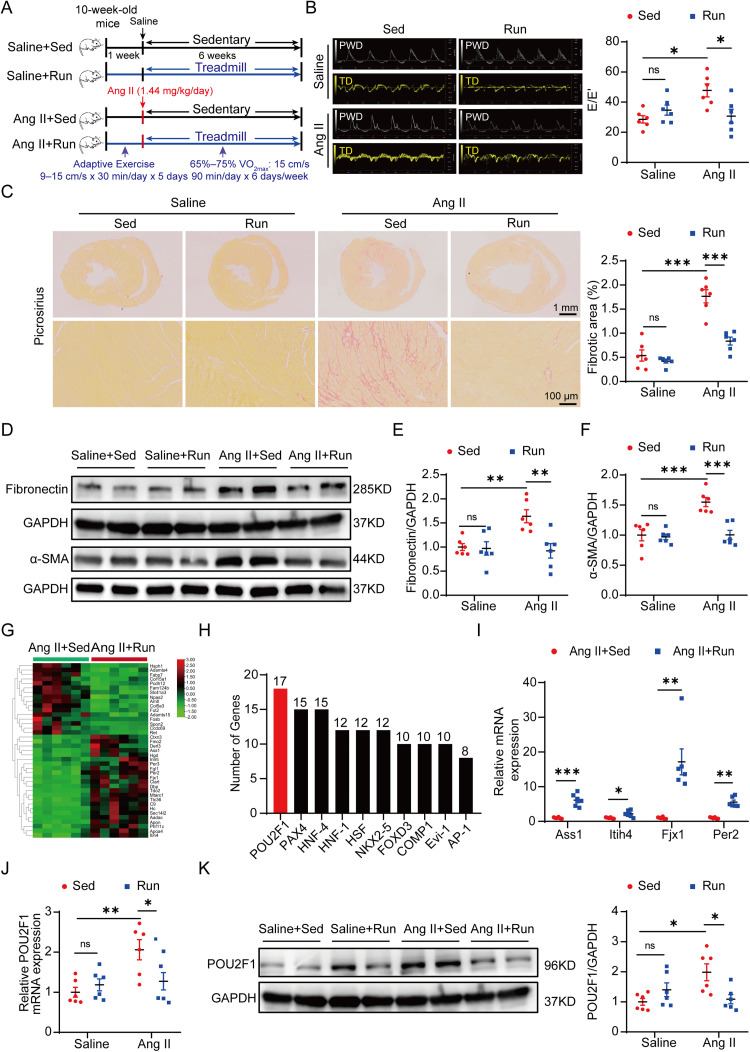
Background: Exercise training protects against heart failure. However, the mechanism underlying the protective effect of exercise training on angiotensin II (Ang II)-induced cardiac fibrosis remains unclear.

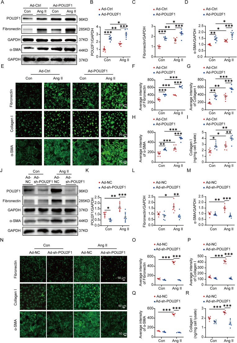
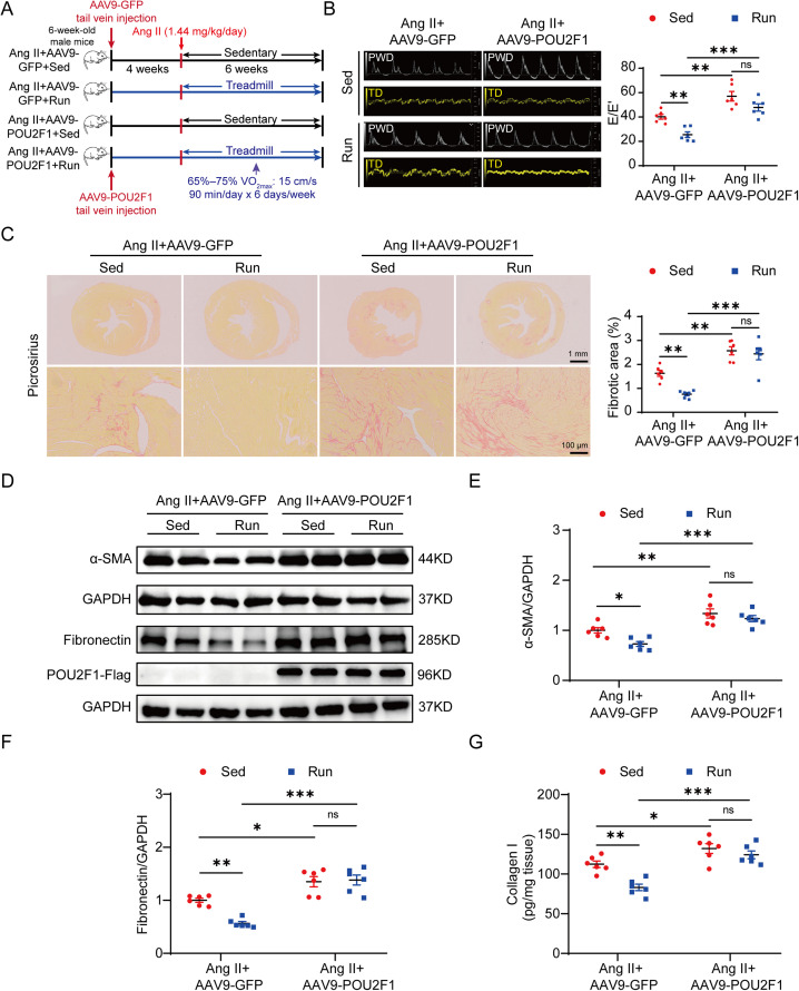
Methods: An exercise model involving C57BL/6N mice and 6 weeks of treadmill training was used. Ang II (1.44 mg/kg/day) was administered to induce cardiac fibrosis. RNA sequencing and bioinformatic analysis were used to identify the key factors mediating the effects of exercise training on cardiac fibrosis. Primary adult mouse cardiac fibroblasts (CFs) were used in vitro. Adeno-associated virus serotype 9 was used to overexpress POU domain, class 2, transcription factor 1 (POU2F1) in vivo.

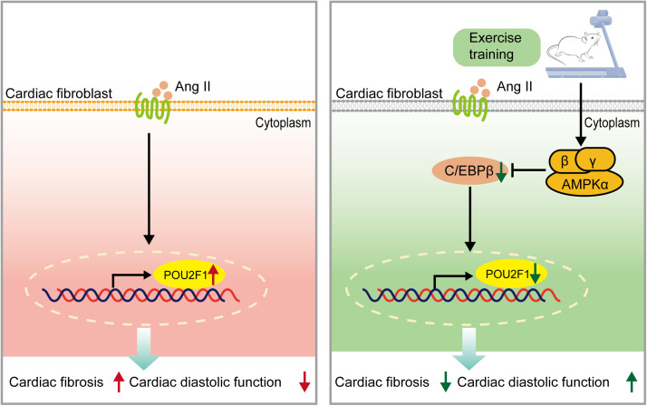
Results: Exercise training attenuated Ang II-induced cardiac fibrosis and reversed 39 gene expression changes. The transcription factor regulating the largest number of these genes was POU2F1. Compared to controls, POU2F1 was shown to be significantly upregulated by Ang II, which is itself reduced by exercise training. In vivo, POU2F1 overexpression nullified the benefits of exercise training on cardiac fibrosis. In CFs, POU2F1 promoted cardiac fibrosis. CCAAT enhancer-binding protein β (C/EBPβ) was predicted to be the transcription factor of POU2F1 and verified using a dual-luciferase reporter assay. In vivo, exercise training activated AMP-activated protein kinase (AMPK) and alleviated the increase in C/EBPβ induced by Ang II. In CFs, AMPK agonist inhibited the increase in C/EBPβ and POU2F1 induced by Ang II, whereas AMPK inhibitor reversed this effect.

Conclusion: Exercise training attenuates Ang II-induced cardiac fibrosis by reducing POU2F1. Exercise training inhibits POU2F1 by activating AMPK, which is followed by the downregulation of C/EBPβ, the transcription factor of POU2F1.

Keywords: AMPK; C/EBPβ; Cardiac fibrosis; Exercise; POU2F1.


Conflict of interest statement

The authors declare that they have no competing interests.

Figures

Image, graphical abstract
Graphical abstract
Fig 1
Fig. 1
POU2F1 is involved in exercise training and the attenuation of Ang II-induced cardiac fibrosis. (A) a pattern diagram of the experimental protocol; (B) representative pulsed wave Doppler images across the mitral flow and tissue Doppler images of the mitral valve ring and measurements of E/E’; (C) picrosirius red staining of heart tissues and quantification of the fibrotic area (scale bars: top = 1 mm, bottom = 100 μm); (D) protein levels of fibronectin and α-SMA in heart tissues were detected using the Western blot; Quantitative analysis of the relative protein expressions of (E) fibronectin and (F) α-SMA in heart tissues; (G) heat map of DEGs; (H) the top 10 transcription factors predicted to bind to the greatest number of DEGs according to TRANSFAC database. The number indicates the amount of DEGs with predicted binding sites for the indicative transcription factor; (I) four genes regulated by POU2F1 and associated with fibrosis were validated by qPCR. (J) qPCR analysis of the POU2F1 mRNA levels in heart tissues; (K) the protein level of POU2F1 in heart tissues was detected using the Western blot. Data are presented as mean ± SEM. n = 6. A two-way analysis of variance followed by Tukey's post hoc test was used in (B), (C), (E), (F), (J), and (K). The t test or Mann–Whitney U test was used in (I). * p < 0.05; ** p < 0.01; *** p < 0.001. α-SMA = α-smooth muscle actin; Ang II = angiotensin II; AP-1 = activator protein 1; Ass1 = argininosuccinate synthase 1; COMP1 = protein kinase domain-containing protein; DEG = differentially expressed gene; Evi-1 = ecotropic viral integration site 1; Fjx1 = four-jointed box kinase 1; FOXD3 = forkhead box protein d3; GAPDH = glyceraldehyde-3-phosphate dehydrogenase; HNF-1 = hepatocyte nuclear factor 1; HNF-4 = hepatocyte nuclear factor 4; HSF = heat shock factor; Itih4 = inter-alpha-trypsin inhibitor heavy chain 4; NKX2-5 = NK2 transcription factor related, locus 5; ns = not significant; PAX4 = paired box homologous gene 4; Per2 = recombinant period circadian protein 2; POU2F1 = POU domain, class 2, transcription factor 1; PWD = pulsed-wave doppler; qPCR = quantitative polymerase chain reaction; Run = running; Sed = sedentary; SEM = standard error of mean; TD = tissue doppler.
Fig 2
Fig. 2
POU2F1 promoted CF differentiation and enhanced extracellular matrix protein synthesis. (A−I) CFs were infected with Ad-Ctrl or Ad-POU2F1 then treated with saline or Ang II for 48 h. (A) the expressions of POU2F1, fibronectin, and α-SMA were determined using the Western blot; (B−D) quantitative analysis of the relative protein expressions of (B) POU2F1, (C) fibronectin, and (D) α-SMA; (E) representative images and quantifications of the immunofluorescence analysis of (F) fibronectin, (G) Collagen I, and (H) α-SMA in CFs; (I) the secretion of Collagen I in the cell culture supernatant of CFs was determined by ELISA; (J−R) CFs were infected with Ad-NC or Ad-sh-POU2F1 to knock down POU2F1 then treated with saline or Ang II for 48 h; (J) Western blot analysis of POU2F1, fibronectin, and α-SMA in CFs. Quantitative analysis of relative protein expression of (K) POU2F1, (L) fibronectin, and (M) α-SMA in CFs; (N) Representative images and quantifications of the immunofluorescence analysis of (O) fibronectin, (P) Collagen I, and (Q) α-SMA in CFs; (R) the secretion of Collagen I in the cell culture supernatant of CFs was determined by an ELISA. Data are presented as mean ± SEM. n = 6. A two-way analysis of variance followed by Tukey's post hoc test was used. * p < 0.05; ** p < 0.01; *** p < 0.001. α-SMA = α-smooth muscle actin; Ad-Ctrl = control adenovirus; Ad-NC = negative control adenovirus; Ad-POU2F1 = POU2F1-overexpressed adenovirus; Ad-sh-POU2F1 = sh-POU2F1 adenovirus; Ang II = angiotensin II; CF = cardiac fibroblast; ELISA = enzyme-linked immunosorbent assay; GAPDH = glyceraldehyde-3-phosphate dehydrogenase; POU2F1 = POU domain, class 2, transcription factor 1; SEM = standard error of mean.
Fig 3
Fig. 3
The protective effects of exercise training in Ang II-induced cardiac fibrosis are abolished by POU2F1 overexpression. (A) a pattern diagram of the experimental protocol; (B) representative pulsed wave Doppler images across the mitral flow and tissue Doppler images of the mitral valve ring and measurements of E/E’; (C) picrosirius red staining and quantification of the fibrotic area (scale bars: top = 1 mm, bottom = 100 μm); (D) the protein levels of α-SMA and fibronectin in heart tissues were detected using the Western blot; Quantitative analysis of relative protein expressions of (E) α-SMA and (F) fibronectin in heart tissues; (G) the protein levels of Collagen I in heart tissues were determined using an ELISA. Data are presented as mean ± SEM. n = 6. A two-way analysis of variance followed by Tukey's post hoc test was used. * p < 0.05; ** p < 0.01; *** p < 0.001. α-SMA = α-smooth muscle actin; AAV9 = adeno-associated virus serotype-9; Ang II = angiotensin II; ELISA = enzyme-linked immunosorbent assay; GAPDH = glyceraldehyde-3-phosphate dehydrogenase; GFP = green fluorescent protein; ns = not significant; POU2F1 = POU domain, class 2, transcription factor 1; PWD = pulsed-wave doppler; Run = running; Sed = sedentary; SEM = standard error of mean; TD = tissue doppler; VO2max = maximal oxygen uptake.
Fig 4
Fig. 4
Exercise training reduced POU2F1 by activating AMPK and downregulating C/EBPβ. (A) schematic of the binding site between C/EBPβ and the mouse POU2F1 promoter region and the structures of the reporter plasmids carrying the full POU2F1 promoter region (POU2F1 wild type) or the −1457 to −1448 bp site deletion fragment (POU2F1 binding site deletion); (B) HEK 293A cells were transfected with the wild-type or mutant plasmid and then treated with control plasmid or C/EBPβ overexpression plasmid, respectively. A dual-luciferase reporter assay was performed (n = 3); (C) ChIP analysis using anti-C/EBPβ antibody or IgG, soluble chromatin (500−600 bp in length) from CFs overexpressing C/EBPβ, and primers targeting the region spanning the C/EBPβ binding sites in the POU2F1 promoter; (D) ChIP analysis using anti-C/EBPβ antibody or IgG, soluble chromatin (500−600 bp in length) from mouse heart tissues, and primers targeting the region spanning the C/EBPβ binding sites in the POU2F1 promoter; (E) the expression of C/EBPβ was determined by qPCR; (F) the expression of POU2F1 was determined by qPCR; (G) representative images and quantitative analysis of the Western blot for (H) C/EBPβ and POU2F1, as well as (I) fibronectin and α-SMA; (J) the secretion of collagen I in the cell culture supernatant of CFs was determined by ELISA; (K) representative images and quantitative analysis of the Western blot for (L) p-AMPK to total AMPK and (M) C/EBPβ to GAPDH in the Ang II-induced cardiac fibrosis model; (N−Q) CFs were pretreated with compound C (1 μmol/L) for 0.5 h and treated with metformin (1 mmol/L) for 0.5 h. Then, Ang II (10−6 mol/L) was added to CFs for 48 h; (N) representative images and quantitative analysis of the relative protein expressions of (O) p-AMPK to total AMPK, (P) C/EBPβ to GAPDH, and (Q) POU2F1 to GAPDH in harvested CFs. (R) ChIP analysis using anti-C/EBPβ antibody or IgG, soluble chromatin (500−600 bp in length) from CFs treated with AngII and/or metformin, and primers targeting the region spanning the C/EBPβ binding sites in the POU2F1 promoter. Data are presented as mean ± SEM. n = 6 in (C−R). The t test was used in (C), (E), (F), and (H−J). A two-way analysis of variance followed by Tukey's post hoc test was used in (B), (L), and (M). A one-way analysis of variance was used in (O−R). * p < 0.05; ** p < 0.01; *** p < 0.001. Ad-Ctrl = control adenovirus; AMPK = AMP-activated protein kinase; Ang II = angiotensin II; bp = base pair; C/EBPβ = CCAAT enhancer-binding protein β; CFs = cardiac fibroblasts; ChIP = chromatin immunoprecipitation; ELISA = enzyme-linked immunosorbent assay; GAPDH = glyceraldehyde-3-phosphate dehydrogenase; IgG = immunoglobulin G; ns = not significant; p-AMPK = phospho-AMP-activated protein kinase Thr 172; POU2F1 = POU domain, class 2, transcription factor 1; qPCR = quantitative polymerase chain reaction; Run = running; Sed = sedentary; SEM = standard error of mean.
Fig 5
Fig. 5
A working model of the role of POU2F1 in the protective effect of exercise training (attenuation of Ang II-induced cardiac fibrosis). AMPK = AMP-activated protein kinase; Ang II = angiotensin II; POU2F1 = POU domain, class 2, transcription factor 1; C/EBPβ = CCAAT enhancer-binding protein β.

Similar articles

Cited by

References

    1. Milutinović K, Stojiljković S, Ćuk J, et al. Athlete's heart. Fizicka kultura. 2018;72:139–147.
    1. Villella M, Villella A. Exercise and cardiovascular diseases. Kidney Blood Press Res. 2014;39:147–153. - PubMed
    1. Lloyd-Jones DM, Hong Y, Labarthe D, et al. Defining and setting national goals for cardiovascular health promotion and disease reduction: The American Heart Association's strategic impact goal through 2020 and beyond. Circulation. 2010;121:586–613. - PubMed
    1. Piercy KL, Troiano RP, Ballard RM, et al. The physical activity guidelines for Americans. JAMA. 2018;320:2020–2028. - PMC - PubMed
    1. Orso F, Fabbri G, Maggioni AP. Epidemiology of heart failure. Handb Exp Pharmacol. 2017;243:15–33. - PubMed