Changes in abdominal adipose tissue depots assessed by MRI correlate with hepatic histologic improvement in non-alcoholic steatohepatitis
- PMID: 36368598
- PMCID: PMC9852022
- DOI: 10.1016/j.jhep.2022.10.027
Changes in abdominal adipose tissue depots assessed by MRI correlate with hepatic histologic improvement in non-alcoholic steatohepatitis
Abstract
Background & aims: Non-alcoholic steatohepatitis (NASH) is prevalent in adults with obesity and can progress to cirrhosis. In a secondary analysis of prospectively acquired data from the multicenter, randomized, placebo-controlled FLINT trial, we investigated the relationship between reduction in adipose tissue compartment volumes and hepatic histologic improvement.
Methods: Adult participants in the FLINT trial with paired liver biopsies and abdominal MRI exams at baseline and end-of-treatment (72 weeks) were included (n = 76). Adipose tissue compartment volumes were obtained using MRI.
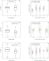
Results: Treatment and placebo groups did not differ in baseline adipose tissue volumes, or in change in adipose tissue volumes longitudinally (p = 0.107 to 0.745). Deep subcutaneous adipose tissue (dSAT) and visceral adipose tissue volume reductions were associated with histologic improvement in NASH (i.e., NAS [non-alcoholic fatty liver disease activity score] reductions of ≥2 points, at least 1 point from lobular inflammation and hepatocellular ballooning, and no worsening of fibrosis) (p = 0.031, and 0.030, respectively). In a stepwise logistic regression procedure, which included demographics, treatment group, baseline histology, baseline and changes in adipose tissue volumes, MRI hepatic proton density fat fraction (PDFF), and serum aminotransferases as potential predictors, reductions in dSAT and PDFF were associated with histologic improvement in NASH (regression coefficient = -2.001 and -0.083, p = 0.044 and 0.033, respectively).
Conclusions: In adults with NASH in the FLINT trial, those with greater longitudinal reductions in dSAT and potentially visceral adipose tissue volumes showed greater hepatic histologic improvements, independent of reductions in hepatic PDFF.
Clinical trial number: NCT01265498.
Impact and implications: Although central obesity has been identified as a risk factor for obesity-related disorders including insulin resistance and cardiovascular disease, the role of central obesity in non-alcoholic steatohepatitis (NASH) warrants further clarification. Our results highlight that a reduction in central obesity, specifically deep subcutaneous adipose tissue and visceral adipose tissue, may be related to histologic improvement in NASH. The findings from this analysis should increase awareness of the importance of lifestyle intervention in NASH for clinical researchers and clinicians. Future studies and clinical practice may design interventions that assess the reduction of deep subcutaneous adipose tissue and visceral adipose tissue as outcome measures, rather than simply weight reduction.
Keywords: central obesity; deep subcutaneous adipose tissue; liver histology; visceral adipose tissue.
Copyright © 2022 European Association for the Study of the Liver. All rights reserved.
Figures


Comment in
-
Shrinking fat, healing liver: unlocking the metabolic dysfunction associated steatohepatitis puzzle.Hepatobiliary Surg Nutr. 2024 Feb 1;13(1):132-135. doi: 10.21037/hbsn-23-569. Epub 2024 Jan 16. Hepatobiliary Surg Nutr. 2024. PMID: 38322227 Free PMC article. No abstract available.
Similar articles
-
Multicenter Validation of Association Between Decline in MRI-PDFF and Histologic Response in NASH.Hepatology. 2020 Oct;72(4):1219-1229. doi: 10.1002/hep.31121. Epub 2020 Oct 9. Hepatology. 2020. PMID: 31965579 Free PMC article. Clinical Trial.
-
Longitudinal correlations between MRE, MRI-PDFF, and liver histology in patients with non-alcoholic steatohepatitis: Analysis of data from a phase II trial of selonsertib.J Hepatol. 2019 Jan;70(1):133-141. doi: 10.1016/j.jhep.2018.09.024. Epub 2018 Oct 4. J Hepatol. 2019. PMID: 30291868 Clinical Trial.
-
Sitagliptin in patients with non-alcoholic steatohepatitis: A randomized, placebo-controlled trial.World J Gastroenterol. 2017 Jan 7;23(1):141-150. doi: 10.3748/wjg.v23.i1.141. World J Gastroenterol. 2017. PMID: 28104990 Free PMC article. Clinical Trial.
-
Change in MRI-PDFF and Histologic Response in Patients With Nonalcoholic Steatohepatitis: A Systematic Review and Meta-Analysis.Clin Gastroenterol Hepatol. 2021 Nov;19(11):2274-2283.e5. doi: 10.1016/j.cgh.2020.08.061. Epub 2020 Aug 31. Clin Gastroenterol Hepatol. 2021. PMID: 32882428 Free PMC article. Review.
-
Non-invasive methods for imaging hepatic steatosis and their clinical importance in NAFLD.Nat Rev Endocrinol. 2022 Jan;18(1):55-66. doi: 10.1038/s41574-021-00584-0. Epub 2021 Nov 23. Nat Rev Endocrinol. 2022. PMID: 34815553 Free PMC article. Review.
Cited by
-
Challenges and opportunities in obesity: the role of adipocytes during tissue fibrosis.Front Endocrinol (Lausanne). 2024 Apr 15;15:1365156. doi: 10.3389/fendo.2024.1365156. eCollection 2024. Front Endocrinol (Lausanne). 2024. PMID: 38686209 Free PMC article. Review.
-
Association of the fat mass index with hepatic steatosis and fibrosis: evidence from NHANES 2017-2018.Sci Rep. 2024 Mar 23;14(1):6943. doi: 10.1038/s41598-024-57388-1. Sci Rep. 2024. PMID: 38521854 Free PMC article.
-
Shrinking fat, healing liver: unlocking the metabolic dysfunction associated steatohepatitis puzzle.Hepatobiliary Surg Nutr. 2024 Feb 1;13(1):132-135. doi: 10.21037/hbsn-23-569. Epub 2024 Jan 16. Hepatobiliary Surg Nutr. 2024. PMID: 38322227 Free PMC article. No abstract available.
-
Frontiers and hotspots of adipose tissue and NAFLD: a bibliometric analysis from 2002 to 2022.Front Physiol. 2023 Dec 21;14:1278952. doi: 10.3389/fphys.2023.1278952. eCollection 2023. Front Physiol. 2023. PMID: 38187139 Free PMC article.
-
Abdominal adipose tissue and type 2 diabetic kidney disease: adipose radiology assessment, impact, and mechanisms.Abdom Radiol (NY). 2024 Feb;49(2):560-574. doi: 10.1007/s00261-023-04062-1. Epub 2023 Oct 17. Abdom Radiol (NY). 2024. PMID: 37847262 Review.
References
-
- Chalasani N, Younossi Z, Lavine JE, Charlton M, Cusi K, Rinella M, et al. The diagnosis and management of nonalcoholic fatty liver disease: Practice guidance from the American Association for the Study of Liver Diseases. Hepatology 2018;67:328–357. - PubMed
-
- Santoro N, Caprio S. Nonalcoholic fatty liver disease/nonalcoholic steatohepatitis in obese adolescents: a looming marker of cardiac dysfunction. Hepatology 2014;59:372–374. - PubMed
-
- Lawlor DA, Callaway M, Macdonald-Wallis C, Anderson E, Fraser A, Howe LD, et al. Nonalcoholic fatty liver disease, liver fibrosis, and cardiometabolic risk factors in adolescence: a cross-sectional study of 1874 general population adolescents. J Clin Endocrinol Metab 2014;99:E410–417. - PMC - PubMed
-
- McCullough AJ. Update on nonalcoholic fatty liver disease. J Clin Gastroenterol 2002;34:255–262. - PubMed
Publication types
MeSH terms
Associated data
Grants and funding
- U01 DK130190/DK/NIDDK NIH HHS/United States
- U01 DK061731/DK/NIDDK NIH HHS/United States
- UL1 TR000006/TR/NCATS NIH HHS/United States
- P30 DK120515/DK/NIDDK NIH HHS/United States
- UL1 TR000058/TR/NCATS NIH HHS/United States
- U01 DK061728/DK/NIDDK NIH HHS/United States
- UL1 TR000454/TR/NCATS NIH HHS/United States
- UL1 TR000004/TR/NCATS NIH HHS/United States
- UL1 TR002345/TR/NCATS NIH HHS/United States
- R01 DK106419/DK/NIDDK NIH HHS/United States
- U01 DK061737/DK/NIDDK NIH HHS/United States
- U01 DK061713/DK/NIDDK NIH HHS/United States
- U01 DK061732/DK/NIDDK NIH HHS/United States
- UL1 TR000150/TR/NCATS NIH HHS/United States
- UL1 TR002548/TR/NCATS NIH HHS/United States
- U01 DK061718/DK/NIDDK NIH HHS/United States
- UL1 TR001442/TR/NCATS NIH HHS/United States
- U01 AA029019/AA/NIAAA NIH HHS/United States
- UL1 TR000436/TR/NCATS NIH HHS/United States
- U01 DK061730/DK/NIDDK NIH HHS/United States
- U24 DK061730/DK/NIDDK NIH HHS/United States
- UL1 TR000439/TR/NCATS NIH HHS/United States
- R01 DK124318/DK/NIDDK NIH HHS/United States
- U01 DK061738/DK/NIDDK NIH HHS/United States
- UL1 TR000424/TR/NCATS NIH HHS/United States
- UL1 TR002378/TR/NCATS NIH HHS/United States
- UL1 TR000448/TR/NCATS NIH HHS/United States
- U01 DK061734/DK/NIDDK NIH HHS/United States
- UM1 TR004528/TR/NCATS NIH HHS/United States
- P01 HL147835/HL/NHLBI NIH HHS/United States
- UL1 TR000040/TR/NCATS NIH HHS/United States
- UL1 TR000077/TR/NCATS NIH HHS/United States
- R01 DK121378/DK/NIDDK NIH HHS/United States
- UL1 TR000423/TR/NCATS NIH HHS/United States
- UL1 TR000100/TR/NCATS NIH HHS/United States
LinkOut - more resources
Full Text Sources
Medical

