. 2022 Nov 7;23(21):13619.
doi: 10.3390/ijms232113619.

Natriuretic Peptides-New Targets for Neurocontrol of Blood Pressure via Baroreflex Afferent Pathway

Affiliations

Natriuretic Peptides-New Targets for Neurocontrol of Blood Pressure via Baroreflex Afferent Pathway

Xinyu Li et al. Int J Mol Sci. .

Abstract

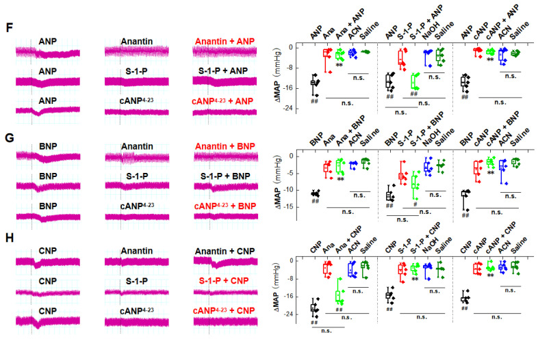
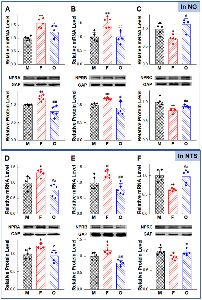
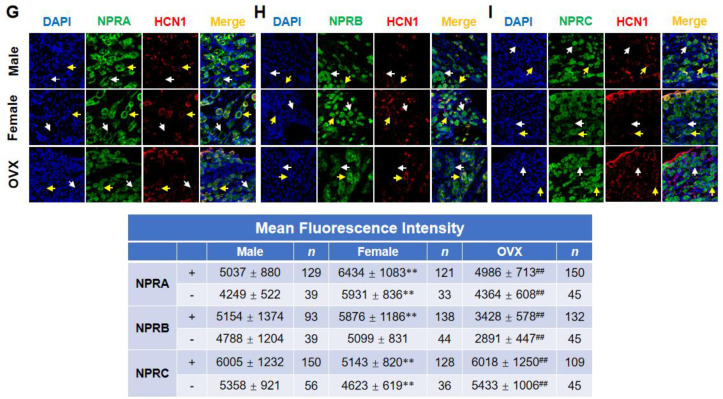
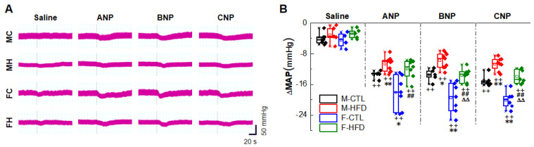
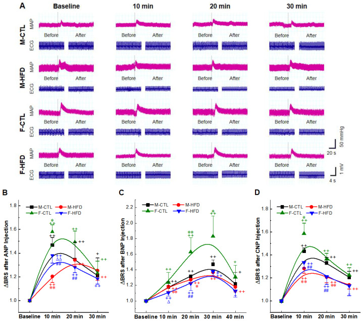
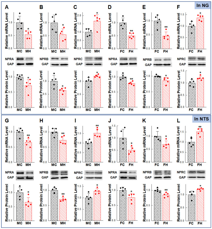
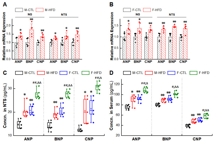
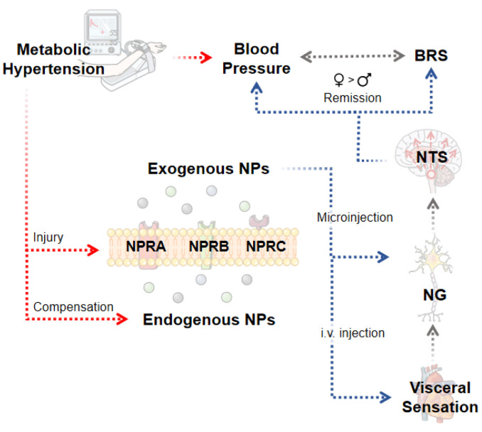
Natriuretic peptides (NPs) induce vasodilation, natriuresis, and diuresis, counteract the renin-angiotensin-aldosterone system and autonomic nervous system, and are key regulators of cardiovascular volume and pressure homeostasis. Baroreflex afferent pathway is an important reflex loop in the neuroregulation of blood pressure (BP), including nodose ganglion (NG) and nucleus tractus solitarius (NTS). Dysfunction of baroreflex would lead to various hypertensions. Here, we carried out functional experiments to explore the effects of NPs on baroreflex afferent function. Under physiological and hypertensive condition (high-fructose drinking-induced hypertension, HFD), BP was reduced by NPs through NG microinjection and baroreflex sensitivity (BRS) was enhanced via acute intravenous NPs injection. These anti-hypertensive effects were more obvious in female rats with the higher expression of NPs and its receptor A/B (NPRA/NPRB) and lower expression of its receptor C (NPRC). However, these effects were not as obvious as those in HFD rats compared with the same gender control group, which is likely to be explained by the abnormal expression of NPs and NPRs in the hypertensive condition. Our data provide additional evidence showing that NPs play a crucial role in neurocontrol of BP regulation via baroreflex afferent function and may be potential targets for clinical management of metabolic-related hypertension.

Keywords: baroreflex afferent function; blood pressure regulation; gender difference; high-fructose induced hypertension; natriuretic peptide.


Conflict of interest statement

The authors declare no conflict of interest.

Figures

Figure 1
Figure 1
Natriuretic peptides (NPs)—mediated blood pressure (BP) reduction by nodose ganglion (NG) microinjection under physiological condition. (A) Real-time monitoring of mean arterial pressure (MAP) changes with the dotted line as the starting point of administration (ANP 1 mg/mL, saline solution, 3 µL). (B) Quantitative analysis of BP changes (the difference between nadir after NPs injection and initial MAP). Averaged data was presented as mean ± SD. ** p < 0.01 vs. male rats; ## p < 0.01 vs. female rats; ΔΔ p < 0.01 vs. saline-injection control rats; n = 5–8, from 6 rats for male and female group, and 5 rats for the OVX group. (C) Effects of different concentrations of ANP (1 mg/mL, 3 µL) microinjection in the male rats. Averaged data was presented as mean ± SD. * p < 0.05, ** p < 0.01 vs. male rats, # p < 0.05, ## p < 0.01 vs. female rats, n = 5 rats for each group. (D,E) Dose-response curve of ANP microinjection. EC50 values were calculated in male, female, and ovariectomized (OVX) rats. Averaged data were presented as mean ± SD. (FH) NG microinjection was performed in male group. ANP/BNP/CNP 1 mg/mL, saline solution; anantin 1 mg/mL, ACN solution; cANP4–23 1 mg/mL, ACN solution; S-1-P 4 mg/mL, NaOH solution; ACN 0.2 mg/mL, saline solution; and NaOH 0.3 mmol/L, saline solution; 3 µL, respectively. Effects of NPRA inhibitor: Anantin, NPRB inhibitor: S-1-P, and NPRC activator: cANP4–23 on the functions of ANP, BNP, and CNP, respectively. The representative bands of MAP fluctuation monitored in real time were shown on the left. Summarized data of microinjection was shown on the right. Averaged data was presented as mean ± SD. ** p < 0.01 vs. NPs-injection group, # p < 0.05, ## p < 0.01 vs. saline-injection control group, n.s., no significant difference. n = 5 rats for each group.
Figure 1
Figure 1
Natriuretic peptides (NPs)—mediated blood pressure (BP) reduction by nodose ganglion (NG) microinjection under physiological condition. (A) Real-time monitoring of mean arterial pressure (MAP) changes with the dotted line as the starting point of administration (ANP 1 mg/mL, saline solution, 3 µL). (B) Quantitative analysis of BP changes (the difference between nadir after NPs injection and initial MAP). Averaged data was presented as mean ± SD. ** p < 0.01 vs. male rats; ## p < 0.01 vs. female rats; ΔΔ p < 0.01 vs. saline-injection control rats; n = 5–8, from 6 rats for male and female group, and 5 rats for the OVX group. (C) Effects of different concentrations of ANP (1 mg/mL, 3 µL) microinjection in the male rats. Averaged data was presented as mean ± SD. * p < 0.05, ** p < 0.01 vs. male rats, # p < 0.05, ## p < 0.01 vs. female rats, n = 5 rats for each group. (D,E) Dose-response curve of ANP microinjection. EC50 values were calculated in male, female, and ovariectomized (OVX) rats. Averaged data were presented as mean ± SD. (FH) NG microinjection was performed in male group. ANP/BNP/CNP 1 mg/mL, saline solution; anantin 1 mg/mL, ACN solution; cANP4–23 1 mg/mL, ACN solution; S-1-P 4 mg/mL, NaOH solution; ACN 0.2 mg/mL, saline solution; and NaOH 0.3 mmol/L, saline solution; 3 µL, respectively. Effects of NPRA inhibitor: Anantin, NPRB inhibitor: S-1-P, and NPRC activator: cANP4–23 on the functions of ANP, BNP, and CNP, respectively. The representative bands of MAP fluctuation monitored in real time were shown on the left. Summarized data of microinjection was shown on the right. Averaged data was presented as mean ± SD. ** p < 0.01 vs. NPs-injection group, # p < 0.05, ## p < 0.01 vs. saline-injection control group, n.s., no significant difference. n = 5 rats for each group.
Figure 2
Figure 2
Gender-related differential expression of natriuretic peptide receptors (NPRs) in the nodose ganglia (NG) and nucleus tractus solitarius (NTS). (AC) mRNA and protein expression of NPRs in NG of adult male, age-matched female, and OVX rats. * p < 0.05, ** p < 0.01 vs. male rats, # p < 0.05, ## p < 0.01 vs. female rats, n = 5. (DF) mRNA and protein expression of NPRs in NTS of adult male, age-matched female and OVX rats. * p < 0.05, ** p < 0.01 vs. male rats, # p < 0.05, ## p < 0.01 vs. female rats, n = 5. (GI) Representative immunostaining for NPRA, NPRB, and NPRC; the hyperpolarization-activated channel specifically expressed on myelinated afferents (HCN1-positive) and neurons were labeled by the antibodies against DAPI (blue), HCN1 (red), and NPRs (green). The yellow arrows pointed to HCN1 positive cells, and the white arrows pointed to HCN1 negative cells. Magnification power was 200×. The bottom table: Quantization results of immunohistochemical staining of NPRs. Data was shown as mean ± SD. ** p < 0.01 vs. male rats, ## p < 0.01 vs. female rats, from 4–5 slices.
Figure 2
Figure 2
Gender-related differential expression of natriuretic peptide receptors (NPRs) in the nodose ganglia (NG) and nucleus tractus solitarius (NTS). (AC) mRNA and protein expression of NPRs in NG of adult male, age-matched female, and OVX rats. * p < 0.05, ** p < 0.01 vs. male rats, # p < 0.05, ## p < 0.01 vs. female rats, n = 5. (DF) mRNA and protein expression of NPRs in NTS of adult male, age-matched female and OVX rats. * p < 0.05, ** p < 0.01 vs. male rats, # p < 0.05, ## p < 0.01 vs. female rats, n = 5. (GI) Representative immunostaining for NPRA, NPRB, and NPRC; the hyperpolarization-activated channel specifically expressed on myelinated afferents (HCN1-positive) and neurons were labeled by the antibodies against DAPI (blue), HCN1 (red), and NPRs (green). The yellow arrows pointed to HCN1 positive cells, and the white arrows pointed to HCN1 negative cells. Magnification power was 200×. The bottom table: Quantization results of immunohistochemical staining of NPRs. Data was shown as mean ± SD. ** p < 0.01 vs. male rats, ## p < 0.01 vs. female rats, from 4–5 slices.
Figure 3
Figure 3
Blood pressure (BP) reduction effects of natriuretic peptides (NPs) under hypertensive condition of high-fructose-drinking-induced hypertension (HFD) rat models. (A) Real-time monitoring of BP changes (NPs 1 mg/mL, 3 µL). The representative recordings of mean arterial pressure (MAP) were collected from male control rats (MC), male-HFD rats (MH), female control rats (FC), female-HFD rats (FH). (B) Quantitative analysis of changes in MAP. Averaged data was presented as mean ± SD. * p < 0.05, ** p < 0.01 vs. M-CTL with the same treatment, ## p < 0.01 vs. F-CTL with the same treatment, ∆∆ p < 0.01 vs. M-HFD with the same treatment, ++ p < 0.01 vs. saline-injection control group, n = 5–11, from 5 rats for M-HFD and F-HFD group, 6 rats for M-CTL group, and 7 rats for F-CTL group.
Figure 4
Figure 4
The baroreceptor sensitivity (BRS) improvement effects of natriuretic peptides (NPs) administration in high-fructose-drinking-induced hypertension (HFD) rats. NPs 1 mg/mL, 25 µg/kg was administrated intravenously (i.v.). (A) The mean arterial pressure (MAP) and heart rate (HR) were recorded in the presence of ANP before and after intravenous injection of 3 µg/kg phenylephrine (PE). The BRS was presented as ΔHR/ΔMAP (bpm/mmHg). (BD) Quantitative analysis results of BRS. Averaged data was presented as mean ± SD. * p < 0.05, ** p < 0.01 vs. M-CTL with the same treatment, # p < 0.05, ## p < 0.01 vs. F-CTL with the same treatment, p < 0.05, ∆∆ p < 0.01 vs. M-HFD with the same treatment, + p < 0.05, ++ p < 0.01 vs. baseline, n = 5 rats for each group.
Figure 5
Figure 5
Abnormal expression of natriuretic peptides receptors (NPRs) in nodose ganglion (NG) and nucleus tractus solitarius (NTS) under hypertensive condition. Expression changes in NPRs were verified in NG (AF) and NTS (GL) of high-fructose-drinking-induced hypertension (HFD) models of adult male and female rats. Averaged data was presented as mean ± SD. GAPDH means the internal control. * p < 0.05, ** p < 0.01 vs. male or female control rats, n = 5.
Figure 6
Figure 6
Upregulation of expressions of natriuretic peptides (NPs) in nodose ganglion (NG) and nucleus tractus solitarius (NTS) of high-fructose-drinking-induced hypertension (HFD) rat models. (A,B) mRNA expression of NPs. Averaged data was presented as mean ± SD. GAPDH means the internal control. * p < 0.05, ** p < 0.01 vs. male or female control rats, n = 5. (C,D) Concentrations of NPs measured with ELISA assay. Averaged data was presented as mean ± SD. * p < 0.05, ** p < 0.01 vs. M-CTL, ## p < 0.01 vs. F-CTL, p < 0.05, ∆∆ p < 0.01 vs. M-HFD, n = 5.
Figure 7
Figure 7
Schematic diagram explaining natriuretic peptides (NPs)—mediated anti-hypertensive effects via baroreflex afferent pathway. This study confirmed that microinjection of NPs in the nodose ganglion (NG) can lower blood pressure under both physiological and pathophysiological status. Meanwhile, exogenous NPs can perform baroreceptor sensitivity (BRS) improvement effect. These anti-hypertensive functions and the expression of natriuretic peptide receptors (NPRs) were closely related to estrogen. Abnormal expression of NPs and its receptors in nodose ganglion (NG) and nucleus tractus solitarius (NTS) in models of hypertension with metabolic disorders suggests a negative feedback regulation.

Similar articles

References

    1. Misono K.S., Philo J.S., Arakawa T., Ogata C.M., Qiu Y., Ogawa H., Young H.S. Structure, signaling mechanism and regulation of the natriuretic peptide receptor guanylate cyclase. FEBS J. 2011;278:1818–1829. doi: 10.1111/j.1742-4658.2011.08083.x. - DOI - PMC - PubMed
    1. De Bold A.J. Atrial natriuretic factor: A hormone produced by the heart. Science. 1985;230:767–770. doi: 10.1126/science.2932797. - DOI - PubMed
    1. Sudoh T., Kangawa K., Minamino N., Matsuo H. A new natriuretic peptide in porcine brain. Nature. 1988;332:78–81. doi: 10.1038/332078a0. - DOI - PubMed
    1. Sudoh T., Minamino N., Kangawa K., Matsuo H. C-type natriuretic peptide (CNP): A new member of natriuretic peptide family identified in porcine brain. Biochem. Biophys. Res. Commun. 1990;168:863–870. doi: 10.1016/0006-291X(90)92401-K. - DOI - PubMed
    1. Goetze J.P., Bruneau B.G., Ramos H.R., Ogawa T., de Bold M.K., de Bold A.J. Cardiac natriuretic peptides. Nat. Rev. Cardiol. 2020;17:698–717. doi: 10.1038/s41569-020-0381-0. - DOI - PubMed

Substances